Page 1 :
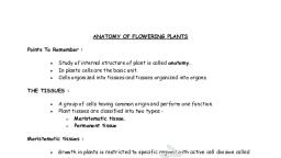
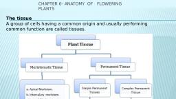
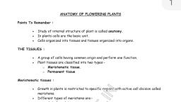
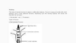
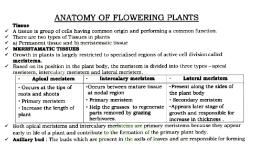
4, PLANT ANATOMY, , , , «+ Plant anatomy deals with the study of gross internal structure of plant., * Basic unit of anatomy is tissue., + Term tissue was used by N. Grew (known as father of plant anatomy)., , * A group of structurally similar or dissimilar cells that perform or help to perform a common function, and have a common origin is called a tissue., , fe, , “, , Tissues can be conveniently grouped in two headings:, A. Meristematic tissues, B. Permanent tissues, , Meristematic Tissue, , + Meristematic tissue is made up of group of immature cells that are preparing to divide or are in, continuous state of division., , + Term meristem was coined by C. Nageli, (Gr. Meristos = divisible)., + Meristem are found in growing regions of plants., , + Meristem are ultimate source of all tissue in plant., , + Chief characteristics of meristem are:, , Scanned with CamScanner
Page 2 :
« Cells may be round, oval, polygonal or rectangular but isodiametric (equal size), ¢ Have thin cellulosic wall with dense cytoplasm & conspicuous nuclei., , e Donothave intercellular spaces and reserve food material., , ¢ Cells are in active state of metabolism., , e Chloroplast and chromoplasts are absent but leucoplast may be present., , e Vacuoles are either small or absent., , e ERis small and nucleocytoplasmic ratio is very high., , Types of meristem, A. On the basis of origin and development, meristems can be promeristem, primary meristem and, secondary meristem., , 1. Promeristem (= Primordial meristem or Ur-meristem or Embryonic meristem):, ¢ Represent primary stages of meristematic cells., ¢ Plant embryo truly represents this kind of meristem. [Ind. Emb], ¢ They give rise to primary meristems., 2. Primary meristems:, ¢ Cells are always in active state of division and give rise to primary permanent tissues., , ¢ They are found below the promeristem at shoot and root apices, at the apex of leaves, in, intercalary parts and intrafascicular cambium in dicot stems., , 3. Secondary meristems:, , ¢ Develops from primary permanent tissue at a later stage and give rise to secondary permanent, tissues., , ¢ Examples: cambium of roots, interfascicular cambium of stem, wound cambium and cork, cambium in stem as well as root., , B. On the basis of position on plant body; Figure: Meristem based on position, meristematic tissues can be divided into apical,, intercalary & lateral meristem., , 1. Apical meristems:, , , , , , Apical meristem, , « These tissues are found at apices of stem and, root., , ¢ Responsible for increase in length, ie, they, form growing points at the apices of roots tercalary meristem and, stems., , 2. Intercalary meristems:, , ¢ These tissue are intercalated in between the, , permanent tissues., 3 ‘ ‘ Lateral meristem :, , ¢ Originate from the apical meristems during, development [Ind Emb, IOM 1997]., , ¢ May be present at the base of internodes (i.e,, , above node) e.g. in grasses (Gramineae) | or at, the base of leaves e.g. in monocots and Pinus or at the base of node e.g. mint or Mentha., , ¢ Aactivity of these meristems also add to the length of the plant or its organs., , | eee SS SSS, , i, i'¢ Axillary and terminal bud are formed from apical meristem., , Scanned with CamScanner
Page 3 :
2, —, , Leaf of pinus is evergreen due to intercalary meristem on leaf base., , Increase in length in grass is due to intercalary meristem |, , The process of conversion of meristematic tissue into permanent tissue is called differentiation and:, the reverse process is called de-differentiation., , “, , Example of de-differentiation: Formation of cambium (2° meristem) from medullary ray), , fe, , 9g, , (permanent tissue) |, , i, !, , Plastochoron: time gap between two successive primordia _, , i &, i, , prmeeresree:, , 3. Lateral meristem:, ¢ Present along the lateral sides of stem and roots., « Responsible for increase in girth of stem and roots., ¢ Example: intrafascicular, interfascicular cambium and cork cambium (phellogen)., , Root and shoot apex organization:, , ,, , ** Several theories have been put forward to explain the activity of apical meristems:, 1. Apical cell theory (By Nageli), « According to this theory, activity of single apical cell leads to development of entire plant body., , ¢ Applicable to higher algal forms, bryophytes and some pteridophytes but is certainly not, applicable to the seed plants (spermatophytes)., 2. Histogen theory (By Hanstein), « According to this theory, cells of root and shoot apices organized into three distinct zones(, called histogen)., , , , , , Embryonic leaf, , Dermatogen: Gives epidermis, , Periblem: Gives cortex and Endodermis, , Plerome: Gives Stele, , Figure: Types of Histogen on Shoot apex, , Scanned with CamScanner
Page 4 :
a. Plerome (central core): form stele., , b. Periblem (several layer surrounding plerome): form hypodermis, cortex and, endodermis., , c. Dermatogen (outermost single layer): form epidermis., , Sa aa a ee as aa aaa nee ee a 3, , In case of root apex Hanstein proposed one more histogen, i.e, Calyptrogen which is responsible for, bie ener Oo Oe i, , 3. Tunica-corpus theory (By Schmidt), , +, , This theory recognizes only two zones in the apical meristems, i.e, outer tunica and central, corpus., , According to this view epidermis is derived from outer layer of tunica and the remaining, tissues are derived from remaining layer of tunica and entire corpus., , Cells of the tunica divide anticlinally only and help to increase the extent of surface., Cells of corpus divide in all planes and contribute to the depth and mass of inner tissues., , oo, x oo,, , ox, , , , , primordia, Axillary bud, , Ground meristem, Procambium, , Protoderm, , Figure: Diagram showing organization of shoot apical cells as Tunica and Corpus, , 4. Quiescent centre concept (By Clowes), , ¢, , +, , +, , +, , Discovered by Clowes in root tip of Zeamays., , It is cup like region of passive cells lying between the root cap and active root meristem., Cells of this region normally remain inactive and serve as reservoir to meristematic zone., Cells of quiescent centre are characterized by having low DNA, RNA and protein contents., , Based on function; meristematic tissue can be divided into following three types., 1. Protoderm: makes epidermal tissue system., , 2. Ground: makes ground tissue and pith., , 3. Procambium: makes primary vascular tissues., , Based on plane of division, meristem can be divided into following types., , 1. Mass meristem: cells divides in three plane increasing volume and growth, e.g. early, growth of embryo, cortex, pith etc., , 2. Plate meristem: cells divides in two plane so that plate like area is increased, e.g. formation, of epidermis, endodermis, , 3. Rib or file meristem: cells divides in one plane to form row of cells causing increase in, length, e.g. lateral root formation, , Scanned with CamScanner