Notes of NEET,JENPUS &ETC BATCH, Biology Neural control.pdf - Study Material
Page 1 :
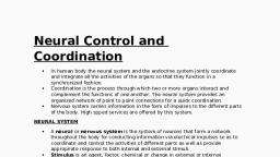
Neural Control and Coordination, , , , , , In all the multicellular animals above the level of sponges, the, system meant to perceive stimuli detected by the receptors, to, transmit these to various body parts, and {o effect responses, through effectors, is called nervous system, In vertebrates, it is, highly specialized and plays at least three vital roles, , (1) Sensory function : It senses certain changes (stimuli), both within body (internal environment) and out side body, (external environment),, , (2) Integrative functions : It analyses the sensory, information, store some aspects, and make decisions regarding, appropriate behaviour., , (3) Motor functions : It may respond to stimull by initiating, muscular contractions or glandular secretions,, , Nervous systems in various animals, , (1) Coelenterata : True nerve cell or ganglion cells occur for, the first time in coelenterates. They are derived from interstitial cells, of epidermis, forming nerve net or nerve plexus below whole, epidermis. A polar neurons are found in coelenterata., , (2) Platyhelminthes : Nervous system of planarians marks, the beginning of a centralized nervous system encountered in, higher animals, That is made up of brain or cerebral ganglia, two, Jateral longitudinal nerve chords, numerous peripheral nerves and, transverse commissures or connectives. This is sometimes called, the ladder type of nervous system., , In Nematoda (eg. ascaris) these system made up of central, nervous system, peripheral nervous system and rectal nervous, system. Rectal nervous system more developed in male, Ascaris, with dorsal, ventral, and lateral nerve cords., , (3) Annelida : Nervous system well developed and, concentrated. It consists of three parts : central nervous system,, peripheral nervous system and sympathetic nervous system, central, N.S, made up of Nerve ring and ventral nerve cord. Nerves are of, mixed type, consisting of both afferent (sensory) and efferent, (motor) fibres, , (4) Arthropoda : The nervous system of prawn or arthropod), is of the annelidan type. However it is somewhat larger and has, ‘more fusion of ganglia, It consists of (i) TWv2 central nervous system, including brain connected with a ventral. ganglionated nerve cort!, through a pair of circum-oesophage.al commissures, (ii) The, Peripheral nervous system including nerves and (ii) The, sympathetic nervous system. In arthropods like cockroach, sympathetic nervous system also known as stomatogastric nervous, system, made up of 4 ganglion and retro-cerebral complex., , , , (5) Moltusea : In gastropodes (e.g. pila) consists of paired, ganglia, commissures and connective uniting them and nerves, Tunning from these central organs to all parts of the body. It has, various type of ganglia as cerebral, buccal, pleuro-pedal, supra, Intestinal and visceral ete, In palecypoda nervous system is greatly, reduced due to sluggish and sedentary mode of life and there is, litle evidence of the brain, But in cephalopoda shows a high grade, of organization attained only by some insects and arachnids, among the other invertebrates.’, , (6) Echinodermata : Echinodermates has simple and, primitive type of nervous system. It has the form of a nerve net,, consisting of nerve fibres and a few ganglion cells, all confined to, the body wall except the visceral nerve plexus situated in the gut, wall. At certain places the nervous tissue is concentrated to form, distinct nerve cords. It is made up of (I) Superficial or ectoneural, nervous system (ii) Hyponeural or deep nervous system (Ii) Aboral, (0 coelomic nervous system and (iv) Visceral nervous system., , (7) Hemichordata : Nervous system is of primitive type, resembling that of coelenterates and echinodermates, with both, dorsal and ventral nerve cord., , (8) Chordates : Nervous system well developed and formed, by ectoderm. It is formed by central nervous system, peripheral, nervous system and autonomic nervous system.
Page 2 :
Development of central nervous system in, human, , , , Nervous system begins developing early in third week of, development from ectoderm. Nervous tissue also develop from, ectoderm except microglial cell, develop from mesoderm. The, central nervous system of vertebrates includes the brain and the, spinal cord. These are derived from a longitudinal mid-dorsal, ectodermal thickening of the embryo, called the medullary or, neural plate. This neural plate or neural groove is converted by, fusion into a closed mid-dorsal longitudinal neural tube ying, above the notochord. Histologically, the embryonic neural tube, exhibits three zones of cell,, , Future neural crest, , Neural plate, , Ectoderm, , , , e ', eked “47 Masada atin, , Notochord \, , Neural groove Endoderm, , Fig : 5.6-1 Stages in the embryonic development of central, nervous system, , (2) Germinal layer : These are actively dividing cells lining, the neural canal, They form the connective tissue lining of neural, canal, called ependyma, and ventricles of brain., , (2) Mantle layer : It consists of embryonic neurons or, nematoblasts, forming the grey matter., , (3) Marginal layer : It consists of nerve fibres, mostly, surrounded by fatty myelin sheaths, and forms the white matter., Neurons and fibres are supporied by a special connective tissue of, ectodermal origin, the neurogiia, cells of which become, increasingly abundant and diversified in higher vertebrates., Development of brain, , ‘The anterior end of embryonic neural tube is already enlarged, forming the embryonic brain, called encephalon. By differential, growth and two constrictions, it is divided into a linear series of, three primary cerebral vesicles, termed the forebrain, midbrain and, hindbrain, These give rise to the three major divisions of the adult, , ‘brain - (1) prosencephalon (forebrain), (2) mesencephalon, (midbrain), and (3) rhombencephalon (hindbrain). These further, become subdivided into 5 subdivisions. Prosencephalon divides, into an anterior telencephalon and posterior diencephalon; the, mesencephalon remain unchanged. The rhombencephalon divides, into an anterior metencephalon and a posterior myelencephalon., Ultimately, telencephalon develops into cerebral hemisphere and, basal ganglia and houses lateral ventricle. Diencephalon develops, into thalamus, hypothalamus, and pineal gland and houses the, third ventricle. Mesencephalon develops into mid brain and houses, cerebral aqueduct. Metencephalon develops into pons and, cerebellum; and myelencephalon develops into medulla oblongata,, houses 4° ventride. The area of neural tube inferior to, myelencephalon gives tise to spinal cord., , , , , , Prosencephalon, {forebrain}, Mesencephalon, (midbrain), Rhombencephalon, ) (hindbrain), Frontal section, Rhombencephalon, (hindbrain), , , , , , (midbrain), rosencephalon, plist), , loping eye, loping, , heart, , Spinal cord, Lateral view of right side, Cerebral hemisphere, Lateral ventridle ‘Teloncephalon, Optic vesicle Diencephalon, ‘Third ventricle 1, neal ened ‘Mesencephalon, nen (midbrain), =} Metencephalon, Fourth ventricle Myelencephalon, , Developing ear, ‘Metencephalon, ‘Mesencephalon, Diencephalon, ‘Telencephalon, Developing eve, , , , Lateral view of right side, Fig : 5.6-2 Development of the brain and spinal cord
Page 3 :
Parts of nervous system, , Nervous system is divided into three parts ~, Nervous system, , Central Nervous Peripheral Nervous Autonomic Nervous, ‘System (CNS) ‘System (PNS) System (ANS), F, Brain Spinal Cranial -~— ‘Spinal Sympathetic Parasympcord Nerves. Nerves lervous _athetic, (All Mixed) System Nervous, ‘System, Sensory Nerves (I, __ Motor Nerves Mixed Nerves, 1, vin) (U,V, VI, XL, XM) (V, Vil, IX, X), , Central nervous system (CNS), , Inall the vertebrates including man, CNS is dorsal, hollow and, non-ganglionated while in invertebrates when present, it is ventral,, id, double and ganglionated. CNS is formed of two parts :, , (1) Brain - Upper and broader part lying in the head., , (2) Spinal cord - Lower, long and narrow part running from, beginning of neck to trunk. CNS is covered by 3 meninges and its, wall has two type of matter., , ‘Types of matter : CNS of vertebrates is formed of two types, of matter —, , , , (i) Grey matter : It is formed of cell-bodies, non-medullated, nerve fibres, neurogla, dendrites of association neurons and motor, neurons., , (i) White matter : It is formed of medullated nerve fibres o, myelinated axon of motor and sensory neurons, which appear, white due to the presence of medullary sheath,, , Meninges : The meninges are connective tissue membranes, which surround the brain and spinal cord of CNS. In the fishes,, there is only one meninx called meninx primitiva (piamater), In, amphibians, reptiles and birds, the brain is covered by two, meninges or membranes : inner pia-arachnoid and outer, , duramater. In mammals, CNS is covered by three meninges ot, membranes or cranial meninges. Brain meninges are continuous, with spinal meninges, , Superior sagittal sinus, , , , cranium, , Skin, Parietal bone of 4, ——Duramater > 8, , Arachnoid mo E, , ‘Subarachnoid., , ech Pia mater 2, , ‘rachnoid &, mein Cerebral, Fels cerebri comes, , Fig: 5.6-3 Meninges of brain, , , , Neural Control and Coordination 947, , ‘The three layers of cranial meninges in order from superficial, to deeper duramater, arachnoid and piamater. Duramater is, nonvascular, tough made up of fibrous connective tissue,, Arachnoid mater made up of reticular connective tissue with, collagen and elastin fibre, while innermost vascular piamater, (nutritive) made up of loose areolar connective tissue. Between, dura and arachnoid mater presence of sub dural space (no CSF in, mammals here}, between Arachnoid and piamater presence of, sub-arachnoid space (with CSF in mammals, CSF also found in, ventricles and central canal). Between duramater and periosteum, presence of epidural space. An extension of duramater between, ‘two cerebral hemispheres is called falx cerebri, Tentorium, an, extension of duramater between cerebrum and cerebellum., , Cerebrospinal fluid : All the ventricles of the brain, central, canal of spinal cord are continuous and lined by a columnar,, ciliated epithelium, the ependyma, They contain Iymphslike, extracellular fluid called the cerebrospinal fluid (C.S.F.). This fluid, is secreted by the choroid plexuses by filtration of blood. The, choroid plexuses consist of loose connective tissue of pia mater, covered internally by a simple cuboidal epithelium of secretory, (glandular) nature, The cerebrospinal fluid slowly flows toward the, fourth ventricle by secretion pressure and passes into the spinal, cord. Some fluid escapes into the subarachnoid spaces through, three pores a median aperture (of magendie) and a paired lateral, aperture (of Luschka) in the roof of the fourth ventricle in the, medulla. From the subarachnoid spaces, the cerebrospinal fluid is, transferred (o the blood of the venous sinuses, Nervous tissue is without, Iymphatic vesees, , The cerebrospinal fluid (CSF) provides ~, , (i) Protection to brain from mechanical shocks, physical injury., , (ii) Optimum physiological fluid environment for neural, functions eg. conduction of nerve impulses, transport of, aminoacids, sugars, O, etc,, , (ii) ‘Relief mechanism for the increase in intracranial pressure, that occurs with each arterial pulse of blood to brain., , {iv} ‘Sink like facility for metabolites of brain,, , (v) The blood CSF barrier for selective transport process, between blood and CSF., , (vi) Nourishment to CNS., , Major site of CSF formation is choroid plexus, and mid, ventricular wall and sub-arachnoid wall also contribute. CSF is cell, free, slightly alkaline, and is isotonic to plasma. Rate of formation, of C.S.F is 20 milh (480 mifday) approx, 1/2 litre per day. Total, amount present in and around CNS is 80-150 ml it means there is, atleast 3 times renewal of C.S.F. every day. CSF contains glucose,, proteins, lactic acid, urea, Na’, K°, Ca’, Mg'*, Cr, HCO, and, some WBC., , Blood brain barter faciltate maintenance of stable internal, environment, It acts as physiological and pathological barter., , , , Q Hydrocephalus : The enlargement of head, a, pathological condition characterized by an abnormal accumulation, of cerebrospinal fluid resulting in headache, vomiting, pain and, sliffness of the neck,
Page 4 :
948 Neural Control and Coordination, , , , , , 1 Increased cerebrospinal fluid may result in Meningitis., , 1 Meningitis may occur due to infection and inflamation of, meninges or injury of meninges., , 1 Infection may be viral, bacterial or both. The most, common cause of meningitis is the infection of Streptococcus, pneumoniae, Neisseria meningitidis and Haemophilus influenzae., , 1 Lumbar puncture is done for drainage of excess of, cerebrospinal fluid during meningitis., , O Cerebro-spinal fluid is formed by choroid plexus (ACP, and PCP)., , ‘There are three choroid plexus in humans —, , , , (i) Lateral choroid plexus : It is in the roof of I and Il, ventricle., , (ii) Anterior choroid plexus : It is in the roof of III ventride, {diocoel)., , (ii) Posterior choroid plexus or pelochoroida, the roof of IV ventricle, , Oxygen and glucose requirements : Brain controls the, functions of our body organs and also provides the qualities of, mind — learning, reasoning, and memory. For these activities, brain, needs a large and constant energy supply. At any given time, the, activites of the brain account for 20% of the body's consumption, of oxygen and 15% of its consumption of blood glucose. Brain, deprived of oxygen for just 5 minutes is permanently damaged., Mental confusion results if brain is deprived of glucose., , Structure of human brain (Encephalon), , It is soft, whitish, large sized and slightly flattened structure, present inside cranial cavity of cranium of the skull. In man, itis, about 1200-1400 gm in weight and has about 10,000 million, neurons, Brain is made up of 3 parts, , (1) Fore brain or Prosencephalon : It forms anterior, tworthied of brain and is formed of three parts., , Itis in, , , , , , Cerebrum, , fe sine, thalamus, |Hypothalamus, Pineal gland, Midbrain, , Brain ster Pons, Medulla oblongata, Cerebellum -—, , Spinal cord, , , , , Diencephalon, , , , , , Posterior, , Fig : 5.6-4 Main parts of human brain visible fram lateral view, , Olfactory lobes : These are one pair, small sized, dubshaped, solid, completely covered by cerebral hemisphere dorsally,, Each is differentiated into two parts —, , (i) Olfactory bulb : Anterior, swollen part, and, , (ii) Olfactory tract : Posterior and narrow part which ends in, olfactory area of temporal lobe of cerebral hemisphere., , Function : These control the smell,, , (2) Itis normal in frog, rabbit and man., , {b) Itis well developed in dog. So power of smell is more in dog,, , (c) These are also well developed in dog fish and name dog, fish is on the basis of well developed olfactory lobes., , Cerebrum : Cerebrum is divided into 5 lobes (a) frontal (b), parietal, (c) occipital, (d) temporal and (e) Insula. A lobe called, insula is hidden as it lies deep in the sylvian fissure. The cerebral, hemisphere are separated from olfactory lobes by rhinal fissure., The median fissure divides the cerebrum into a right and a left, cerebral hemisphere., , A few sulci are well developed and form three deep and wide, fissures which divide each cerebral hemisphere into four lobes, anterior frontal lobe, middle parietal lobe, posterior occipital lobe, and lateral temporal lobe eg. Fissure sulcus lying between the, frontal and parietal lobes is central fissure or sulcus, that is lying, between the parietal and occipital lobes is parieto-occipital fissure, and that demarcating frontal and parietal lobes from the temporal, lobe is lateral or Sylvian fissure. Each cerebral hemisphere is with a, fluid-filled cavity called lateral ventricle or paracoel., , ‘Two cerebral hemispheres are interconnected by thick band of, transverse nerve fibres of white matter called corpus callosum. The, peripheral portion of each cerebral hemisphere is formed of grey, ‘matter and is called cerebral cortex, while deeper par is formed of, white matter and is called cerebral medulla, Cerebral cortex is the, highest centre for many sensations and activities and is with a, number of sensory areas. Cerebral cortex 2-4 mm thick., , , , , , Histology of cerebrum : The whole brain possess grey, matter outside and white matter inside around ventricle., , () Grey matter : In cerebrum grey matter is very much, developed, it is on an average 2-4 mm. thick but at poles its, thickness is 1.3 mm. It is thickest at pre central gurus (4.5 mm, thick). Grey matter of cerebrum is called cortex or pallium., Phyllogenetically or evolutionarily cortex is divided into 3 parts —, , (a) Allocortex or paleocortex : It is the cortex of olfactory, area of frontal lobe and olfactory bulbs. In lower vertebrates, (cartilagenous fish) olfactory lobes occupy most of the part of, cerebrum. So in these animals sense of olfaction is very-very much, developed. Sense of olfaction is oldest sense., , (b) Mesocortex : It is relatively not much older in development., , (Q) Neocortex or neopallium or, neencephalon = It is most recent cortex and is developed, maximum only in human. tt is in prefrontal cortex or prefrontal, region (organ of mind), precentral and postcentral qyrus etc. The, neocortex is having 6 layer of neurons while remaining cortex, possess only 5 layers., , isocortex or, , The cerebral cortex is having area of about 2200 cm? while, the cranial cavity is only 1450 cm®, so to accomodate cerebrum, there appears foldings in the cortex. The ridges are called gyrus, (or gyri) or convolution while the depression are called sulcus, {sulci in plural)., , (ii) White matter : It is inner part of brain, White matter is, aggregation of myelinated and unmyelinated axons of many, neurons. Its fibres are divided into 3 categories
Page 5 :
ural Control and Coordination 949, , , , , , , , (a) Commissural fibres : These neurons connect gyri of 2, hemispheres, such as corpus callosum. habenular commissure,, anterior commissure, posterior commissure,, , (b) Associate fibres : They connect gyri of same hemisphere., , (c) Projection neuron : They are infact ascending and, descending nerve tract, they connect one part of brain to another part, of brain or fo spinal cord. (In spinal cord they were called as column),, , Anterior, , , , , , , , , ~ Frontal, Longitudinal fssure, , Gus Precentral a, Parietal, lobe, , Occipital lobe, , IM peace Fight hemisphere, , Left hemisphere, , (a) {b), Fig : 5.6-5 (a) Details of gyrus, sulcus and fissure, (b) Posterior, superior view of cerebrum, Associated structures of cerebrum : Cerebrum has, following specific structure,, (i) Sub cortex : Nuclei on white matter. It is cluster of grey, , Neurons in depth of white matter, they are formed in whole brain, and are named differently., , , , Basal ganglia or central nucleus : These are several, ‘groups of nuclei in each cerebral hemisphere., , Corpus siriatum : Corpus striatum is the largest nucleus,, consist of caudate nucleus and lenticular nucleus. The lenticular, nucleus is sub-divided in putamen (outer shell) and globus pallidus, (ball). Other structure, functionally linked to and some times, considered part of basal ganglia are :, , (a) Claustrum : It is the name given to grey matter present, between insula and putamen,, , , , , , , Hippocampus (in temporal lobe) ~, Dentate gyrus:, , Posterior, , Sagittal section, , (b) Epistriatum or Amygdaloid body : It is structure, present at the end of caudate nucleus,, , {c) Red nucleus and substantia nigra of mid brain., (d) Sub thalamic nuclei of diencephalon., , , , , , , Longitudinal fissure, , Septum pellucidum—_|, Interal capsule, , Compa siatum, , Caudate nucleus, Thala tamien, Subthalamic nucleus 5 Globus pallidus, Hypothalamus: 9 — Third ventricle, , Optic tract, Fig: 5.6-6 Basal ganglia, Function of basal ganglia, , () Caudate and putamen control large automatic movements, of skeletal muscle like swinging of arm while walking, , (ii) Globus pallidus control muscle tone for specific body, movernents,, , (ii) Corpus callosum : It is the band of white neurons, present between both cerebral hemisphere and connect them on, medial surface. It is present only in mammal. It has anterior part, genu, middle part trunchus and last part splenium., , Below corpus callosum there are two fused band of white, neurons called fornix. There anterior part is called cohmn and, posterior part is called crura, Between column and genu a, membrane is called septum lucidum or septum pellucidum., ‘Septum lucidum encloses a space called V; or Pseudocoel, because, it is not possessing C.S.F. i.e. why it is called pseudocoel., , (iv) Limbie system : Limbic system present on inner border, of cerebrum and floor of diencephalon, It is also called emotional, brain or animal brain. Limbic system controls emotion, animal, behaviour like chewing, licking, sniffing, docility, tameness,, affection (animals) rage, pain, pleasure, anger, sexual feelings,, fear, sorrow grooming, It has following structure ~, , , , Anterior nucleus of thalamus, , —Cingulate gurus (in frontal lobe}, Anterior commissure, , ‘Mammilary body in hypothalamus, ‘Septal nuclei, , Olfactory bulls, , ‘Amygdala, , Parahippocampal avrus, {in temporal lobe}, , Anterior, , Fig : 5.6-7 Components of limbic system and surrounding structure
 Learn better on this topic