Notes of NEET,JENPUS &ETC BATCH, Biology Morphology of animal.pdf - Study Material
Page 1 :
RANA TIGRINA (The Common Indian Frog), , ‘Systematic position, , Phylum = Chordata, , Sub phylum - Vertebrata or Craniata, Class = Amphibia, , Order - — Salientia or Anura, Genus - Rona, , Species - — tigrina, , Habitat, , Rana tigrina is the most widely distributed species in Northern, India. Generally frogs are found in ponds, tank, pools, dches, etc, However, they may leave their aquatic habitat to come on land to, hunt for their prey, which are mostly insects., , Habits, , (1) Locomotion : (a) Jumping and leaping, (b) Swimming, Absence of neck is helpful in swimming in water and jumping on, land., , (2) Feeding : The adult frog is carnivorous. Tadpole (larva of, frog) is herbivorous., , (8) Croaking : The male frog croaks louder than the females, because of the presence of two vocal sacs in male frog, The vocal, sacs act as resonators. The croaking is mating call to attract the, female frog,, , (4) Hibernation (Winter sleep) : During hibemation frog, , respires through skin (cutaneous respiration) only., , (5) Aestivation (Summer sleep) : During this period frog, takes rest in shady, cool and moist place and recuperates its, energy., , _Morphology of Animals, , (6) Protective Colouration : The frog is capable of, changing its body colour with change in its surroundings. It an not, nly avoid its enemies but can catch its prey unnoticed., , (7) Breeding : The male frog jumps on the female frog and, holds her tightly with the help of his fore-timbs. Gripping of the, female by the male is also very much aided by the presence of, nuptial pads, This sexual embrace is called the amplexus., Fertilization is external. During development, a fish like tailed, tadpole is produced, which respires with the help of gills and feeds, upon vegetable matter., , (8) Moutting : The frog sheds off almost once a month its, skin during its active life in the form of small casting. This, phenomenon is known as moulting or ecdysis., , External morphology, , The dorsal surface of frog is spotted olive green and ventral, pale yellow; this protective colouration help to camouflage, ic., escape the detection by enemies,, , Body division : The body of frog has two main divisions, head and trunk; absence of neck and tail helps both in jumping on, land and swimming in water., , (1) Head : Head is anterior flattened triangular part with, a, wide transverse terminal mouth, a pair of small dorsal extemal, nares, two dorsc-lateral eyes, a mid-dorsal light coloured brow spot, or third eye or pineal body and obliquely placed pigmented, circular tympanum ot ear drum, Eyes are provided with nictitating, membrane for protection, , (2) Trunk : Trunk is the large, oval, flattened main part of, the body. It is differentiated into hard anterior thorax and soft, posterior abdomen. The trunk is provided with a pair of fore and, hind limbs. The fore limbs are shorter and stouter, end in four, digits thumb or pollex is absent. The hind limbs are much larger, and muscular than the fore limbs, end in five digits
Page 2 :
Morphology of Animals 467, , , , , , ‘Sexual dimorphism, , The male and female frogs can be differentiated externally, The male frog possesses vocal sacs, which are most developed, during the breeding season. During the breeding season an, amplexusory or nuptial pad is developed on first finger of each, hand of the male frog., , , , Hind Limb Web, Fig : 2.4-1 External morphology of frog, , Internal morphology, ‘Skin (Integument), , Histologically, the skin consists of outer epidermis and inner, dermis,, , (2) Epidermis : Epidermis is ectodermal in origin and is, made up of stratified squamous epithelium. It consists of stratum, germinativum (S. malpighi}, S. spinosum, S. granulosum, S., lucidum and S. comeum (horny layer). S. comeum is protective in, function and is periodically cast off (moulting). This phenomenon,, is known as ecdysis., , (2) Dermis : It is punctured at places by ducts of cutaneous, glands. Dermis is region of skin that lies below the epidermis. It has, a loose connective tissue or stratum spongiosum on the outside, and compact dense connective tissue or stratum compactum on, the inner side. Stratum spongiosum has lymph spaces, nerve fibres,, blood capillaries, mucous glands, poison glands, and, chromatophores. Mucous glands secrete mucus which makes the, skin moist and slippery. Chromatophores provide colouration to, skin. They are of three types-melanophores (brownish black),, lipophores (reddish and yellowish) and guanophores (whitish), Digestive system, , It consists of alimentary canal and digestive glands., , Alimentary canal : The alimentary canal of frog is a long, and coiled tube with varying diameter extending between mouth, and cloaca,, , (1) Mouth : The mouth is a terminal wide opening bounded, by two bony jaws. Upper jaw is fixed while lower jaw can move up, and down with the help of hinge joint., , (2) Buccopharyngeal cavity : Teeth are present on, premaxillae, maxillae of upper jaw and vomers of the roof of, buccopharyngeal cavity. The lower jaw of frog is toothless, Teeth, fare homodont, acrodont and polyphyodont. These are not used, for chewing instead they help to hold the prey. The tongue is large, muscular, sticky, bilobed at the tip and free from behind and is, used for capturing the prey. Frog has no salivary glands, Various, apertures like opening of eustachian tube, vocal sac (only in male),, gullet and glottis are present in the buccopharyngeal cavity, , (3) Oesophagus : Because of the absence of neck in frog,, the oesophagus is only a short tube. The oesophagus leads to the, ‘stomach,, , (4) Stomach : It is divisible into two parts. Cardiac stomach,, the anterior larger part is present near the heart. The opening of, the oesophagus into the cardiac stomach is guarded by a cardiac, sphincter, a powerful narrow tapering part, which is separated from, the duodenum by a muscular constriction, the pyloric constriction, externally, which indicates the position of pyloric sphincter, which, controls the entry of food into duodenum., , (5) Small Intestine : It is the longest part of the alimentary, , canal suspended by mesentery, and is divisible into duodenum, and ileum,, , , , () Duodenum = It is a ‘U’ shaped structure, It receives the, hepatopancreatic duct from the liver and pancreas., , , , (ii) Heum : It is a narrow tube which is coiled in order to, accommodate itself in a limited space. The internal lining of the, ileum is thrown into a large number of finger like branched, projections known as villi which increases the absorptive surface, area,, , (6) Rectum (Large Intestine) : Posteriorly, it opens into the, cloaca through an aperture known as anus which is guarded by an, anal-sphincter. The rectum stores the faecal matter and water is, absorbed by its wall, , (7) Cloaca : It is the last part of the alimentary canal, which, receives the rectum in both the sexes, but in female frog, the cloaca, also receives the ureters and oviducts, while in the male the, urinogenital ducts are received in addition to the rectum,, , The urinary bladder also opens into the cloaca. The cloaca, opens out through a cloacal aperture,, , Digestive glands, , (1) Liver : It is the largest gland of the body. It consists of, three lobes-right, left and median. Liver secretes a greenish alkaline, fluid bile, which is temporarily stored in gall bladder before being, released into the duodenum. Bile helps in digestion of food by, changing its pH from acidic to alkaline and by emulsifying the fat., , (2) Pancreas : It is diffused creamish gland that lies in the, loop between stomach and duodenum. It has lobules that secretes, pancreatic juice. Pancreatic juice is alkaline, It contains amylolytic,, proteolytic and lipolytic enzymes. Pancreatic juice is poured, alongwith bile into duodenum thraugh hepatopanereatic duct., , , , , , (3) Gastric Glands : They secrete gastric juice having HCI, and pepsinogen. Oesophagus also has glands which produce, propepsin. Both propepsin and pepsinogen are changed into, active pepsin., , (4) Intestinal Glands : Their secretion is called intestinal, juice or succus entericus. It contains enzymes peptidases, maltase,, lipase, ete.
Page 3 :
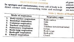
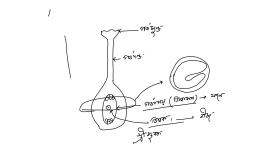
468 Morphology of Animals, , , , , , feuaespenee Roof of bucco-pharyngeal cavity, eavity, , , , Urinary bleder, Fig: 2.4-2 The digestive system of frog, , Respiratory system, , Frog can perform respiration through three methodscutaneous, buccopharyngeal and pulmonary., , (1) Cutaneous Respiration : Skin of Frog is thin and richly, supplied with blood capillaries. It is kept moist by mucus and, water. Oxygen from air diffuses into blood through moisture over, the skin, CO, similarly, diffuses in the reverse direction from, blood to air through moist surface of skin. Cutaneous respiration, occurs in water, during hibemation and even aestivation. So it is, the most vital mode of respiration, , (2) Buccopharyngeal Respiration : Exchange of gases, take place in the lining layer of buccopharyngeal cavity. Throat is, raised and lowered altemately by petrohyal and sternobyal muscles, respectively. This expels out foul air and brings in fresh air., Buccopharyngeal respiration is performed when oxygen requirement, is less, during rest over land and while ‘floating’ in water., , (3) Pulmonary Respiration : It occurs when oxygen, demand is high. The rate is not more than 20/min, as compared to, ‘8O/min for buccopharyngeal respiration. Throat is lowered, air, from outside enters buccopharyngeal cavity, it is called aspiration., ‘Throat is raised, glottis opens and air passes into lungs through, laryngotracheal chamber, it is called inspiration. Lungs are two, ‘ovoid pinkish elastic bags having a large number of small shallow, chambers or alveoli. Blood capillaries overlie the epithelial lining of, the alveoli. Exchange of gases occurs between air and blood, capillaries. Lungs contract and foul air is pushed into, ‘buccopharyngeal cavity by lowering of throat. Throat is now, raised, extemal nares opened and the foul air is passed out. The, process is called expiration., , Laryngotracheal chamber, , It is voice box of frog. Laryngotracheal chamber possesses, , two vocal cords, an opening of glottis into pharynx and bronchus, , , , into lungs. It is supported by three cartilages, two arytenoid and, one cricoid., , Circulatory system, , The circulatory system of frog consists of blood vascular, system and lymphatic system., , (1) Blood Vascular system : It is of closed type as the, blood flows in the blood vessels. It represents single circulation. It, means both the oxygenated and the deoxygenated blood enters, the heart and get mixed in the ventricle. Blood vascular system, comprises blood, heart and blood vessels., , {i) Blood : It is mobile connective tissue consisting of blood, plasma (fluid) and blood corpuscles (cells). Three types of blood, ompuscles are present in the plasma, viz, erythrocytes (RBCs-Red, blood corpuscles), leucocytes (WECs-white blood corpuscles) and, thrombocytes-spindle cells. RBCs are nucleated, oval and biconvex, and have haemoglobin (respiratory pigment). WBCs are amoeboid, shaped and are protective in function. Thrombocytes are spindle, shaped and help in blood clotting., , (ii) Heart : The heart lies enclosed by a thin, transparent, two, layered sac, pericardium. It is a three-chambered structure made of, two upper auricles and a single lower ventricle. Two additional, chambers connected to the heart of frog are sinus venosus and, truncus arteriosus. Sinus venosus is a triangular chamber attached, dorsally to heart formed by the union of three main vena cava. A, short sac like truncus arteriosus is present on the ventral side of the, heart over the larger right auricle Truncus arteriosus is further, divided into a long basal thick walled conus arteriosus or, pylangium and a short distal thin walled bulbous aorta or, synangium. A large twisted spiral valve further divides incompletely, the cavity of pylangium into a left dorsal cavum pulmocutaneum, and tight ventral cavum aorticum. A definitely arranged network of, arteries and veins forms the arterial and venous systems., , , , Right cart trunk, , temic tr, rs Right pulmocutaneous trunk, , , , leler-uriculae septum, , , , Venice", , Fig : 2.4-3 Heart of frog, (2} Portal system : In some vertebrates there is a, characteristic arrangement where the veins does not directly take, blood towards the heart but divides into a capillary set and supply, the organ. This is known as portal system. In frog there are two, portal systerns ~, (i) Renal portal syste, , (ii) Hepatic portal system
Page 4 :
Morphology of Animals 469, , , , Portal System :, , () Renal Portal System : A branch from femoral called, femororenal combine with sciatic vein to form renal portal vein. It, proceeds towards kidney of its side, receives a dorso-lumbar and, then enters the kidneys breaking up into capillaries for quicker, extraction of nitrogenous waste products and salt. The purified, blood is then collected from the kidney by renal veins., , (i) Hepatic Portal System : Ii is formed of two veins,, hepatic portal vein and anterior abdominal vein. Hepatic portal, vein is formed by fusion of a number of veins from digestive tractrectal, intestinal, splenic, duodenopancreatic, gastric and, ‘oesophageal. Anterior abdominal is formed by the union of pelvic, branches from femorals. It recieves vesicular vein from urinary, bladder and a number of small veins from abdominal wall. Near, liver it is joined to hepatic portal vein by a small branch. The two, then enter liver, break up into capillaries for disposal of their, contents, Purified blood is taken out by hepatic veins into, posteaval., , , , , , , , Posterior venacava,, , Hepatic portal system, , Fig : 2.4-4 Portal system of frog, , (3) Lymphatic system : Lymph flows through the lymphatic, system. Lymph is like blood but it is without RBCs and, thrombocytes. It is colourless. Lymph acts as “middle man’. In, addition to the lymph, lymphatic system comprises lymph, capillaries, (closed at the tip), Iymph sinuses (spaces filled with, Iymph) and two pairs of lymph hearts. The flow of lymph is from, the lymph capillaries —> lymph sinuses —» Iymph hearts —> veins, Nervous system, , The nervous system of frog is composed of central nervous, system, peripheral nervous system and an autonomic nervous system., , (1) Central nervous system ; It comprises the brain and, spinal cord. The brain is lodged in the siull, while spinal cord is,, enclosed by the vertebral column., , (2) Peripheral nervous system : The nerves arising from, the central nervous system constitute the peripheral nervous, system, The nerves originate from the brain and spinal cord and, are known as cranial nerves and spinal nerves respectively, , (3) Autonomic nervous system : It includes the nerves and, ganglia that control and coordinate such organs which are not, under voluntary control. It comprises sympathetic nervous system, and parasympathetic nervous sustem., , Brain : The brain is the anterior most part of the central, nervous system. Its situated in the cranial cavity and is covered by, , a delicate, pigmented and vascular membrane, the plamater. The, cranial cavity is internally lined with a tough membrane, the, Guramater, The space in between duramater and piamater is filed, with a shock absorbing cerebrospinal fluid. The brain is divided, into forebrain, midbrain and hindbrain. The forebrain includes, olfactory lobes, paired cerebral hemisphere and unpaired, diencephalon. The mid brain, the broadest, bears dorsolaterally a, pair of large rounded optic lobes (corpora bigemina). The hind, brain consists of a poorly developed cerebellum and medulla, oblongata. Medulla oblongata passes out through the foramen, magnum and continues into spinal cord, which is contained in the, ‘neural canal of vertebral column., , , , —— Oectory nerve, , ;——— Olfactory lobe, _ Cerebral hemisphere, Anterior choroid plexus, ie elasma,, Pinesl beni Diencephalon, Pineal sta lobe, (Cerebellum, Infuncibularn, Hypophyss, Posterior choroid plexus, va Medll oblongata, irs, Hs, , Spinal cont. —, , , , Fig : 2.4-5 Brain of frog, (a) Dorsal view, (b) Ventral view, , Cranial nerves : The nerves which connect the brain and, leave the brain box (cranium) are known as cranial nerves, The, number of cranial nerves is definite in a particular group of, animals. In frog, the number is ten pairs. The serial number of a, nerve is also definite., , Sense organs, , Sense organs receive stimuli (changes in the environment), from outside or inside of the animals and pass impulses to the, nervous system. Frog has following five types of sense organs —, , (1) Tango-receptors (organs of touch) : These are nerve, cendings and touch corpuscles which are found in the skin. Nerve, ending are also present in the viscera (soft internal organs)., , (2) Olfacto-receptors (organs of smell) : They are present, in the nasal epithelium of the nasal chambers., , (3) Gustato-receptors (organs of taste) : These are found, as taste buds which are present in the epithelium of the tongue and, the buccopharyngeal cavity., , (4) Photo-receptors (organs of sight) : Eyes., , (5) Statoacoustic receptors (Hearing and balancing, organs): Ears,, , Out of these, eyes and intemal ears are well organized, structures and the rest are cellular aggregations around nerve, ending. Eyes in a frog are a pair of large, spherical structures, situated in the orbit in the dorsolateral side of head protected by, eyelids. The eye ball is composed of three concentric layers,, sclerotic, choroid and retina. The comea is transparent and permit, entry of light into the eye.
Page 5 :
470 Morphology of Animals, , , , , , Ears are statoacoustic organs meant for equilibrium and, hearing. Ear of frog consists of only two parts, middle and internal, ears; external ear is absent. Tympanum is a dark, flat circular patch, of skin found externally behind eye. Middle ear contains bony, columella auris and carilagenous stapedial plate, which passes, vibrations to intemal ear or membranous labyrinth., , Endocrine system, , The chemical coordination of various organs of the body is, done by hormones secreted by the endocrine glands. The important, endocrine glands found in a frog are pituitary, thyroid, parathyroid,, ‘thymus, pineal body, pancreatic islets, adrenals and gonads., , , , Excretory system, , Frog excretes urea as nitrogenous waste. It is therefore,, ureotelic animal. Urinary or excretory system consists of a pair of, kidneys (mesonephric), a pair of ureters, a urinary bladder, cloaca, and cloacal aperture., , (1) Kidneys : The main organ of excretion are paired kidneys, which are compact, dark red and bean like structures situated little, posteriorly in the body cavity on both sides of vertebral column., Each kidney is composed of several structural and functional units, called uriniferous tubules or nephrons. Each nephron has a, Malpighian capsule, a neck and a coiled tubule which opens into a, transverse collecting tube, , (2) Ureters : They are fine transparent tubes which arise, singly from back of kidney, proceed posteriorly and pass into, cloaca. In inale each ureter contains a swelling called seminal, vesicle for temporary storage of sperms, Ureters of male are also, called urinogenital ducts., , (3) Urinary Bladder : Its distensible transparent bilobed sac, which is connected to cloacal chamber below the opening of, ureters. Urine coming into cloacal chamber from ureters is passed, into urinary bladder for storage. As the urinary bladder gets filled,, the sphincter guarding opening of urinary bladder relaxes and, urine flows into cloacal chamber and from here to the outside, through cloacal aperture., , Other Excretory Organs : Liver is the organ where urea is, formed from ammonia released during deamination of excess, amino acids. It also synthesises bile pigments (biliverdin, bilirat, from decomposition products of haemoglobin. Bile is passed into, duodenum for elimination of waste bile pigments. In rectum a, small quantity of salt is excreted by its wall into faecal matter, e.g.,, calcium phosphate., , , , Reproductive System, , ‘Sexes are separate. Male frog is generally larger with narrower, body. It has vocal sacs in the throat region for louder croaking. In, breeding seacon male frog becomes brightly coloured. Its inner, fingers develop nuptial or amplexusory pads. Female frog is, generally smaller with broader body. During breeding season its, abdomen swells up. Nuptial pads and vocal sacs are absent., , Male reproductive system : Male reproductive organs, consists of a pair of elongated or ovoid yellowish testes, vasa, efferentia, kidneys, urinogenital ducts and cloaca., , (1) Testes : Testes are found attached to the anterio-ventral, surface of kidneys by a double fold of peritoneum called, mesorchium. Testis is composed of a large number of small tubes,, the seminiferous tubules. The wall of seminiferous tubules is made, up of germinal epithelium, which forms sperms by spermatogenesis., , (2) Vasa efferentia : These are 10-12 in numbers and after, arising from testes run through the mesorchium and enter the, kidneys of their side. In kidneys, vasa efferentia open into Bidder's, canal which is connected to the urinogenital duct through, collecting tubules., , (3) Urinogenital duct : The urinogenital duct comes out of, the kidneys and finally opens into the cloaca. Before opening into, cloaca, it dilates to form a seminal vesicle to store the sperms, temporarily. In some frogs the serninal vesicles are not found., , , , (4) Cloaca : The cloaca is a small median chamber that is, used fo pass faecal matter, urine and sperms to the exterior,, , Fat body, , Vasa efferent, , , , Right tess } Left testi, , , , , , , , ‘Adrenal gland, , Kidey, , Urinogenial duct, , Reectum, Urinary bladder, , , , , , Cloaea, , Fig : 2.4-6 Male urinogenital organs of frog, , Female reproductive system : Female reproductive system, of frog is formed of a pair of ovaries, a pair of oviduets and cloaca., , (1) Ovaries : A pair of ovaries, situated near kidneys,, comprises the female reproductive organs. The ovaries are, attached to the dorsal abdominal wall, supported by a peritoneal, fold called mesovarium. The wall of ovary consists of an outermost, germinal epithelium. The cells of the germinal epithelium give rise, to the ovarian follicle. In each follicle, one cell becomes larger than, the others cells to form ovum or female gamete by oogenesis., During the breeding season, the wall of the ovary ruptures to, release the ova into the coelom, A mature female can lay 2500 to, 3000 ova at a time., , , , (2) Oviduets : These are paited, white glandular, long, coiled, tubes Iying one on either side of the body cavity. Each oviduct, consists of ovarian funnel, ovarian tubule and ovisac
 Learn better on this topic