Notes of NEET,JENPUS &ETC BATCH, Biology Digestion.pdf - Study Material
Page 1 :
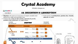
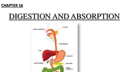
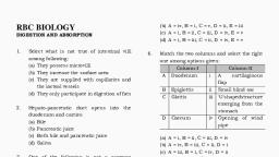
Digestion and Absorption, , , , , , Animals are not able to sunthesise their own food, therefore, they depend on ready-made food for their nutritional, requirements. The term nutrition refers to the sum total of all the, Processes related with the conversion of the raw foodstuff into the, stuff of the body to supply energy for different metabolic activities, and also for the repair and growth. In other word we can define, nutrition as the process by which an organism derives energy to, work and other materials, required for growth and maintenance of, the various activities of life., , Food intake : Different organisms obtain food in different, ‘ways but cary out similar chemical reactions to utilize it. To take, food, protozoans use pseuclopodia, flagella or cilia; sponges and, mussels, a current of water; Hydra, tentacles beset with stinging, cells; planarians and earthworms, a muscular pharynx; flukes and, Jeeches, oral sucker; insects and other arthropods, mouth parts of, various kinds; and seastars and sea urchins, tubelect, Sharks, capture prey with the jaws; frog ané lizard with the tongue; birds, with beaks of sorts; rabbit and hare use forepaws, lips and teeth;, cattle, lips and teeth; camivores, claws and teeth; giraffes, tongue;, elephants, proboscis (trunk); humans, monkeys and apes use, hands., , Digestion, , ‘The process by which complex food is converted into simplest, food with the help of digestive enzymes (Hydrolytic enzymes) is, called digestion. Hence process of digestion is a hudrolutic process., , ‘Types of digestion, , , , (2) Intracellular : When the process of digestion occurs, within the cell in the food vacuole. Examples : Protozoa, Porifera,, Coclenterata and free living platyhelminthes,, , (2) Extracellular : When the process of digestion occurs, outside the cell, Examples : Coelenterates and phylum, platvhelminthes to phylum chordata,, , Digestive system of human, , Digestion in vertebrates occurs in the digesive tract or, alimentary canal, The various paris involved in digestion can be, broadly grouped in two groups ~, , (i) Digestive tract or alimentary canal, , (ii) Digestive glands, , Digestive tract or alimentary canal, , On the basts of the embryonic origin, the alimentary canal of, vertebrates can be divided into three parts ~, , (1) Fore gut / Stomodaeum : Eclodermal. It includes buccal, cavity / oral cavity, pharynx, oesophagus, stomach and small part, of duodenum, , (2) Mid gut / Mesodacum : Endodermal. It includes small, Intestine, and large intestine,, , (3) Hind gut / Proctodaeum : Ectodermal, It includes anal, canal and anus, Parts of alimentary canal and its histology, Mouth, , ‘The mouth is a transverse slit bounded by two movable lips or, labia, upper lip and lower lip. Upper lip has small ridges on the, sides, @ tubercle in the middle and a vertical groove (philtrum), above,, , Vestibule, , Itis a narrow space between lips and gums in front and gums, and cheels on the sides. Its lining contains mucous glands. In the, vestibule, a small median fold of mucous membrane, the superior, labial frenulum, connects the middle of the upper lip to the gum, and usually a similar but smalier inferior labial frenulum connects, the middle of the lower lip to the gum,
Page 2 :
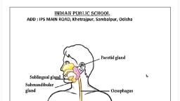
‘762 Digestion and Absorption, Mouth (oral cavity) —_— Supetior in, + contains teeth and tongue ‘Superior labial, Parcdid ves { _ contains teeth and tongu =, {Salivary gland) Sublingual gland = Gingivae (gums), Submandibular gland +-— (salivary gland) yi. _ Palatoglessal arch, (Salivary aland} <palete Se De Feuces,, esophagus eee Palechanygta, as — Palatine tonsil, { Tongue, ai | ingualfrenulum, ‘i et Submandibular, -|——Panereas opening, Transverse colon ~Gingivae (gums), 5 Descending eaten “Inferior labial frenulum, _—Sigmoid colon Interior up, —— Reeth Fig : 5.1-2 Structures of the Oral cavity, —Anus, , , , Fig : 5.1-1 Human Alimentary canal, Buccopharyngeal cavity, , It includes anterior buccal cavity lined by stratified squamous, epithelial cells and posterior pharyngeal cavity lined by columnar, epithelial cells It is distinguished into three region. Pharynx is a, vertical canal beyond the soft palate. The food and air passages, cross here. Pharynx may be divided into three parts;, Nasopharynx, Oropharynx and Laryngopharynx., , Main structures of Buccopharyngeal cavity are —, , (1) Palate : The roof of buccal cavity is called Palate. In, crocodiles and mammals horizontal shelf like processes of, premaxilla and maxilla and the palatine bones of upper jaw fuses, and forms a secondary palate which separates the buccal cavity, from nasal cavity. Palate is distinguished into three regions ~, , (i) Hard palate : Anterior, bony portion formed of maxilla, and palatine bones in human and premaxilla, maxilla and palatine, bones in rabbit. Hard palate have transverse ridges called palatine, rugae. Such rugae or ridges are more developed in camivorous, mammals because their function (sto firmly grip the food and, prevent it from slipping out the cavity, , (li) Soft palate : Posterior sott part, made up of connective, tissue and mus, , , , (iii) Vellum palatifuvula : Posterior most part of soft palate,, which hangs in the region of pharynx. It closes the internal nostrils, during degglutition., , (2) Palatine glands : Numerous mucous glands. Chiefly, present in soft palate, secretes mucous for lubrication., , (3) Naso-palatine duct : One pair, present in rabbit,, extends from nasal passage to the buccal passage, contains, , Jacobson's organ concerned with olfaction., (4) Vibrissae : A tuft of hairs on upper lip of rabbit,, , (5) Hare-eleft : A clelt on the upper lip of rabbit, which, makes it bilobed., , (6) Tongue (linguae) : Ectodermal, sinale, pinkish, oval,, elongated highly muscular (mesodermal) and protrsible present, on the floor of buccopharyngeal cavity the cells present are, stratified squamous epithelial cells. A furrow termed the sulcus, terminalis divides the oral part and pharyngeal part of the tongue, ‘The limbs of the sulcus terminalis run laterally and forward from a, ‘median pit, named the foramen caecum., , , , Epigtottis, , , , , , Palatine tonsil, , Firm papillae, Fungitorm ‘ies a, papillae salt, Fig :5.1-3 Locations of papillae and areas of taste on the, tongue, , Pestetior part of tongue (endodermal) is zttached with, hyoid, middle one with the floor of buccopharyngeal cavity with, the help of frenulum lingum and anterior part is free. The tongue is,, provided with two specialized structure viz, lingual papillae and, lingual gands or weber's gland. Lingual glands are the mucous, glands, which secretes mucous, Lingual papillae are numerous,, minute projections chiefly present on the dorsum of the tongue. All, these lingual papillae can be grouped as simple lingual papillae, and taste papillae. Toste papillae are of following types —, , (i) Circumvallate : Circular largest 8-12 in number, present, in the posterior part of the tongue extending from one side to, another. They possess taste buds. These are the largest of all the, papillae,, , (ii) Fungiform : Mushroom shaped (Fungi — shaped),, numerous, present at the anterior margins and tip of the tongue., ‘They have 200 taste buds.
Page 3 :
Digestion and Absorption 763, , , , (ii) Foliate : Leaf like flat, less 8-10 in number, present at the, posterior margin of the tongue. They are absent in human and, found in rabbit., , (iv) Filiform = Conical shaped, smallest and most numerous, , distributed throughout tongue. They are without taste buds,, Hence, in human taste is recognized with the help of circumvallate, and fungiform taste papillae. In man the anterior end of tongue, feels sweet taste, posterior part feel bitter taste, sides feel sour taste, and a small part behind the anterior end feel salty taste., , , , , , Fig : 5.1-4 Dorsal surface of human tongue, showing three kinds, of papillae and some other associated structures, , Functions of tongue : Important function of tongue are es, follows ~, , (i) Acts as universal toothbrush, as it helps in tooth cleaning., , (ii) Helpsin speaking,, , (ii) Helps in degglutition., , (iv) Helps in misting saliva with food., , (v) Acts as a curry comb in many animals, hence help in body, cleaning., , (vi) Helps in taste detection, , (vii) In dog helps in regulation of body temperature. The, phenomenon é called as “Panting”, , (vill) In freg and other animals, it helps in prey capturing, , (7) Teeth : Teeth is a living structure. On the basis of, embryonic origin, teeth in vertebrates are of following two types ~, , (i) Horny/ectodermal/epidermal/false teeth ; The teeth, which develops only from ectoderm. Examples ~ Cyclostomes,, tadpole larva of frog, prototherian mammals etc., , (ii) True teeth : The teeth which develops from both, ectoderm and mesoderm. Examples ~ Fishes, amphibians, reptiles,, euthe Tammals ete., , Differentiation of teeth : Morphologically, teeth can be, distinguished as homodont or heterodont,, , (i) Homedont : When all the teeth are structurally and, functionally similar, Examples ~ Vertebrates except metatherian, and eutherian mammals., , (ii) Heterodont : When the teeth are different in structure, and functions. They are distinguished into four types incisors,, canines, premolars and molars. Examples — metatherian and, eutherian mammals., , , , , , , , {a) Incisors : These are the front teeth bome by the, premaxilae in upper jaw and tips of dentaries in lower jaw. They, are singe-rooted monocuspid and long, curved and sharp-edged, They are adapted for cutting or cropping and biting,, , (b) Canines : There is one pointed canine in each maxillary, of upper jaw and each dentary of lower Jaw next to the incisors,, They are meant for piercing, tearing and offence and defence., ‘They are single rooted and monocuspid.,, , (c) Premolars : They have one root (only in upper frst PM, ‘two tools) and two cusps (bicuspid). They are meant for crushing,, stinding and chewing., , (a) Molars : They have more than two toots upper molars, have three roots and lower molars have two roots) and 4 cuspid,, , Attachment of teeth : On the basis of attachment of teeth, at their bases with the jaw bones, teeth can be differentiated into —, , (i) Aerodont : Teeth are attached to the free surface or, summit of the jaw bone, as in 2 shark or frog. Such teeth are apt to, break off casily but are replaced., , {ii) Pleuredont : In this condition, common in urodeles and, lizards, teeth are attached to the inner side of jaw bone by their, base as well as one side,, , (ii) Thecodont : Such teeth are characteristic of mammals., Teeth have well developed roots implanted in deep individual pits, or sockets called alve rr theca, in the jaw bone. These type of, teeth also present fossil toothed bird, (Archeaeopteryx), , , , , , in crocodilians,, , , , ‘daw bone, , , , Acrodont Peurodont hasan, , Fig : 5.1-5 Methods of attachment of teeth on jaws, , Succession of teeth : According to their replacement, {succession}, teeth can be divided into 3 categories: polyphyodont,, diphyodont and monaphyodont., , (i) Polyphyodont : In lower vertebrates, teeth can be, replaced an indefinite number of times during life, eg., - Fishes,, amphibia, reptilia,, , (ii) Diphyodont : In most mammals teeth develop during life, in two successive sets, a condition known as diphyodont. Teeth of, the first set are known as deciduous teeth or milk teeth or lacteal, teeth whereas the second set is called permanent teeth., , (ii) Monophyodont : In some mammals such as platypus,, marsupials, moles, sirenians, toothed whale etc. only one set of, teeth develops known as monaphyadont condition,, , , , ‘Types of cheek teeth, , (i) Bunodont : Crown with small, blunt and round cusps as in, man, monkey, pig etc. found in mixed diet mammals., , , , (ii) Secodont : With sharp cutting edges for tearing flesh as in, carnivores
Page 4 :
ii) Lophodont : Only one cusp is present with transverse, ridges called lophos, e.¢., Elephant., , (iv) Selenodont : With vertical crescentic cusps as in grazing, ‘mammals lie cow, sheep and goat. Selenodont teeth are two types —, , (a) Brachyadont : Normal low crowned selenodont teeth, with large rocts are termed brachyodont. e.g. Ground squirrel,, cattle,, , (b) Hypsodont : In large grazing mammals teeth are, elongated, prism shaped with high crown and low roots. e.g. Harse., , Structure of teeth : Teeth divided into three parts ~, , (i) Root : Inner most, attached to the bone with the help of, cement (hyaluronic acid),, , (ii) Neck : Middle, small, covered with gum. Gum provides, strength to the teeth., , (ii) Apex or crown : External exposed part of teeth. Longest, part, white in colour., , A small cavity present inside teeth called as pulp cavity or, dentine pulp cavity. It contains blood vessels, lymphatic vessels,, nerve fibres, connective tissue ete. and provides nutition to, odontoblast cells or osteoblast cells. The odontoblast cells are, mesodermal in embryonic origin forming immediate covering of, the pulp cavity. The cells secrete dentinalivory. Bulk of tooth in a, mammal is formed of dentine. Dentine is a layer of inorganic, substances (62-69%), which surrounds the odontoblast cells. It, is mesodermal in origin. Enamel, secreted by, Ameloblast/Erameloblast cells, forms the outermost covering. It is, ectodermal and made up of 92% of inorganic substances, hence, considered as hardest part of the body. The inorganic substances, present are [Ca,(PO,),,Ca(OH),H,0] Calcium phosphate (85%),, Calcium hydroxide and Calcium Carbonate. Cement/Cementum, aitaches the tooth root to the bone., , , , , , —Pulp in pulp cavity, , =——Cementum, = ET Root canal, , Fig : 5.1-6 Structure of tooth, , Dental formula : Each mammalian species is characterized, by its own specific dentition with a definite number and, arrangement of teeth. Hence, dentition is of taxonomic, importance. It is expressed by a densal formula as below —, , eg pm. =8x2=28 or briefly,, , 2033_2+043+3,2 16 _9¢, , 1023 140+2+3°2 127, , (i = incisors; ¢ = canines; pm = premolars; m = molars), , , , 16_, , , , , , , , , , , , , , , , , , , , , , , , , , , , Fore | 383 Car, , anda | gag", , Dog Ste x22 ‘Squire!, , Gore) 2332-06 |, , Man = 2 S203 ‘Elephant, , Cow 0.033 Human set, 31a3 7 | (mike, , , , Gesophagus (food tube), , (1) Morphology : Single, ectodermal, dorsal to trachea,, approximately 25 em long. passes through thoracic cavity and, opens into stomach present in abdominal cavity. Oesophagus, anteriorly opens into pharynx through gullet and posteriorly into, stomach through cardiac orifice, , (2) Histology : Serosa is absent but outermost layer of, connective tissue is called as tunica adventitia. Muscular layer are, striated/voluntary in anterior region and unstriated/involuntary in, posterior part. The epithelial lining is made up of nan-keratinized, stratified squamous epithelial cells, Goblet cells are present., , esophagus lack digestive glands but multicellular glands are, found, which extends upto submucosa, Due to the presence of, these submucosal mucous glands, submucosa of oesophagus is, thickest than other parts of alimentary canal., , Function : Conduction of food., , Stomach, , (1) Structure : Single oval, elongated, unilobed present, ‘within abdominal cavity below diaphragm. It consists of three parts, a cardiac/fundie (anterior), cospusfoody (middle, chief part) and, pyloric (posterior part) in human, whereas in rabbit stomach is,, bilobed and consists of three parts as cardiac (Anterior), fundic, (middle, chief part) and pylorus (posterior). Two types of valves, are present in the stomach wiz, Cardiac sphincter valve between, ‘oesophagus and stomach and pyloric sphincter valve between, stomach and duodenum. In naw born baby cardiac sphincter is, much less developed that is why regurgitation of gastric contents is, very common. Inner surface of stomach is raised into numerous, longitudinal folds called gastric rugae. In case of ruminant, mammals (cud chewing mammals) cesophagus consists of only, skeletal or voluntary muscles., , , , , , , , , , Oesophagus, Lower Oesophageal 0 Funds, sphincter —_\ — Serosa, Body — Longitudinal layer, Lesser curvature (Circular layer, Pylorus., , 5 — Oblique layer, , , , , , AV /— Greater curvature, QV, » Pyloric \ ei otineiaol, sap) sphincter pytaric Pyloric antrum, Duodenum mal, Fig : 5.1-7 Human Stomach
Page 5 :
Digestion and Absorption 765, , , , , , (2) Histology : Outermost layer is serosa, Muscular layer is, three layered with outer longitudinal, middle circular and inner, oblique. Muscles are involuntary and unstriated. Epithelial lining is, made up of simple columnar epithelial cells and specialized cells, present in the gastric glands. The nomenclature of gastric glands is, according to the parts of the stomach. Various types of gastric, siands and the cells present in them are as follows ~, , () Anterior part : Cardiac gastric glands in rabbit and, human, Cells present are mucous neck cells secreting mucous,, , (ii) Middle part : Fundic gastric/Main gastric glands in rabbit, ‘and compus in human has at least four distinct types of cells ~, , (a) Peptic or zymogenic or chief or central cells :, Secretes two digestive proenzymes pepsinogen and prorennin., , (b) Oxyntic or parietal cells : Secretes HCI and castle's, intrinsic factor required for the absorption of vitamin By», Hyperacidity is abnormally high degree of acidity due to the, secretion of large quantity of HCI Le. gastric juice., , (c) Mucous neck cells : Secrotes alkaline mucous,, , (d) Argentaffin cells or Kultchitsky or enterochromaffin, cells : Responsible for the secretion of vasoconstrictor seratonin., , (ii) Posterior part : Pyloric gastric glands in rabbit and, human-cells are mucous neck cells secreting mucoiis and some, cells, called “gastrin” or “G” cells, secrete a hormone, named, gastrin, which increases the motility of gastric wall and stimulates, gastric glands for active secretion., , , , , , , , , Gastric pit, , Surface mucis cell, , Se meg BE (secretes mucus), , epithelim 28) Mucus neckcell, , eh DR iaseas aces, propria, , Porietal cell fcerctes, } hydrochloric acid, * and intrinsic factor), , site Chiet cell [secretes, pepsinogen and, ‘gastric lipase}, , G call fsocretes the, ‘hormone gastrin), , Fig : 5.1-8 LS, Gastric gland, , Functions, , (1) Storage of food., , (2) Trituration or churning of food to mix with gastric juice,, , (3) Functions of gastric juice (discussed along with gastric, juice), , Stomach of ruminants (cud-chewing mammals) : The, stomach of cates have four parts, as rumen (paunch),, reticulum(honeycomb), omasum psalterium) and abomasum., (rennet). Some authors believe that first three chambers are parts of, , the oesophagus, the fourth chamber is the real stomach secreting HCI, and engymes, The embryological studies have proved that all the, chambers are parts of the real stomach, Camel and deer lack, omasum, Reticulum is the smallest part and its cells are provided, with waier pockets for the storage of metabolic water., , In the rumen, food undergoes mechanical and chemical, breakdown. Mechanical brealdown results from through churning, brought about by muscular contractions and aided by cornified, surface of vill. Chemical breakdown is caused by symbiotic, microorganisms (bacteria and ciliates) that release enzyme, cellulase, which act on cellulose and simplify it into short-chain, fatty acids, such as acetic acid, butyric acid, propionic acid, This is, called microbial digestion,, , , , , , erophagis, , Abomasum,, , etl, Duodenum, , Fig: 5.1-9 The compound stomach of a ruminant, , Small intestine, , (1) Structure : Endodermal, longest part of alimentary canal, present in the abdominal cavity, supported by a peritoneal, membrane called mesentery. Wall of jejunum and ileum has, circular or spiral intemal fold called fold of kerckring or valvulae, cconniventes. Also numerous firger like projection called vill project, from the wall of lumen, increasing intemal surface area about ten, time, The distal end of ileum leads into the large intestine by ileoceecal valve in man but in rabbit sacculus rotundus and ileo-coecal, valve both are present., , (2) Parts : It is approximately 3 metres in human. It is, divisible into three parts duodenum, jejunum and ileum,, , Table : 5.1-2 Parts of small intestine, , , , , , ‘Duodenum Jejunum eum, (Proximal part) (Middte part) (Posterior part), 25.em. Long About 1 mlong and about 4 | About 2 m long, Forming U-shaped | ™ wide. and (aaa ee, loop before | Wall is thicker and more |, leading to | vascular. Wall is. thinner, Jeluniats peneress | Via thickerand tongue-ike, | on less vascular, lies in the loop. Sey, , Plicae best developed. gece, , Peyer'spathes arelockig: | 5. jas, developed., Peyer's patches, are present.
 Learn better on this topic