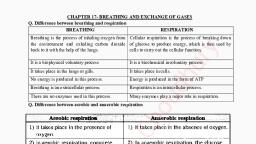
Notes of NEET,JENPUS &ETC BATCH, Biology Breathing and respiration - Study Material
Page 1 :
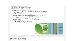
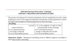
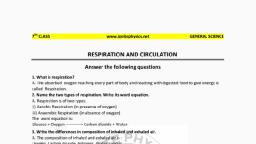
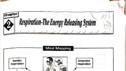
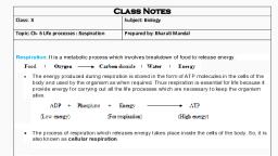
Breathing and Exchange of Gases, , , , Cells continually use oxygen (O,) for the metabolic reactions, that release energy from nutrient molecules and produce ATP, (Adenosine Tri Phosphate). At the same time, these reactions, release carbon dioxide. Since an exce amount of CO, produces, acidity that is toxic to cells, the excess CO, must be eliminated, quickly and efficiently, The two systems that cooperate to supply, 0; and eliminate CO, are the cardiovascular system and the, respiratory system. The respiratory system provides for gas, exchange, intake of O, and elimination of CO,, whereas the, cardiovascular system transports the gases in the blood between, the lungs and body cells., , Respiration, —E, , Respiration is a process which involves intake of oxygen from, environment and deliver it to the cells. it include stepwise oxidation, of food in cells with incoming oxygen, elimination of CO,, produced in oxidation, release of energy during oxidation and, storing it in the form of ATP. It takes place in three basic steps —, , (1) Pulmonary ventilation : The first process, pulmonary, (pulmo = lung) ventilation, or breathing, is the inspiration (inflow), and expiration (outflow) of air between the atmosphere and the, lungs., , (2) External (pulmonary) respiration : This is the, ‘exchange of gases between the air spaces of the lungs and blood in, pulmonary capillaries. The blood gains O, and loses CO;., , (3) Internal (tissue) respiration : The exchange of gases, between blood in systemic capillaries and tissue cells is known as, internal (tissue) respiration. The blood loses O, and gains CO,, Within cells, the metabolic reactions that consume O, and give off, CO, during production of ATP are termed cellular respiration,, Respiratory surface, , The surface at which exchange of gases (CO, and O;) takes, Place is called respiratory surface, Respiratory surface must be, , vascular and have enough area for gas exchange. For example —, plasma membrane in protozoa, body wall (skin) in annelids,, alveocapillary membrane in men., Respiratory medium, , Oxygen is dissolved in air and water. Thus water and air are, source of oxygen for animals and called respiratory medium. Water, and air are external respiratory medium. Inside the body an, internal respiratory medium is also found. This internal respiratory, medium is tissue fluid., , Types of respiration : Its of two types, , (1) Aerobic respiration : It occurs in the presence of, molecular oxygen. The oxygen completely oxidizes the food into, carbon dioxide and water, releasing large amount of energy, The, organisms showing aerobic respiration, are called aerobes. It is, found in most of animals and plants. Aerobic respiration is of two, ‘main types direct and indirect, , CoH:O5+ 60, + 6CO, +6H,0+ 2830kI, ‘Guutse”cegenCatondiaude War Eneay, , {i) Direct respiration : It is the exchange of environmental, ‘oxygen with the carbon dioxide of the body cells without special, respiratory organs and without the aid of blood, It is found in, aerobic bacteria, protists, plants, sponges, coelenterates, flatworms,, roundworms and most arthropods., , (i) Indirect respiration : It involves special respiratory, organs, such as skin, buccopharyngeal lining, gills and lungs, and, needs the help of blood, The respiration in the skin,, buccopharyngeal ining, gills and lungs is respectively called, ‘cutaneous buccopharyngeal, bronchial and pulmonary respiration., Cutaneous respiration tales place in annelids, some crustaceans,, eel fish, amphibians and marine snakes, It occurs both in water, and in ait. Buccopharyngeal respiration is found in certain, amphibians such as frog and toad. It occurs in the air. Bronchial, respiration is found in many annelids, most crustaceans and
Page 2 :
816 Breathing and Exchange of Gases, , , , molluscs, some insect larvae, echinoderms, all fishes and some, amphibians. It occurs in water only. Pulmonary respiration is found, in snails, pila, some amphibians and in all reptiles, birds and, mammals, It takes place in air only., , (2) Anaerobic respiration : It occurs in the absence of, molecular oxygen and is also called fermentation. In this, the food, is only partially oxidised so only a part of energy (5%) is released, and of energy remains trapped in the intermediate compounds. It, is found in lower organisms like bacteria and yeast. It is also found, in certain parasitic worms (Ascaris, Taenia) which live in deficient, medium. The organism showing anaerobic respiration, are called, anaerobes. These involve one of following reactions., , , , CHO, —PY* 4 2CHgOH+ 2CO, +118 kd, ‘Glucose Bhatt, , (Fementaton of ase, , (CoH ,0, —B esse _, 2CH SCHOHCOOH + Energy, tance acted, , Certain body tissues of even aerobes also show anaerobic, metabolism e.g., during the vigorous contraction of skeletal muscle, fibres. In this, the glucose is metabolised into the lactic acid in, anaerobic conditions. The rapid formation and accumulation of, lactic acid are responsible for muscle-fatigue. The mammalian, RBCs shows anaerobic respiration as these lack the mitochondria., In lens of eye and comea of eye respiration is anaerobic because, these structures are non vascular. Anaerobic respiration appeared, first in primitive organisms because there was absence of O, in, primitive atmosphere., , Respiratory organs, , (1) Skin : Respiration by skin is called cutaneous respiration., Skin isthe only respiratory organ in most annelids (earthworm and, leeches) and an additional respiratory organ in amphibians (Toads, and frogs). Skin should be thin, moist, naked, permeable and well, vascular for respiration. For cutaneous respiration animal should, have large surface area then its volume and should have relatively, inactive life to minimize the use of oxygen, Some marine annelids, such as sandworms (nereis) have parapodia (locomotory, appendages) for respiration. In frog 100% cutaneous respiration, uring hibernation, In all marine snalwes 20% respiration by skin., , (2) Tracheae : In insects, peripatus centipedes and millipedes, tracheae are found for respiration. Tracheae are complex system of, whitish, shining, intercommunicating air tubules. Tracheae are, ectodermal air tubes. In cockroaches, three paits of longitudinal, tracheal trunks are present all along the length of body which are, further connected with each other with the help of transverse, branches. The main tracheae give off smaller tracheae whose, branch repeatedly form a network of trachioles throughout the, body. Trachea intemally lined by chitinous cuticle called intima,, which spirally thickened to form taenidae. Trachioles without, taenidae, trachioles lined by trachein protein. From each tracheal, trunk three branches come out. The dorsal branch is supplied to, , the dorsal muscles where as ventral one to nerve cord and ventral, muscles and middle one to the alimentary canal., , “Teachiole Mule, , Chitin of, , , , Fig: 5.2-1 Trachea of cockroach, , (3) Book lungs and book gills : Spiders ticks, mites and, scorpion (belongs to class arachnida) have book lungs for, respiration. In scorpion 4 pairs of book lungs are present. A book, lung is a chamber containing a series of thin vascular, parallel, lamellae arranged like the pages of book. Book gills are found in, marine king crab or horse shoe crab., , (4) Gills : Aquatic animals such as prawn, unio, fishes, sea, stars and tadpoles respire by gills. Respiration by gills called, bronchial respiration. Gills are of two types —, , (i) External gills : External gills are found in arenicola (lug, worm), larvae of certain insects e.g. damsel fly and some, amphibians e.g. necturus, siren, proteus, frog tadpole first develop, external gills which are replaced by internal gills later., , (ii) Internal gills : The internal gills may be phyllobranch, (prawn), monopectinata (pila) eulamellibrach (unio),, lamellibranch, fillibranch (pisces). In all fishes, gills are hemibranch, or demibranch and holobranch. In gills, gill lamellae are found, which have capillary network. Water is drawn into gills + blood, flowing in the capillaries of gill Jamellae absorb oxygen from water, and release CO, —> water containing CO, is thrown out from, , ills. The 80% of O, of incoming water is absorbed., , Table : 5.2-1 Oxygen content of respiratory media, , , , , , , , , , , , , , , , , , , , , , Respiratory media ‘Oxygen content, Air 209.5 mii, Fresh water at 25°C 58 mill, Fresh water at 5°C 9.0 mii, ‘Sea water at 5°C 64 mil, , , , , , Frog breathes by, called, , (5) Buceopharyngeal, buccopharyngeal lining of buccopharyngeal cavity. This is, buccopharyngeal respiration., , lining +
Page 3 :
Breathing and Exchange of Gases 817, , , , , , , , , , , , , , , , air conditioner and makes the temperature of in going air nearly, equal to body. It also acts asa filter not give entry to dust particles,, , (c) Olfactory region : It is upper region. It is lined by, olfactory epithelium. This is also called Schneiderian epithelium., the organ of smell and detect the odour of, inspired air. Inspiration is stopped if odour of air is foul or, offensive. According to new researches pheromone receptors are, , , , (li) Nasal conchae : Lateral wall of nasal cavity have three, shelves like structures called conchae or turbinate, 3 pairs of nasal, conchae are found. Nasal conchae are covered with mucus, membrane, They increase the surface of nasal chamber. Both the, chambers of nasal cavity open into nasopharynx by their apertures, called intemal nostrils or conchae. Adjacent to internal nostril there, are opening of eustachian tube, Names of these three canchae and, ‘names of the bones that form them are given below., , (a) Superior conchae : The dorsal most chochae is, supported mainly by nasal bone called nasoturbinate, It is the, , (b) Middle conchae : Ethmoid bone called ethmoturbinate., , (c) Inferior conchae : The ventral most conchae supported, by maxilla bone called maxilloturbinate. It is a separate bone itself., , (ii) Pharynx : It is the short vertical about 12 cm long tube., ‘The food and air passages cross here. It can be divided in 3 parts ~, , , , , , , , tharymx : Nasopharynx is only respiratory upper, Part in which internal nares open. There are 5 opening in its wall, , Table : 5.2-2 Respiratory organs of animals, ‘Animals Respiratory organs ‘ i, , Protists, Bacteria Direct respiration through plasma _ fies or mosquitoes., , membrane, Porifera Piasina membrane of each cell, Coelentarates General body surface Olfactory region, Plaivhelminthes (Fasciola | Anaerobic, hepatica, tapeworm), Nematodes (Ascaris) Anaerobic found in nasal cavities., ‘Annelids (Earthworm and | Skin, Leechs), Nereis Parapodia, Insects Trachea, Centipedes Trachea, Milipedes Trachea, ‘Spider and Scorpion, | Book lungs, ticks, mites, Marine king crab Book gills, Prawns, Unio and Pla | Gills, , “Echinodermata Dermal branchiae, Tube feet,, , Respiratory tree, Bursae smallest conchae., Fishes, Tadpoles Gills, Frogs, Toads Buccopharyngeal ining, Lungs, Skin, Reptiles, Mammals Langs, Birds AirsacsiLungs, Lung fh ‘Air bladder., Urochordata (Herdmania)_| Test, Marine turtle Giocal respiration Ne, ‘Mollusca (Unio) Mental, , SSS, , Respiratory system of human, , Human respiratory system is derived from endoderm, Human, respiratory system may be divided into two components —, , (1) Respiratory tract or conducting portion, , (2) Respiratory organs, , (1) Respiratory tract or conducting portion : It is the, passage for the air. In this part gaseous exchange does not takes place., Itis also called dead air space. It is divided in following parts , (i) Nose (Latin-Nasa) (Greek-Rhine) : Cavity of nose is called, ‘nasal cavity. Nasal cavity is divided into two parts by nasal septum, called mesethmoid. Each part is called nasal chamber. Each nasal, chamber opens out side by extemal nares, Nasal septum has two, part. First part is small and is made of cartilage (hyaline). Second, Part is major and it is bony. Vomer is the main bone. Each nasal, chamber has three region,, , (a) Vestibular region : Vestibular region also known as, vestibule is lined by non Keratinized squamous epithelium, it is, ectodermal in origin and have sebaceous gland, sweat gland and, hair. Vestibule is also found in inner air larynx, mouth and vagina., It acts like a seive to check the entry of large dust particles and, other things,, , (b) Respiratory region : Middle region lined by respiratory, epithelium which is ciliated pseudostratfied columnar epithelium. It, Contains mucus and serous cells. Mucus cells produce mucus and, serous cells produce watery fluid. Respiratory epithelium is highly, vascular and appears pink or reddish. Respiratory region acts as a, , , , two internal nares, two eustachian tube opening and one opening, into oropharynx., , (b) Oropharynx : Middle part is called oropharynx, In this, Part oral cavity open mown as fauces, Two pair tonsils the palatine, and lingual tonsils are found in the oropharynx., , {c) Laryngopharynx or hypopharynx : Lowest partis called, Jaryngopharynx. It leads into two tubes. One at the front is wind pipe, oor trachea and one at the back is food pipe or oesophagus. Both oro, and laryngo pharynx is both a respiratory and a digestive pathway,, , Pharyngeal tonsil, , Opening of auditory _ Inferior nasal, , , , , , , , , , , , (Eustachian) tube conchae, Nasopharynx_ Hard palate, Soi palaie Orel, Palatine tonal retail, aces 3 1, Oropharynx ‘ongue, Lingual tonsil te, 1. Mandible, Hyoid bone, , , , > —Thyroid cartilage (Adam's apple), , , , Oesophagus, , , , —— Cricoid cartilage, “— Trachea’, Fig : 5.22 Sagittal section showing the regions of the pharynx, Nasopharynx is lined by ciliated pseudostratified epithelia,, oropharynx and laryngopharynx are lined by non keratinized, epithelium. Pharynx is lined by non keratinized stratified squamus, epithelium. This epithelium is cillieted in nasopharynx. Mouth, Serves as an altemate route for air when nasal chambers are, blocked. Foramen by which pharynx opens into larynx called
Page 4 :
818 Breathing and Exchange of Gases, , , , , , glottis. In general it remains open. During swallowing itis closed. It, provides passage for air. Pharynx leads into the oesophagus, through an aperture called gullet. In general condition it remains, closed and opens at the time of swallowing. During swallowing, epiglottis closes the glottis., , (iv) Larynx or Voice box : It is found both in frogs and, rabbits, Larynx does not help in respiration. It is present on tip of,, trachea and is made up of 9 cartlages such as thyroid (single) has, a prominence called pomum admi or adam's apple, cricoid, Je), arytenoid (paired), are piece of hyaline cartilage. While, epiglottis (single), carniculate (paired), cuneiform (paired), santorini, are piece of elastic cartilage. Clinically, the cricoid cartilage is the, landmarks for making an emergency ait way,, , Larynx is a short tubular chamber and opens into the, aryngopharynx by a slit like aperture called glottis. Glottis always, remains open except during swallowing. Larynx is more prominent, in men than women due to male hormone. Before puberty, the, larynx is inconspicuous and similar in both sexes, Larynx is a voice, producing instrument. For this purpose larynx have two types of, ‘vocal cord, In birds voice producing organ is syrinx, found at lower, end of tracheae., , ‘ Epighottis, Hyoid bone, ——Thyrohyoid membrane, , , , , , , , ), | |) Thyroid cartilage, (Adam's apple), , icothyroid ligament, Cricoid cartilage, Cricotracheal ligament, , yr ; Thyroid gland, troche cantage, , Fig : 5.2-3 Larynx (Anterior view), , , , , , Epiclottis—, Hyoid bone ——, Thyrohyold membrane, , Comiculate cartilage, “Thorold cartlage, (Adam's apple), , Aryienoid carage, , , , , , , , , , , , Cricold cartilage, Cricotracheal ligament, , ‘Thyroid gland, Parathyroid glands (4), Tracheal cartilage ———= ——>, , Fig : 5.2-4 Larynx (Posterior view), , (a) False vocal cord or vibrating fold or anterior vocal, cord : These are folds of mucus membrane. Gap between them is, called tema vestibuli, These are not responsible for sound, production. In elephants only true vocal cords are present and are, responsible for this trumpet sound., , (b) True vocal cord or posterior vocal cords : They are, made up of yellow elastic fibres. Gap between them is called rema, glottides or peep hole. In males the length of true vocal cord is 2.25, ‘cm and in female is 1.75 em, Sound produced by rabbit is called, , , , , , quaking. Hippopotamus lacks rue vocal cords. Pitch is controlled, by the tension of vocal folds., , ee ot, , pms, a Vocal folds(true vocal cords), , , , Fig : 5.2-6 Movement of vocal folds together (adduction), , (v) Trachea : It is a tubular structure of about 12 em, in, length and 2.5 em in diameter. The wall of trachea is made of, fibres, cartilage muscles and the mucus membrane. In middle of, thorax at the level of 4" and 5* thoracic vertebra divides it into two, branches called right and left primary bronchi. Further division of, primary bronchi is given in form of arrow diagram., , , , , , geeo L, , a5 4 i Mojo pinay bronchi, 3E Secondary bronchi, apts ie, ESES | teracorseqrena ton, Pa ED | tempore, , a t, , Resear ‘bronchiole, , Diameter decrease, , zg Alveolar duct, eas aun, gfe J, £3 4 velar sc, , ‘Air sac or alveoli (gaseous exchange), Table : 5.2-3 Different epithelium lining in respiratory tract, , ‘Vestibular region of nose ‘Skin having hair, , , , , , , , Respiratory region of nose Ciliated pseudostratified, , ‘Olfactory region of nose Olfactory (Schneiderian) epithelium, Pharynx (Oropharynx, | Non-keratinised stratified, Layngopharyn squamous, , “Trachea and bronchi (Upper) Pseudostratified ciliated columnar, , epithelium with mucus cells, , ‘Lower bronchi (Secondary / | Lined by simple ciliated columnar, Terian) | epithet, , Terminal bronchioles and | Simple ciliated ‘columnar, beginning of respiratory | epithelium without mucus cells, bronchiole, , Rest of respiratory bronchioles, | Non ciliated cuboidal epithelium, alveolar duct, , Alveoli ‘Non ciliated squamous:, , Alveoli of frog's lungs Columnar ciliated velium:
Page 5 :
Breathing and Exchange of Gases 819, , , , ‘Three special types cell are found in Bronchioles epithelium, , (a) Kultchitsky cells or argentaffin cells : They secrete, serotonin and histamine. Histamine dilate while serotonin constrict, the bronchioles,, , (6) Clara cells : They secrete a phospholipid named, diapalmityl lecithin which acts as a surfactant. This surfactant, Prevents the collapse of bronchioles lacking carfilagenous rings,, Collapsing of lungs is called atelectesis. Poitle in 1956 proved the, existence of surfactant. Surfactant is formed by clara cells only at, later stage of foetal life. Some times at birth some infants are, devoid of surfactant so there is great respiratory difficulty because, lungs refuse to expand. In this condition death may occur, This is, called respiratory distress syndrome (RDS) or hyaline membrane, disease (HMD) or glassy lung disease., , (c) Dust cells : They are phagocytes which eat foreign, particles (dust),, , , , , , Respiratory organs, , In men the respiratory organ are a pair of lung. Some snakes, have unpaired lungs. Respiration by lungs is called pulmonary, respiration. Lungs are found in all vertebrates except fishes, In, fishes such as protopterus, neoceratodus and lepidosiren air, blacider is found, which is modified lung. Respiration in men and, rabbit is pulmonary,, , Lungs : Lungs lie in thoracic cavity on both side of heart in, mediasternum space. Base of lung is attached to diaphragm. Right, lung is divided into 3 lobes viz. Superior, Middle, Inferior and left, lung is divided into two lobes Superior and Inferior. In rabbit, the, left lung is divided into two lobes left anterior and left posterior, where as the tight lung has four lobes anterior azygous, right, anterior, right posterior and posterior azygous. Lungs of reptiles are, more complex than those of amphibians, In birds lungs are, supplemented by elastic air sacs which increase respiratory, efficiency. The narrow superior portion of lung is termed the apex, or cupula., , , , , , , , Visceral pleura, , Pasi pl, , Pleural cavit, , Right suit, bronchus Y=, , Right primary,, , bronchus, , Right tertiary 79, , bronchus,, , Right bronchiole’ Maggs, , Right terminal” We, , bronchiole, , Fig : 5.2.7 Branching of airway from the trache, tree (Anterior view), , Each lung is enclosed in two membrane called pleura. Pleura, are layers of peritoneum of thorax. Inner membrane is called the, visceral pleuron. It is firmly bound to surface of lungs. The outer, membrane is called parietal pleuron. It is attached to chest wall or, wall of thoracic cavity. A narrow space exists between the two, pleura. It is called pleural cavity. In pleural cavity a watery fluid is, , Lett primary, bronchus, , , , , , the bronchial, , , , found called pleural fluid. Pleural fuid is glycoprotein in nature, and secreted by pleura. Pleural fluid lubricate the pleura so that, they may slide over each other without friction, This fluids reduces, friction between the membrane. When the lungs expand and, contract in respiration. Pressure inside pleural cavity is negative — 5, mm Hg, Pleurisy is inflamation of pleura and cause collection of, fluid in pleural cavity. It results painful breathing (dyspnea). The, surface of lung lying against the ribs, known as coastal surface. The, ‘mediastinal (medial) surface of each lung contains a region — the, lus, through which bronchi, pulmonary blood vessels, lymphatic, vessels and nerve enter and exit., , , , Terminal bronchiole, , , , , , , , , Pulmonary venule. Pulmonary arteriole, Lymphatic vessel, oe ¥ Respiratory bronchiole, connective tissue / ae, , Py., , >> Alveolar ducts, , , , Pulmonary, {alveolar} capillary Ny, , , , , Dl Alveolar sae, , va, Visceral pleura: P|, Alea —, Fig : 5.2-8 Diagram of a portion of a lobule of the lung, , , , Pulmonary volumes and capacities, , ‘The apparatus commonly used to measure the volume of air, exchanged during breathing and the rate of ventilation is a, spirometer (spiro=breathe) or respirometer. The record is called a, spirogram. There are 4 respiratory volumes and capacity., , Respiratory volumes, , (1) Tidal volume (TV) : Volume of air inspired or expired in, relaxed or resting position - 500 mi. It consists of 150 ml of dead, space volume and 350 mi of alveolar volume., , (2) Inspiratory reserve volume (IRV) : By taking a very, deep breath, you can inspite a good deal more than 500 mi. This, additional inhaled air, called IRV is about 3000 ml., , (3) Expiratory reserve volume (ERV) : If you inhale, normally & then exhale as forcibly as possible, you should be able, fo push out 1100 mi. of air in addition to 500 ml. of T.V. The extra, 1100 ml. is called ERV,, , (4) Residual volume (RV) : Even after expiratory reserve, volume is expelled, considerable ait remains in the lung, this, volume, which can not be measured by spirometry, it is called, residual volume is about 1200 ml, , (5) Dead space : Portion of tracheobronchial tree where, gaseous exchange does not occur called dead space. It is also, called conductive zone. Dead space is 150 ml., , , , (6) Functional residual capacity (FRC) : It is the amount, of air that remains in the lungs after a normal expiration, It is about, 2300 ml, , FRC = ERV + RV, , = 1100 + 1200 = 2300 ml.
 Learn better on this topic