Notes of NEET,JENPUS &ETC BATCH, Biology Body fluid.pdf - Study Material
Page 1 :
This system is concerned with the circulation of body fluids to, distribute various substances to various body parts., Functions of Circulatory System, , (1) Transport of various substances such as nutrients, waste, products, respiratory gases, metabolic intermediates (Such as lactic, acid from muscle to liver), vitamins, hormones ete., , (2) Regulation of body pH by means of buffer, body, temperature homeostasis, water balance etc., , (3) Prevention of diseases by means of antibodies and, antitoxins., , (4) Support or turgidity to certain organs like penis and nipples., Types of Circulation, , Circulatory system in various groups of animals can be, dlassified as follows :, , (1) Intracellular circulation : Occurs inside the individual, cells where the distribution of substances is through cyclosis of cell, cytoplasm. Example ~ Protozoans., , (2) Extracellular circulation : When the distribution of the, substances occurs inside the body through extracellular or, intracellular fluids. This is of following types —, , , , (i) Extra organismic circulation : Canal system in porifera,, water vascular system in Echinoderms and gastrovascular system, in coclenterates., , , , (ii) Intra-organismic circulation : It involves circulation of, body fluids, Its of following types ~, , (a) Parenchymal circulation : In platyhelminthes, the fluid, filled spaces present in the mesodermal parenchyma tissue, between bedy wall and internal organs are used in the distribution, of substances., , (b) Coelomic circulation : Coelomic fluid is concerned with, the transport of substances. Example ~ pseudocoelomic fluid in the, roundworms and haemolymph in Arthropods,, , , , Body Fluids and Circulation, , {c) Blood vascular system : It contains blood and a, pumping structure (heart) for circulation of materials inside the, body. Itis open circulatory system and closed circulatory system., , Table : 5.3-1 Differences between open and closed, circulatory system, , , , S.N. | Open circulatory system, , 1. | In open circulatory system, blood flows through lame, open spaces and channels, called lacunae and sinuses, ‘among the tissues., , Closed circulatory system, , In closed circulatory system, ‘blood flovss through a closed, system of chambers called, heart and blood vessols., , , , , , 2, | Tissues are in direct contact | Blood does not come in direct, , , , , , with the blood. contact with tissue,, , 3, | Blood flow is very slow and | Blood flow is quite rapid and, ‘blood has very low pressure. | blood has a high pressure,, , 4. | Exchange of gases and | Nutrients end gases. pass, nutrients takes place directly | through the capillary well to, bbenween blood and tissues. | the tissue fluid from where they, , are passed on to the tissues., , , , 5, | Less efficient as volume of, blood flowing through a, tisoue cannot be controlled, as blood flows out in open, space., , 6. | Open circulatory system is, found in higher invertebrates, like most arthropods such as, prawn, insects, et, and in, some molluscs (snails, dams,, , More efficient as volume of, blood can be regulated by the, contraction and relaxation of, the smooth muscles of the, ‘blood vessels,, , , , Closed circulatony system is, found in echinodems, some, ‘molluscs, (squids) annelids and, all vertebrates., , , , oyster), , 7. | Respiratory pigment, if | Respiratory pigment Is present, present, is dissolved in| and may be dissolved in, plasma; RBCs are not | plasma but is usually held in, resent. RBCs.
Page 2 :
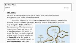
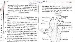
Body Fluids and Circulation 847, , , , Blood circulation in vertebrates, , Blood circulation was discovered by William harvey. Incase of, vertebrates, blood circulation is of closed type, which can be, sgTOUped into two categories:, , , , (1) Single circulation (2) Double circulation, Table : 5.3-2 Differences between single and double, circulation, , , , Double circulation in mammals can be divided into three parts, , (i) Cardiac circulation : The amount of blood present in, the heart. Its value is 8%,, , (i) Pulmonary or lesser circulation : The amount of, blood present in the surrounding of lungs and pulmonary blood, vessels. Its value is 12%,, , (iii) Systemic or greater circulation : The amount of, , blood which circulates in the rest part of the body. Its value is 80%,, I can be divided into three parts , Arterial circulation - 15%,, Capillary circulation - 5%, , S.N. | Single circulation, 1, _| Blood flows only once, , through the heart in a, , complete cycle, , Auricle —> Ventricle, , Tissues <— Gills, , Double circulation, Blood flows in two circuit, pulmonary and systemic., , Pulmonary, , , , , , ‘Venous circulation - 60%, Right Let, ao 8, sna ets, ae, , ventricle ene, , Pulmonary artery Dorsal aorta, , Heart pumps both deoxygenated, and oxygenated blood to lungs and, body respectively, hence called, arteriovenous heart, , Biood is oxygenated in lungs., , le Venacava, , , , , , hence called Venous, Heart, , 3, | Blood is oxygenated in, sil, , 4, | Less lficient as gill, capillaries slow down, the blood flow. So, the, body receives blood at, a low pressure which, decreases the rate of, ; supply to the celis, te keeps the, , Siremic veins and, , , , eseraks| 0,, , , , More efficient as blood flows at, higher pressure, especially in birds, and mammals, which increases the, raie of food and O; supply to the, calls and also rapid removal of, ‘wastes from them ie. provides a, higher metabolic rate., , , , Fig: §.3-1 Distribution of blood volume in different part of, circulatory system, , Heart, , , , , , , , , , , , , , metabolic rate low. The form, structure and function of heart exhibits much, 5, | Found only in fishes, [Found in dipnol, amphibians, variation, The characteristics of heart of fishes, amphibians,, petromyzon replies, brs and mammals. reptiles, birds and mammals is presented in the following table., Table : 5.3-3 Heart of vertebrates, S.No. | Class of vertebrates Characteristics Example, 1 | Pisces (= Branchial | Thick, muscular, made of cardiac muscles, | Labeo, heart}, Cyclostomata has two chambers (i) auricle and (ii) Scoliodon, , Ventricle. The heart is called venous heart, since it pumps deoxygenated blood to gills, for oxygenation, This blood goes directly, from ails to visceral organs (single, Circulation), A sinus venosus and conus, arteriosus is present. Lung fishes have only, ‘one auricle and one ventricle, , , , , , 2. | Amphibians, Lung fish | Hear consis of Frog, (i) Two auricles Toad Neoceratodus or, (i) Undivided ventricle Dipnot, , (ti) Sinus venosus, , (iv) Truncus arteriosus, , (conus + proximal part of aorta) Right, auricle receives blood from all the visceral, ‘organs (deoxygenated) via precaval and post
Page 3 :
848 Body Fluids and Circulation, , , , ‘caval. ‘Pulmonary arlery carries., deoxygenated blood to lungs for, oxygenation. This blood returns to left, auricle via pulmonary vein (Double circuit, irculation), , (v) SA. node in sinus venosus, , (oi) Truncus arteriosus divided into, synangium, pylangium, , , , 3. | Repites, , Hear consists of, (0) Left and right auricle, , ) Incompletely divided ventricle, (Ventricle in crocodiles, gavialis, and alligator, is completely divided), (ii) Sinus venosus, (iv) Conus arteriosus divided into right, systemic, let systemic and pulmonary arch., (Double circulation), (v) Foramen panizzae at crossing of right-left, systemic arch., (vi) Only SA node in tight auricle, , Lizards, Snakes, Turtles, , ye, , jaar + auricle, , Venttile WY, , , , Exhibit double circulation, , Heart consists of,, , () Left and right auricle, , (i) Left and right ventricle, , (ii) Complete separation of arterial and, ‘venous circulation, , (iv) Only sight systemic arch is present, , (v) Sinus venosus and tuncus, arteriosus, , absent, (ui) Two pace maker SA node and AV node, (vii) Mitral vaive present., , Pigeon, , Palo:, onary arch, , aes, , , , , , 5. | Mammals, , ‘Same as bird except that mammals have left, , Rabbit, man’, , , , , , systemic arch,, , , , , , , , Shape and position : Reddish, roughly conical, highly, muscular, mesodermal hollow organ of the size of one's fst. Its, average weight in males is about 300 gm. and in females about, 250 gm. It lies behind the stemum in the mediastinum space of, thoracic cavity in between the two lungs. The broader base faces, upward and backward. The narrower apex is directed downward,, forward and slightly towards left, lying between 5 and 6" ribs andl, rests on the diaphragm. The heart is about 12 cm (5 inch) long, 9, ‘cm (35 inch) wide and 6 em (2.2 inch) thick., , , , Fig: 5.3-2 Position of heart in our chest cavity, , Protective covering : Heart is enclosed in a tough, 2, layered fibroserous sac, the pericardium. The outer layer is nondistensible fibrous pericardium and inner layer is thin serous, pericardium which further consists of outer parietal layer (attached, to fibrous pericardium) and inner visceral layer (adhered to the, heart)., , Outer fibrous pericardium, Pectin | Outer parietal, Inner serous pericardium - layer, , Inner visceral, layer, Between the parietal and visceral layers, occurs a narrow, potential space, the pericardial cavity which is derived from coelom, and is filed with serous pericardial fluid for frictionless movement, and protection from shock and mechanical injury., , Histology : The heart wall consists of connective tissue, blood, vessels and cardiac muscle fibres in 3 different layers ~ Epicardium,, Myocardium and Endocardium,, , (1) Epicardium : The outermost epicardium, also called, visceral layer of the serous pericardium, is the thin, transparent, outer layer of the wall. It is composed of mesothelium and, connective tissue, Visceral pericardium, is joined to the, myocardium by connective tissue,
Page 4 :
Body Fluids and Circulation 849, , , , (2) Myocardium : Middle, highly vascular layer, composed, of cardiac muscle fibres are joined together by intercalated dis., The connective tissue in myocardium acts as cardiac skeleton., Myocardium is thickest where the endocardium is thinnest., , (3) Endocardium : Innermost layer lining the cavity of heart, and consisting of endothelium of squamous cells resting on thin, basement membrane of loose connective tissue., , , , 3. Diagram to show the layers of the pericardium, , External structure : Human heart is 4-chambered and is, divided by septa into two halves — right and left. Each half has one, darker, thin walled auricle in the broader upper region and one, lighter, thick-walled ventricle in the narrower lower region,, , Sinus venosus and conus/truncus/bulbus arteriosus are, accessory chambers in the heart of lower vertebrates (fishes and, amphibians). In rabbit, sinus venosus is formed in the embryo but, later it becomes a part of wall of right auricle., , In frog, sinus venosus spreads upon most of the dorsal, side of heart and conus arteriosus lies obliquely upon the ventral, surface of right atrium., , Brachiocephalic, trunk, , Superior >, , vena cava, Ascending, , , , , , , , , , , Great, cardiae, , \, Interventricular, sulcus, , Fig: 5.3-4 External features of human heart, , , , Internal structure, , (1) Aurietes : Atria are thin walled. They act as reservoirs for, blood entering the heart. Right auricle is bigger than left auricle, and both are separated by a myomembranous partition called, Inferatrial or interauricular septum, During embryonic stage, at the, place of this septum, there are present septum primum and septum, secondum having a gap (aperture) called foramen ovale between, them. From the opening of inferior vena cava upto foramen ovale,, there is a flap called Eustachian flap which prevents the blood in, the foetal heart go to hungs because in foetal life, lungs are not, functional purification of blood is done by placenta,, , , , At the time of birth, there is closure of foramen ovale but there, temains depression on posterior part of the right surface of, interauricular septum in rabbit. In man this depression is present, fon both the side because of least regenerative power in human, being. The depression towards right atrium is called fossa ovalis, and depression towards left atrium is called fossa lunata,, , ‘The inner surface of auricles is smooth, A network of muscular, ridges called musculi pectinati or trabeculi.pectinati occurs, intemally in the region of the auricular appendages and give comb, like appearance., , Q PFO (Patent Foramen Ovale) or septal defect : In, case there is no dosure of foramen ovale, then disease is called, PFO. In this condition, there is mixing of blood after birth which, ives bluish appearance to the body called as Cyanosis. Such child, is called Blue Baby,, , (2) Ventricles : The right and left ventricles are, demarcated by an interventricular septum which is obliquely, curved towards right, so that the left ventricle is larger than right, one. However, the cavity of left ventricle is relatively smaller, and nearly circular because the myocardium of left ventricle is 3, times thicker than right ventricle whose cavity is larger and, somewhat crescentic., , ‘The walls of the ventricles are intemally raised into a number of, thick, muscular, column shaped projections called cohimnae carnae, or trabecular camae; and a few large muscular elevations called, Papillary muscles or musculli papillares which are 3 in right, ventricle and 2 in left ventricle. These muscles act as anchors for, chordae tendinae,, , , , ‘Numerous, strong, inelastic thread like tendons are present in, the mammalian heart but absent in frog,, , Q Regurgitation : If there is weakening of papillary, muscles or breaking of chordae tendinae, then AV valves revert, into auricles. So, blood goes in opposite direction, it is called, Tegurgitation. Sometimes, there is narrowing of valves, So, there, remains gap between the valves which causes regurgitation., , Q Moderator band : Right ventricle contains a prominent, muscular trabeculum called moderator band which extends from, the interventricular septum to anterior papillary muscle,
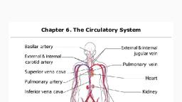
Page 5 :
850 Body Fluids and Circulation, , , , , , , , , , , , , , , , ‘Superior vena cava., Right pulmonary artery, Pulmonary valve, , Right pulmonary veins, , Opening of superior vena cava,, , Opening of coronary sinus, , Tricuspid valve, Right ventricle ——, , Inferior vena cava, , Fi, , , , Major blood vessels associated with heart : The blood, vessels that enter or leave the heart are called Great Blood Vessels., , (1) Superior vena cava or precaval : Brings deoxygenated, blood from head and upper parts of the body into the right auricle, through an opening which is single in human and cat and two in, rabbit as there are 2 precavals — right and leit in rabbit., , (2) Inferior vena cava or post caval : Drains, deoxygenated blood from middle and lower parts of the body into, the right auricle through a single opening which is bordered by a, membranous, falciform fold which is a remnant of the foetal valve, of Eustachian,, , (3) Coronary sinus : Returns deoxygenated blood from, heart wall into right auricle through a single opening,, , (4) Pulmonary vein : Four pulmonary veins, two from each lung,, ‘camy oxygenated blood from the lungs and open into the leit auricle, through four openings. In rabbit, the pulmonary veins open in the left, auride through 2 openings., , (5) Pulmonary aorta/arch : Arises from upper left corner of, tight ventricle through a single opening and divides into right and, Jeft pulmonary arteries which carry deoxygenated blood to the, Jungs for oxygenation., , (6) Systemic aorta : Arises from upper right comer of left, ventricle through a single opening and has 3 regions ~ ascending, aotta, arch of aorla and descending aot. It distributes oxygenated, blood to various body parts except lungs., , (1 Ligamentum arteriosum : During foetal life, because, the lungs ate non-functional hence blood of pulmonary aorta, comes into systemic aorta through a small duct called ductus botalli, or ductus arteriosus soon after birth, deposition of elastin fibre blocks, this duct, forming a new structure called ligamentum botalli or, ligamentum arteriosum., , PDA (Patent Ductus Arteriosus) : If the ligamentum, arteriosus remains open, the condition is called PDA. In this case,, there is mixing of blood which leads to blue baby., , Left common carotid artery, Left subclavian artery, , Brachiocephalic trunk, , Arch of aorta, Jgamentum arteriosum, , Left pulmonary artery, Pulmonary trunk, , a — Left pulmonary veins, Left atrium, , CChordae tendinae, Left ventricle, Interventricular septum, , Descending eorta, , 5.3-5 Intemal anatomy of human heart, , Valves : Various membranous structure in a hollow organ or, passage that temporarily closes in order to permit flow of flood in, one direction only,, , (1) Eustachian valve : Present on the opening of inferior, vena cava (post caval) in the right auricle in rabbit, whereas in, Jnuman, the vestige of eustachian valve is present over the opening, of post caval vein. It allows the passage of blood in right auricle,, , (2) Haversian valve : Present in human but absent in rabbit., It is present over the opening of precaval vein and allows the, passage of blood in right auricle., , (3) Thebesian or coronary valve : Present over the, ‘opening of coronary sinus in right auricle in mammals and allows, the passage of blood in right auricle., , (4) Right A.V. valve or Tricuspid valve : Present between, right auricle and right ventricle, It consists of 3 membranous flaps, or cusps., , , , , , , Superior vena cava, (Deoxygenated) Sip) AN ‘Oxygenated blood, WYER ee Pt, Aorta Pulmonary, Pulmonary val Eman, {Oxygenated blood), Left ati, Right atrium Le, “Tricuspid valve oe bs, Inferior vena cava| A Left venttide, (Deoxygenated) ceil, , Right ventricle, Fig : 5.3-6 Path of blood through the heart, (5) Left A.V. valve or Bicuspid or Mitral valve : Present, between left auricle and left ventricle, It consists of 2 flaps or cusps., ‘The bicuspid valve resembles mitre or topi of bishop, hence, also, called as Mitral valve.
 Learn better on this topic