Notes of NEET,JENPUS &ETC BATCH, Biology Animal tissue.pdf - Study Material
Page 1 :
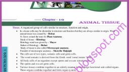
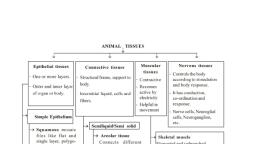
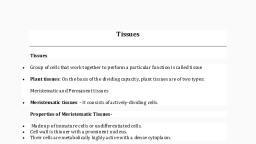
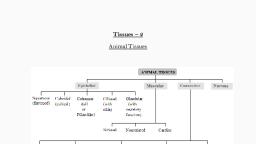
A tissue may be defined as a group of one or more types of, cells having a similar origin and specialized for a specific function, or functions along with the intercellular material., , Branch of biology dealing with the study of tissue is called, histology. The term ‘tissue’ was introduced by Bichat and also, known as ‘Father of histology’, Mayer coined the term ‘histology’, and the founder of histology is Marcello Malpighi. Following types, of tissues are found in animals :, , Epithelial Tissue, , An epithelium is a tissue composed of one or more layers of, cells that caver the body surface and lines its various cavities, It, serves for protection, secretion and excretion. The word, ‘epithelium’ (G. epi = upon, thele = nipple) was introduced by, Ruysch. They are located on the outer surfaces of organs, including, the skin, They form the linings of tracts, cavities and vessels, Epithelial tissue evolved first in animal kingdom. It originate from, all the three primary germ layers., , Structure, , Cells are arranged in one or more layers, cells are compactly, arranged and there is no inter cellular matrix between them., Neighbouring cells are held together by intercellular junctional, complexes like desmosomes, tight junctions, interdigitations et., The cells of lowermost layers always rest on a non living basement, membrane or basal lamina, Basement membrane is made up of no, cell product of epithelial tissue. It is formed of, mucopolysaccharides, glycoprotein and collagen or reticular fibres., Blood vessels are absent in the epithelia tissues. However, nerve, endings may penetrate the epithelium. It possesses very high, capacity of renewal (mitotic cell division). The following types of, modifications and junctions are found in the plasma membrane of, adjacent epithelial cells o keep the cells together., , , , , , Fig: 2.3-1 Diagram to show an epithelium with Its basement, membrane resting upon underlying connective tissue, , Microvilli : These are simple and minute cytoplasmic, processes arising from free exposed surfaces of the cell. They, absorb material. e.g. Intestine., , Stereocilia : These are non-motile cytoplasmic processes., eg, Epididymis, vas deference., , Kinoellia : It is contractile motile fibrous processes arising, from basal granules. e.g. Oviduct, Fallopian tube., , Tight junctions (Zona occludens) : At certain places the, plasma membranes of adjacent cells are tightly packed or even, fused together. e.g. Brain., , Desmosomes : Desmosome is present in epithelial tissue., They consist of thickened area and several fine tonofibrils, extending from each plasma membrane into cytoplasm of, respective cells, Macula adherens is a kind of desmosome. e.g., Vagina, Urinary bladder., , Gap junction : At place, the adjacent cells form ion-rich gap, junctions for intercellular communication and chemical exchange., ‘These junctions probably do not provide physical support., , Interdigitations : These are interwoven finger-like processes, of plasma membranes of adjacent cells.
Page 2 :
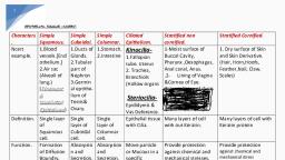
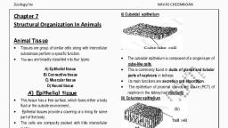
“Ani, , , , , , Intercellular bridges : These are minute projections that, arise from adjacent cell membranes. The intercellular bridges make, contact with one another., , , , epithelial cel, , Functions, , Epithelial tissues have a wide spread distribution throughout, the body and serve several important functions —, , (1) Generalized protection is the most important function of, membranous epithelium. It is the relatively tough and impermeable, epithelial covering of the skin that protecis the body from, mechanical and chemical injury and also from invading bacteria, and other disease causing micro-organisms., , (2) Epithelial structures specialized for sensory functions are, found in the skin, nose, eve and ear., , (3) Glandular epithelium is specialized for secretory activity,, secretory products include hormones, mucous, digestive juices and, sweat., , (4) The epithelium lining of the gut and respiratory tracts, allows the absorption of nutrients from the gut., , (5) Itis the specialized epithelial lining of kidney tubules that, makes the excretion and concentration of excretory products in the, urine., , (6) Ciliated epithelium moves fluid, mucus and other materials,, in the organs it lines,, , (7) Germinal epithelium of the seminiferous tubules and, ovaries produces spermatozoa and ova respectively., , (8) The ability of epithelium to regenerate quickly helps in the, healing of wounds., , (9) Pigmented epithelium of retina darkens the cavity of, eyeball, , (10) The epithelia check the absorption of harmful or, , unnecessary materials,, , (11) Epithelium of alveoli of the lungs brings about exchange, of gases between blood and air., , (12) Epithelium also produce exoskeletal structures such as, scales, feathers, hair, nail, claws, homs and hoofs., , ‘Types of epithelial tissue, , Mainly based on the location and functions of tissue it Is of, following types , (1) Simple epithelium : It simple in structure and basically, formed by single layer cells,, , (i) Simple squamous epithelium : It consists of only one, layer of flat, scale like cells, usually polygonal cells which are, closely fitted together like the tiles of a mosaic. It is also known as, pavement epithelium. e., It forms lining of blood vessels, lymph, vessel, heart, peritoneum, pleura, Bowman's capsule, thin segment, of loop of Henle and lung alveoli,, , , , Fig : 2.3.3 Simple squamous epithelium, , (i) Simple cuboidal epithelium : The simple cuboidal, epithelium is composed of one layer of cuboidal shaped cells, resting on a basement membrane. The nuclei are situated centrally,, e.g. the cuboidal epithelium is present in the small salivary and, pancreatic ducts, thyroid vesicles, parts of membranous labyrinth,, PCT, DCT, ovaries, seminiferous tubules of testes, ciliary bodies,, choroid, iris of eyes, thin bronchioles and sweat gland of, mammalian skin., , , , Fig : 2.3-4 Simple cuboldal epithelium, , (iii) Simple columnar epithelium : It consists of a single, layer cells, many of which have modified structure. Three common, modifications are goblet, cilia and microvili, Simple columnar, epithelium is present in the stomach and intestine e.g. inner lining, of gall bladder and bile duct. It also occurs in the gastric gland,, intestinal glands, pancreatic lobules.
Page 3 :
Animal Tissues, , , , , , , , Micro, , Mucus, , Cyloplasm, Goblet cell,, , , , , Nucleus, Cell membrane, , Absorptve ell, Basement membrane, , .3-5 Simple columnar epithelium, , (jv) Simple ciliated epithelium ; It bears numerous, delicate hair like outgrowths called cilia arising from basal granules, which helps to create a current to transport the materials. The, ciliated epithelium is of two types :, , (a) Ciliated columnar epithelium : It lines respiratory tract, (Lower end of bronchi), fallopian tubes (oviducts), ventricles of, ‘brain (ependyma), central canal of spinal cord, tympanic cavity., , , , , , Replacement cel, , Basement membrane, , Fig: 2.3-6 Simple columnar ciliated epithelium, , {b) Ciliated cuboidal epithelium : It occurs in certain parts, of nephrons of the kidneys., , (v) Pseudostratified epithelium : It always consist of single, layer of irregularly shaped colurnnar cells, touches the basement, membrane. The long cells have oval nuclei however,, , Mucus, , , , , , , Cxtoplaam, E Short cell, Faiement membrane, Fig: 2.3-7 Pseudostratified epithelium, , ‘Short cells have rounded nuclei although epithelium is one, cell thick, but it gives the appearance of a stratified epithelium,, hence it is called pseudostratified epithelium. Mucus secreting, goblet cells are numerous and cilia are present. It is of two types ~, , (a) Pseudostratified columnar ciliated epithelium : It is, found in the lining of trachea and bronchi (Upper), , {b) Pseudostratified columnar epithelium : It is found in, certain segments of human male urethra and parotid salivary, gland, vasa deferentia and epididymis., , , , (c) Stratified squamous epithelium : The cells in the, deepest layer are columnar or cuboidal with oval nucle. Its called, germinative layer. The cells of this layer divide by mitosis to form, new cells, , (2) Compound epithelium : It is complex in structure and, basically formed by two or more than two layers of cells., , (i) Stratified squamous keratinised epithelium :, Stratified squamous epithelium is characterized by multiple layers, of cells with typical, flattened squamous cells, at the free or outer, surface of the sheet. The, presence of keratin in, these cells contributes 10, the protective qualities of, skin covering the body, surface, Keratin is dead, and waterproof soit, protects the underlying tissues from abrasion and infection e.9,, epidermis of the skin of land vertebrates., , , , , , , , , , , , seep, SNe ia), seats], , , , , Basement, membrane, , , , 8 Stratified squamous, Keratinised epithelium, , , , (i) Stratified squamous non keratinised epithelium : Its, free surface is moist, and the outer epithelial cells, unlike those, found in the skin, do not contain keratin. This type of epithelium, serves a protective function. It is found lining the oral cavity, {buccal cavity), pharynx, oesophagus, anal canal, lowerpart of, urethra, vocal cords, vagina, cervix (lower part of uterus) and, cornea of eyes., , Squamous ayers, , Intermediate layers, , , , Basement membrane, Fig : 2.3-9 Stratified squamous non keratinised epithelium, , (iii) Stratified cuboidal epithelium : It consists of two or, more rows of low cuboidal-shaped cells which are arranged, randomly over a basement membrane. It is found in the sweat, gland duets, larger salivary and pancreatic ducts., , iv) Stratified columnar epithelium :Itis protecive epithelium, having multiple layers of columnar cells, only the most superficial, cells are truly columnar in appearance. Epithelium of this type is, rare. It is found in male urethra and in the mucous layer near the, anus. It also lines mammary gland ducts and epiglottis., , Cyteplasm, , ‘Nuclous, , Germinative, layer, , , , Basement, membrane, , , , (b), Fig: 2.3-10 (a) Stratified columnar ciliated epithelium, (b) Stratified columnar epithelium
Page 4 :
Animal Tissues 423 |, , , , () Stratified columnar ciliated epithelium : It lines the, Jarynx and upper part of the soft palate., , (3) Specialized epithelium : This type of epithelium are, specialized to perform specific activity hence, specialized in, structure also. They are as follows ~, , (i) Transitional epithelium (Urothelium) : It is often ten or, ‘more layers thick. It lacks germinative layer, basement membrane., Stratified transitional epithelium is typically found in the body areas, such as the wall of urinary bladder, ureter and renal pelvis. It is, located in all the hollow viscera subjected to stress and protects, organ wall from tearing,, , (i) Neurosensory epithelium : Olfactory mucosa, called, Schneiderian membrane, lining of internal nares, retina of eyes, and epithelial covering of tongue containing taste buds are, examples of neurosensory epithelia. The sensory cells bear, at their, free ends, slender “sensory hairs” to receive specific stimuli. Basely,, these cells are connected, by means of synapses, with fine fibrils of, sensory nerves,, , (ii) Pigmented epithelium : The epithelial cells of the basal, , layer of retina contain pigment. Hence, this layer is often refered, to as a pigmented epithelium. e.g. — Pigmented layer of retina, iris, and skin., iv) Germinal epithelium : Specialized cuboidal cells, capable of producing gametes as found in gonads. Germinal, epithelium produces gametes e.9., ova (Female gametes) and, sperms (Male gametes), , Glands, , , , , , Glandular epithelium are specialized for secretory activity. A, cell, tissue or organ which secretes a useful chemical material is, known as gland. Glands are made up of cuboidal epithelial cells, which are more secretory. All glands arise as folding of epithelia., ‘The golgi body in gland cells are larger and more secretory. Most, of the glands of body are merocrine types. It originate from all, three germinal layers. (ecto, meso and endoderm). Liver is the, largest gland of the body and lined by glandular epithelium., , ‘Types of glands, , (A) On the basis of number of c, , (1) Unicelludar gland : It consist of unicellular gland cells, which are called as goblet cells or chalice cells. They secrete, mucous and found in mucosa of intestine and stomach. Mucous, lubricates the food for easy peristalsis. Their life span is about 2-3, days, , (2) Multicellular gland : It consist of many cells and are, generally located in underlying connective tissue e.g. gastric and, intestinal glands., , (B) On the basis of presence or absence of ducts, , (1) Exocrine gland : These are those glands which discharge, their secretory products into ducts. It is also called ducted glands or, glands of external secretion. e.g. Salivary glands, Mammary glands, and Tear glands., , (2) Endocrine gland : They are often called ductless gland,, because they discharge their secretory products (hormones), directly into the blood. eg, Pituitary gland, thyroid, parathyroid, and adrenal glands,, , , , (3) Heterocrine gland : These are those glands which are, partly endocrine and partly exocrine in function, e.g, Pancreas., Structural classification of exocrine glands, , Mullticellular exocrine glands are classified by structure, using, the shape of their ducts and the complexity (branching) of their, duets system as distinguishing characteristics. Shape indude tubular, and alveolar (Sac like). Simple exocrine glands e.g. intestinal, glands, mammalian sweat glands, cutaneous glands of frog etc., have only one duct leading to surface. Compound exocrine glands, have two or more ducts e.g. liver, salivary glands ete., , , , , , , , , , , , , , , , , , , , , , , , , , , , , , , , , , , , , , Table : 2.3-1, Type Example., ‘Simple tubular Intestinal glands, erypis of Lleberluhn, in ileum,, ‘Simple colled tubular Sweat glands in man, Simple branched tubular | Gastic (stomach) gland, and Uterine, gland., ‘Simple alveotar ‘Mucous gland in skin of frog, Polson, gland of toad and seminal vesicle,, Simple branched alveolar | Sebaceous glands, ‘Compound tubular Brunners gland, bulbourethral gland, nd liver., Compound alveolar Sublingual and submandibular salivary, sland., Compound tubulo alveolar | Parotid salivary glands, Mammary, gland and Pancreas,, , , , , , , , Classification of glands on the basis of their mode of, secretion, , (1) Apocrine gland : Apocrine glands collect their secretory, products near the apex or tip, of the cell and then release it into a, duct by pinching off the distended end. This process results in, some loss of cytoplasm and damage to the cell. e.g. Mammary, glands. (Modified sweat gland), , (2) Holocrine gland :, , release it, These cells self destruct to complete their functions. e.g,, Sebaceous glands. In case of rabbit sebaceous glands are found in, , (3) Merocrine gland : Meroctine glands (Ecerine or Epicrine, glands) discharge their secretory product directly through the cell or, plasma membrane, without injury to the cell wall and without loss, of cytoplasm. e.g. Sweat glands, exocrine region of vertebrate, pancreas, salivary glands and intestinal glands ete,, , Secretion
Page 5 :
se z, Classification of glands on the basis of nature of product, , (1) Mucous gland : Secret slimy mucous e.g. goblet cells,, palatine gland, gland of uterus, some gastric gland and gland of, colon., , (2) Serous gland : Produce watery secretion. e.g. pancreas,, parotid, salivary gland, sweat gland and intestinal gland., , (3) Seromucous gland’: Secrete mixed liquid. e.g. Most, gastric gland, sublingual, submaxillary salivary aland, pancreas., , (4) Cytogente gland : They produce cells e.g. Testis and, ovary., , Connective tissu, , , , It connects and supports all the other tissues, the intercelular, element predominating, The cellular element is usually scanty. In, function this tissue may be mechanical, nutritive and defensive. It is, a tissue made up of matrix (abundant intercellular substance or, ground substance) and living cells that connects and support, different tissues. All connective tissues in the body are formed by, mesoderm., , Structure, , There are large intercellular spaces between the cells., Intercellular spaces are filled with large amount of extracellular, ‘materials formed of insoluble protein fibres lying in an amorphous,, transparent ground substance called matrix. Ageing of an animal, ‘body is associated with deterioration in its connective tissues., Functions, , (1) Their chief function is to bind other tissues together in the, organs., , (2) Certain connective tissues such as adipose tissues store fat, , (3) Skeletal connective tissues like bones and cartilages, provide the body with a supporting skeletal frame work., , (4) Fluid connective tissues such as blood and Iymph transport, various materials in the body., , (8) Plasma cells synthesize antibodies, viz., macrophages., Lymphocyies ingest cell debris, harmful bacteria and for, ‘matter, Thus these cells of connective tissues are protective in, function., , , , (6) The jelly-like ground substance of connective tissues acts, as shock absorber around some organs such as eye balls and, kidneys., , (7) The bone marrow produces blood cells., , (8) Areolar tissue acts as packing material in various organs., , (9) Collagen fibres of connective tissue help in repair of, injured tissues., , ‘Types of connective tissues, , Connective tissue proper possess soft viscous semisolid or, semi-fiuid matrix. It is divided into following types :, , (1) Areolar Tissue : Areolar tissue is loose connective tissue,, ‘possess transparent gelatinous, highly vascular and sticky matrix, which have variety of cells and fibres. It allows movement of part, , ‘connected by it (Muscle and their compound). Areolar tissue, mainly consist of different types of cells and fibres., , , , Fig: 2.3-12 Areolar connective tissue, , (i) Cells of areolar tissue : It has following types , Fibroblast : These are the most abundant cells, produces, fibres, called as fibroblasts in their young active phase and, fibrocytes when old and inactive. It synthesize proteins (Collagen,, elastin and reticulin). These are undifferentiated mesenchyme stem, cells, capable to give rise other cells of connective tissue. Collagen, and elastin are formed by fibroblasts,, , Histiocytes or Macrophages or Clasmatocytes : These, are polymorphic cells. These are amoeboid cells and are main, phagocytes of connective tissue. They are having most active, lysosomes and phagocytise dead cells and pathogens., Macrophages remove the dead and damaged cells and clean the, body so called scavenger cell. All types of macrophages take part in, phagocytosis., , , , \crophat, , , , Monocyte Alveolar Dust Microglia Astrocytes Reticular Kupfer Splanose, (Blood) Phagocyte cells (Brain) (Spinal cells cells cells, , (Alveoli) (Lung) ‘cord) (Reticulo (Liver) (Spleen), endathelial cells, , Reticular cells : Present only in the reticular tissue and, , , , stellate in appearance, Infact they are modified fibroblast, producing reticular fibres., , Mast cells : Mast cells were discovered by Paul Echrlich, They are large, irregular ovoid cells found in areolar tissue. and, their number increases during allergies. It produces or secretes, histamine (vasodilator), serotonin (vasoconstrictor) and heparin, (anticoagulant). Histamine dilate the blood vessels in allergic and, inflammatory conditions. Heparin checks the clotting of blood, inside the blood vessels. Serotonin act as vasoconstrictor to arrest, bleeding., , Lymphocytes : These are the smallest, less numerous and, spherical or ovoid cells resembling lymphocytes of blood and, lymph. These actively move about by pseudopodia. Their function, is to form and carry antibodies. That is why, they are seen in large, numbers at sites of inflammation.
 Learn better on this topic