Notes of NEET,JENPUS &ETC BATCH, Biology Anatomy of flowering plan - Study Material
Page 1 :
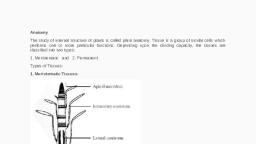
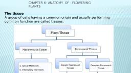
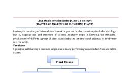
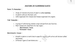
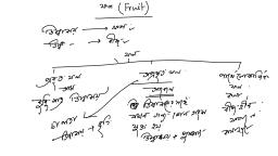
Anatomy of Flowering Plants, , , , AA group of cells performing a particular function is collectively, called as tissue. A tissue may be defined as, “a group of similar or, dissimilar cells having common origin and performing specific, function",, , Tissues are mainly divided into three categories : Meristematic, tissues or Meristems, Permanent tissues and Secretory tissues,, , , , Meristematic tissues or Meristems, , , , The word “Meristem” originated from “Meristos” (Greek =, continuous division) and the term meristem was introduced by, Nageli (1858). A group of cells which are much active and capable, of showing continuous divisions and redivisions, is called as, meristematic tissue. The various characteristic features of the, meristems are discussed below, , (1) They contain immature and young cells and are capable of, repeated divisions., , (2) Intercellular spaces are not present in meristematic tissue,, (3) They contain a homogeneous thin cellulosic wall,, , (4) They contain large nuclei associated with abundant, cytoplasm,, , (5) They are metabolically very active but they do not store, food material and further no plastids in them, , (6) Vacuoles are small or absent,, (7) Meristematic cells are isodiametric in shape., , (8) Undifferentiated tissue in which cells divides continuously, SeeS9G,, , ‘Types of meristems, , ‘The meristems may be classified on the basis of their mode of, otigin, position or function, , According to origin and development : On the basis of, origin, meristematic tissues are of three types :, , (1) Promeristem or Primordial meristem : The, promerister originates from embryo and therefore, called primordial, cor embryonic meristem, Itis present in the regions where an organ or, a part of plant body is initiated. A group of inital cells that lay down, the foundation of an organ or a plant part, is called promeristem, It, ‘occupies a small area at the tips of stem and root. The promeristem_, gives tise to all other meristems including the primary meristem., , (2) Primary meristem : A primary meristem originates from, promeristem and retains its meristematic activity. It is located in the, apices of roots, stems and the leaf primordia. Primary meristem, gives tise to the primary permanent tissue., , , , (3) Secondary Meristem : They always arise in permanent, tissues and have no typical promeristem. Some living permanent, cells may regain the meristematic nature. This process in which, permanent tissue regains meristematic nature is called, dedifferentiation, The secondary meristems are so called because, they originate from permanent cells. The phellogen or cork, camblum arising from epidermis, cortex or other cells during, secondary growth, is an important example of secondary meristem., The secondary meristems produce secondary tissues in the plant, body and add new cells for effective protection and repair., , , , , , According to position : On the basis of their position in the, plant body meristems are classified into three categories, , (1) Apical meristem : This meristem is located at the, growing apices of main and lateral shoots and roots. These cells, are responsible for linear growth of an organ. Solitary apical cells, occur in ferns and other Pteridophytes while apical initials are, found in other vascular plants
Page 2 :
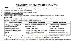
(2) Intercalary meristem : These are the portions of apical, meristems which are, separated from the apex, during the growth of, axis and formation of, permanent tissues. It is, present mosily at the, base of node (eg,, Meriha viridis, Mint),, base of intemode (e.g,, siem of many monocots, viz, Wheat, Paddy,, Grasses, Pteridophytes, like Equisetum) or at, the base of the leaf @ ib), (eg, Pinus). The Fig 2.244 Vartous meristematic, intercalary meristems ultimately disappear and give rise to, permanent tissues., , , , , , (3) Lateral meristem ; These meristems occur laterally in, the axis, parallel to the sides of stems and roots. This meristem, consists of initials which divide mainly in one plane (periclinal) and, results in increase in the diameter of an organ, The cambium of, vascular bundles (Fascicular, interfascicular and extrastelar, cambium) and the cork cambium or phellogen belongs to this, category and are found in dicotyledons and gymnosperms., , According to function : Haberlandt in 1890 classified the, primary meristem at the apex of stem under the following three, types, , (1) Protoderm : It is the outermost layer of the apical, meristem which develops into the epidermis or epidermal tissue, system,, , (2) Procambium : It occurs inside the protoderm. Some of, the cells of young growing region which by their elongation and, differentiation give rise to primary vascular tissue, constitute the, procambium., , (3) Ground meristem : It constitutes the major part of the, apical meristem which develops ground tissues like hypodermis,, cortex, endodermis, pericycle, pith and medullary rays., , According to plane of cell division : On the basis of their, plane of cell division meristem are classified into three categories :, , (1) Ma all, planes, so mass of cells is formed. e.g, formation of spores, cortex,, pith, endosperm., , meristem : The cells divide anticinally, , , , (2) Plate meristem : The cells divide anticlinally in two, planes, so plate like area increased. eg., formation of epidermis, and lamina of leaves., , (3) Rib or File meristem : The cells divide anticlinally in, one plane, so row or column of cells is formed. e.g,, formation of, lateral root., , ‘Structure and organisation of apical meristem, , (1) Vegetative shoot apex : Shoot apex was first recognized, by Wolff (1759) shoot apex is derived from meristem present in, plumule of embryo and occurs at the tip of stem and its branches, as terminal bud. It also occurs in the inactive state in the axils of, leaves as lateral buds. The tip of the shoot apex is dome-shaped, and from its flanks at the base of the dame divide to form one or, more leaf primordia. This continues throughout the vegetative, phase. Many theories have been put forward to explain shoot, apex, such as :, , (i) Apical cell theory : This theory was proposed by Nagel, (1858). According to this theory, shoot apical meristem consists of, single apical cell. This theory is applicable in case of higher algae,, bryophytes and in many pteridophytes but not in higher plants, (ie., gymnosperms and angiosperms)., , (i) Histogen theory : It was proposed by Hanstein (1870), According to this theory, the shoot apical meristem consists of, three distinct meristematic zones or layers (or histogens)., , {a) Dermatogen : Outermost layer and it forms epidermis, and epidermal tissue system., , (b) Periblem : [tis the middle layer which gives rise to cortex, and endodermis., , (c) Plerome : The innermost layer forms pith and stele., , (ii) Tunica corpus theory : This theory was proposed by, Schmidt (1924). According to this theory, the shoot apex consists, of two distinct zones., , (a) Tunica : It is mostly single layered and forms epidermis., The cells of tunica are smaller than corpus. The tunica shows only, ‘anticlinal division and itis responsible for surface growth., , , , Fig : 2.2-2 LS. Vegetativ, , , , (b) Corpus : It represents the central core with larger cells,, Corpus shows divisions in all planes and it is responsible for, volume growth,, , Popham and Chan (1950) introduced the term mantle for, tunica and core for corpus.
Page 3 :
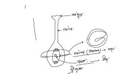
Anatomy of Flowering Plants 381, , , , (2) Root apex : A group of initial cells, present at the, subterminal region of the growing root tip, which is protected by a, root cap, is called root apical meristem or root apex. It is, ‘embryonic in origin and formed from the radicle part of embryo., However, in adventitious roots it is produced from derivatives of, toot apex. According to Hanstein (1870) root apex of most of the, dicotyledons also consists of three meristematic zones - plerome,, periblem and dermatogen (fourth meristem calyptrogen to form, root cap only in monocots), , (i) Dermatogen : It gives tise to epiblema or piliferous layer, or thizodermis., , (ii) Periblem : It gives rise to cortex including endodermis..., , (iii) Plerome : It gives rise to vascular tissue including pith., , Regarding the apical organisation of root following theories, have been put forward., , Korper-Kappe theory : It was proposed by Schuepp (1917)., This theory is comparable with the tunica and corpus theory of, shoot apex. Korper means body and Kappe means cap., , , , Fig : 2.2-3 LS. Root apical meristem, , Quiescent centre theory : It was proposed by Clowes, (1961) in maize. According to him, in addition to actively dividing, cells, a zone of inactive cells is present in the central part of the root, apex called quiescent centre., , , , The cells in this region have light cytoplasm, small nuclei,, lower concentration of DNA, RNA and protein., , (3) Reproductive apex : During reproductive phase, the, vegetative apices are, converted into reproducti, apices. Before conversion,, the apex stops producing, leaf primordia. The summit, of the, remained inactive during, the vegetative phase, starts, dividing, As a result of cell, apical, meristem undergoes change in shape and increase in size, The, apex may develop into a flower or an inflorescence., , , , , , apex — whicl, , , , 2.2-4 LS. Reproductive, , , , apex (diagrammatic), divisions, the, , , , Permanent tissues, , Permanent tissues are made up of mature cells which have, lost the capacity to divide and have attained a permanent shape,, size and function due to division and differentiation in meristematic, tissues. The cells of these tissues are either living or dead, thinwalled or thick-walled, Permanent tissues are of following types, Simple permanent tissues, , Simple tissues are a group of cells which are all alike in origin,, form and function, They are further grouped under three, categories :, , (1) Parenchyma : Parenchyma is most simple and, , cialized tissue which is concerned mainly with the vegetative, activities of the plant., , , , ‘Nucleus, , , , , fitercellular space, , Vacuoles, , Fig: 2.2.5 Parenchyma in T.S., The main characteristics of parenchyma cells are, , (i) The cells are isodiametric, living, thin walled, soft, possess a, distinct nucleus, having well developed intercellular spaces,, vacuolated cytoplasm and cellulosic cell wall., , {ii) The shape may be oval, spherical, cylindrical, rectangular, and stellate (star shaped) in leaf petioles of banana and canna and, some hydrophytes., , (iii) This tissue is generally present in roots, stems, leaves,, flowers, fruits and seeds., , {iv) If they enclose large air spaces they are called as, aerenchyma; if they develop chlorophyll, they are called as, chlorenchyma and if they are elongated cells with tapering ends,, they are called as prosenchyma., , Functions : They perform the following functions, , {i) Storage of food materials. e.g., Carrot, Beetroot etc., , (ii) Chlorenchyma helps in photosynthesis, Aerenchyma, helps in floating of the aquatic plants (Hydrophytes) and also, helps in gaseous exchange during respiration and photosynthesis,, eg, Hydrilla, , (iii) In turgid state they give rigidity to the plant organs., , {iv) In emergency they behave like meristematic cells and help, in healing of the various plant injuries., , {v) Sometimes they store secretory substances (ergastic, substance) such as tannins, resins and gums and they are called as, joblasts.
Page 4 :
382 Anatomy of Flowering Plants, , , , (2) Collenchyma : The term collenchyma was coined by, Schleiden (1839). It is the tissue of primary body. The main, characteristics of collenchyma are given below :, , (i) The cells of this tissue contain protoplasm and are living, without intercellular spaces. The cell walls are thickened at the, comers and are made up of cellulose, hemicellulose and pectin,, , (i) They are compactly arranged cells, oval, spherical or, polygonal in outline. The tissue is elastic, extensible and have, capacity to expand., , (ii) Collenchyma occurs chielly in the hypodermis of, dicotyledonous stems (herbaceous, climbers or plants e.g., Cucurbita, Hellanthus) and leaves. They are usually absent in, monocots and in roots., , ‘Thickening on comers, ‘due to deposition of, callulose and pectin, , , , Fig : 2.2-6 (a) Collenchyma L.S. (b) and (c) T'S. of the same, , Types of collenchyma : Majumdar (1941), collenchyma into three types on the basis of thickening, , divided, , (i) Angular collenchyma : When the thickening of the cells, is confined to the comers of the cells. eg, Tagetes, Tomato,, Datura, Potato, etc., , (ii) Plate or Lamellar collenchyma : When the thickenings, are present in the tangential walls. e.g, hypodermis of sunflower stem., , (ii) Lacunar or Tubular collenchyma : Ifthe thickened cell, ‘wall is associated with intercellular spaces of the adjacent cells. e.g, leaf petioles of compositae (asteraceae) and malvaceae etc., hypodermis of Cucurbita stem, Salvia, Malva., , Functions, , (i) Provide mechanical support to petiole, pedicels, branches, of stem, roots and fruits., , (ii) If they contain chlorophyll they help in photosynthesis., , , , iil) It is present at the margins of some leaves and resists, tearing and bending effect of the wind., , (3) Sclerenchyma : It was discovered and coined by, ‘Mettenius (1805)., , The main features of sclerenchyma are, , (i) It consists of thick-walled dead cells., , (ii) The cells vary in shape, size and origin., , {iii) In the beginning the cells are living and have protoplasm:, but due to deposition of impermeable secondary walls (lignin) they, become dead, thick and hard,, , ‘Types of sclerenchyma : They are of two types :, , (i) Selerenchymatous, fibres : These are greatly, elongated and tapering at both, the ends. The fully developed, fibre cells are always dead., They are polygonal in, transverse section and walls are, highly lignified, Intercellular, spaces are absent and lumen is, highly obliterated. The walls, show simple and oblique pits., They provide mechanical, strength to the plant. Some of, the longest fibre yielding plants, are Linum usitatissimum (Flax, cor Alsi), Corchorus, Cannabis,, te. The fibres are present in, hypodermis of monocot stem,, in pericycle of many dicots, in, secondary wood and vascular, bundle sheath in monocot, stems. There are three different, kinds of fibres, , , , Fig : 2.2-7 Selerenchymatous, fibres (a) LS. (b) T.S,, , (a) Bast fibres : The fibres present in the pericycle (e.g,, Cannabis sativa / Hemp or Bhang), Linum usitatissimum ond, phloem (e.g,, Corchorus capsularis (Jute), Hibiscus cannabinus, (Patsan), Calotropis, Nerium, Sunn hemp etc.). These fibre are, algo known as extraxvlary fibres., , (b) Wood fibres : Those fibres which are associated with, wood or xylem have bordered pits are known as wood fibres., Thick walled wood fibres having simple pits are called libriform, fibres whereas thin walled wood fibres having bordered pits are, called fibre-tracheids. A specific type of wood fibre is produced by, Quercus rabra and is called gelatinous or mucilagenous fibres., , (c) Surface fibres : The fibres present over surface of plant, organs are called surface fibres. e.g., Cotton fibres found in the, testa of seeds, mesocayp fibres of Coconut (Cocus nucifera)., , {ii) Stone cells or Sclereids : They are lignified, extremely, thick walled so that the lumen of the cells is almost obliterated and, may be spherical, oval, cylindrical, T-shaped and even stellate, ‘They are generally found in hard paris of the plant, e.g., endocarp, of Walnut and Coconut. The sclereids provide mechanical support, and hardness to the soft parts,
Page 5 :
Kind of sclereids : They are of five types :, , (a) Brachysclereids or stone cells : These are small and, more or less isodiametric in shape. They occur in the cortex, pith,, phloem, and pulp of fruits (e.g., Pyrus), , (b) Macrosclerelds or rod cells : These are rod-shaped, elongated sclereids usually found in the leaves, cortex of stem and, outer seed coats,, , , , Ramiform pit, (b) ), , Reduced:, lumen, , , , fe), , th), , Stone cells, , , , : 2.2.8 Stone cells (a, b) from pulp of pear, (c, d) from stem, cortex of Hoya, (e, f) from petiole of Camelia, (g) from stem, cortex of Trochodendran, (h) from mesophyll cells of fig leaf, , (c) Osteosclereids or bone cells : These are bone or, barrel-shaped sclereids dilated at their ends. e.g,, leaf of Hakea., , (d) Astrosclereids or stellate cells : These are star-shaped, sclereids with extreme lobes or arms. e.g., leaf of Nymphaea, , (e) Trichosclereids or internal hairs : These are hair-like, sclereids found in the intercellular spaces in the leaves and stem of, some hydrophytes,, , Complex permanent tissues, , A group of more than one type of cells having common origin, and working together as a unit, is called complex permanent tissue., ‘The important complex tissues in vascular plants are : xylem and, phloem. Both these tissues are together called vascular tissue., , (1) Xylem : The term xylem was introduced by Nageli, (1858). Xylem is a conducting tissue which conducts water and, mineral nutrients upwards from the root to the leaves, It is also, know as hadrome (Haberlandt)., , On the basis of origin xylem is of two types :, , (i) Primary xylem : It is derived from procambium during, primary growth, It consists of protoxylem and metaxylem,, , (ii) Secondary xylem : It is formed from vascular cambium_, during secondary growth., , Anatomy of Flowering Plants 383, , , , Xylem is composed of four types of cells :, , (i) Tracheids : Term “Tracheids” was given by Sanio (1863), ‘The tracheids are elongated tubelike cells with tapering or rounded, or oval ends with hard and lignified walls, , The cells are without protoplast and are dead on maturity, Tracheids possess bordered pits. Maximum bordered pits are, formed in gymnospermous tracheids. They also possess various, kinds of thickenings, e.g., annular, spiral, scalariform, reticulate or, pitted tracheids, All the vascular plants have tracheids in their, xylem. The main function of tracheids is to conduct water and, minerals from the root to the leaf. They also provide strength and, mechanical support to the plant., , Simple perforation, S| plate, , , , ‘Simo, = pale ee, , , , Fig : 2.2-9 Xylem-(a) Tracheids, (b) Tracheae, (c) and (e), , Xylem parenchyma (d) Wood fibres (wood selerenchyrma), , (ii) Xylem vessels or Tracheae : Vessels are rows of, elongated tube-like cells, placed end to end with their end walls, dissolved. Vessels are multicellular with wide lumen. The, vessels may be annular, spiral, scalariform, reticulate or pitted, Vessels are absent in pteridophytes and gymnosperms (except, Ephedra, Gnetum, Selaginella, Pteridium). In angiosperms, (porous wood) vessels are always present (Vessels are absent in, family - Winteraceae, Trochodendraceae and Tepacenpaceae, of Angiosperm ie. Lotus, Wintera, Trochodendron). It also, provide mechanical support to the plant and help in, conduction. On the basis of distribution and size of vessels,, porous wood is of two types, , , , (a) Diffuse porous wood (Primitive) : Ves, are uniformly distributed throughout the growth or annual ring, eg, Pyrus, Azadirachta, Eucalyptus, Mangifera sp., Betula. They, are characteristics of plants growing in tropical region,, , (b) Ring porous wood (Advanced) : Large vessels are formed, in early wood when the need of water is great and small vessels are, formed in late wood e.g. Quercus, Morus, Cassia, Delbergia, Tilea sp., , , , f same size
 Learn better on this topic