Notes of Class 9th, Science Tissue - Study Material
Page 1 :
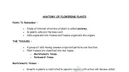
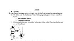
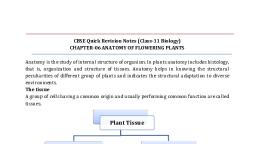
Tissues, , TISSUES, , PLANT AND ANIMAL TISSUES, Plants and animals have different structures and, functions. Thus, the tissues which they possess, are, also different. A comparison between plant and, animal tissue is given in the table. (Refer page no. 48), , It is a well-known fact that all living organisms are, made up of cells. They are either unicellular (e.g.,, bacteria, diatoms, yeasts, protozoans, etc.) or, multicellular (e.g., man, lion, dog, cockroach, neem,, peepal, etc). Each unicellular organism (e.g., Amoeba), is made up of single cell, which acts ", as a site for, diverse life activities such as intake of food, (digestion), intake of oxygen (respiration), excretion,, reproduction, etc. That means, all the activities of the, organism are performed by the single cell. The, multicellular organism, on the other hand, is, composed of a millions of different types of cells., These cells cluster together in order to perform, specific functions of the body., The multicellular organisms usually develop from, single 'initial cell' (i.e., zygote, spore or any other cell)., This initial cell divides by the process of cell division., The cell division continues and a large number of cells, are formed. These cells then undergo cellular, differentiation. Differentiation is the process of, qualitative changes in the cells to perform different, functions in the living organisms. The process of cell, division and cell differentiation leads to the, development of specific organs consisting of specific, groups of cells to perform specific functions in the, body. The cluster of cells (or group of cells) speciallypositioned and designed to perform a particular, function efficiently is called a tissue., A tissue may be defined as a group or collection of, similar or dissimilar cells that perform or help to, perform a common function and have a common, origin., , PLANT TISSUES, Like all other organisms, in all plants (with the, exception of some lower plants), cells of similar origin, are grouped together to perform some specific, functions. Such a group of cells is called tissue. Each, tissue, has a specific function to perform but at the, same time different types of tissues of a plant, function in co-ordination with one another., On the basis of their ability to divide, plant tissues are, of two main types:, (i) Meristematic tissues which consist of, undifferentiated actively dividing cells and, (ii) Permanent tissues, consisting of differentiated, cells which have lost the ability to divide., Meristematic tissues (Meristems), Meristematic tissues are found at all growing points of, a plant, such as the tip of roots, stems and branches, where growth in length occurs. These meristems are, called apical meristems. These tissues occur right, from the birth of the plant and are, therefore, called, primary meristems. Characteristics of meristematic, tissues. :, (i) The cells of these tissues are similar in structure, and have thin and elastic primary cell walls made, up of cellulose., , Table: Differences between the tissues of plant and animals, , Plant tissues, 1. Most of the plants are stationary and remain fixed at, one place, i.e., they do not move from place to place., They need comparatively less energy. Therefore, most, of the tissues of plants are supportive in function and, provide mechanical strength. These tissues are thickwalled, lignified and dead, i.e., they do not contain living, protoplasm. Examples, sclerenchyma, xylem, tracheid’s, and vessels, cork, etc., , 2, , Animal tissues, Animals are not stationary and move from one place, to another in search of food, mates and shelter. They, need more energy as compared to plants. Therefore,, animal tissues are made up of living cells. Examples,, nervous tissues, muscular tissues, connective tissues,, etc.
Page 2 :
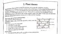
2. Growth of plants is indefinite. Plants grow throughout, their life with the help of certain tissues located in, certain regions in the body. For example, they possess, meristematic tissues in root apex and shoot apex which, divide throughout the life and add new cells to the, body. These cells differentiate to become permanent, tissues which stop dividing. Thus, there are dividing and, non-dividing tissues in plants located at specific regions., 3. The structural organization of plants is not complex as, compared to that of animals. Plant tissues are also not, much complicated., (ii) The cells are rounded, oval, polygonal or, rectangular in appearance., (iii) They have large and prominent nuclei., (iv) Vacuoles small or are absent., (v) Cells are closely packed., (vi) Intercellular spaces are absent., (vii) The cell division occurs actively to produce new, cells., (viii) New cells are produced, take up specific role and, lose the ability to divide. They form the, permanent tissue by the process of, differentiation, as a result of division, they get, transformed into mature permanent tissues., Based on their location in the plant body, meristems, are of three types:, (a) Apical meristem, (b) Lateral meristem, (c) Intercalary meristem, , Growth of animals is definite. It is not confined to, certain regions in the body. The animal organs grow, more or less uniform. Thus, the animals do not, possess dividing and non-dividing regions in the body., In other words, they do not possess specific regions of, dividing and non-dividing tissues., , The structural organization of organs and organ, systems of animals is much more complicated as, compared to that of plants., (i), , Occurs on the sides almost parallel to the long, axis of the root, stem and its branches., (ii) Brings about an increase in the width or girth of, roots and stems, also called secondary growth., (iii) Lateral meristem is of two types, i.e., beneath the, bark in the form of cork cambium and in vascular, bundles of dicots in the form of vascular, cambium. The activity of this cambium results in, the formation of secondary growth., Intercalary meristem, (i), , Occurs at the base of the internodes (in, monocots) or at the base of nodes (mint plant) or, at the base of leaves (Pinus)., (ii) Brings about elongation/growth of that part of, the plant where they are present., , Apical meristem, (i) Occurs at the tips of roots and shoots., (ii) Brings about an increase in length of the plant., Examples: root apical meristem and shoot apical, meristem., , To study the tissues responsible for growth of plants, in certain specific region., Materials required: Two glass jars, two onion bulbs,, water, scale, record file., Produce: Take two wide mouth glass jars and fill them, with tap water. The mouth of jars should b4e such, , Lateral meristem, 3
Page 3 :
that they can hold the onion bulbs. Now, place one, onion bulb at the mouth of one jar (jar a) and another, onion bulb at the mouth of another jar (jar B). Place, the bulbs in such a way that stem and root parts of, each bulbs remain in contact with water. Allow the, roots to emerge out and grow in water. Observe the, growth of roots on day 1, 2 3 on your record book. On, fourth day, cut the tips of roots (upto1 cm) in jar B., Again place the onion of jar B with roots hanging in, water. Now observe the growth of roots in both eth, jars for a few more days. Measure and record the, growth of roots in both the jars for five more days., Record your observations., , (iii) They take permanent shape to perform, permanent functions., (iv) They may be living or dead., (v) The cells may be thin walled or thick walled, The permanent tissues can be grouped, into following categories on the basis of functions, they perform., , Simple permanent tissues, These permanent tissues are called simple because, they are composed of similar type of cells which have, common origin and function. They are further, grouped under three categories - parenchyma, collenchyma and sclerenchyma., (i) Parenchyma: The cells of these tissues may be, spherical, irregular or columnar in appearance. They, have thin cell walls but large vacuoles. The cell wall is, made up of cellulose or calcium pectate. Each cell has, a prominent nucleus. All the soft parts of the plant, body are composed of parenchyma. It forms the basic, packing tissue of plant body. It is the most abundant, tissue of plants. Parenchyma is widely distributed in, various plant organs, viz, roots, stem, leaves, flowers, and fruits. They constitute the major, vegetative, ground tissue. They occur in epidermis, cortex, pith,, pericycle, and mesophyll of leaves, pulp of fruits and, endosperm of seeds. Parenchymatous cells are also, found in xylem and phloem. In storage tissues, the, parenchymatous cells enlarge to store nutrients and, water. These are called storage parenchyma. In, aquatic plants, large air cavities are present in the, ground tissue. These cavities store gases and provide, buoyancy to aquatic plants to help them float. Such, parenchyma are called as aerenchyma., , Observation: It is observed that, - The roots of onion bulb in jar A kept on growing, and, thus, were longer than the roots of onion bulb, in jar B., - In jar B, the roots stopped growing when the tips, of growing roots were cut on 4th day., Conclusion: The roots tips stopped growing in jar-B, because the dividing cells, located “at roop tips, were, remove. It is concluded that growth of roots occurs, due to the activity of dividing cells present at the root, apex (apical meristem). The cells of meristematic, tissue have the ability to divide continuously and add, new cells to the plant body. The daughter cells, derived, from, dividing, meristematic, tissue, differentiated to become permanent tissue and, stopped dividing, Permanent tissues, , Functions of parenchyma:, - The main function of parenchymatous tissue is, storage of food, e.g., starch in the parenchyma of, cortex of potato tuber., - In fleshy stems and leaves, parenchyma cells serve, as water storage tissue, e.g., Euphorbia, Opuntia., - Parenchyma forms the framework of all the plant, organs and tissues like cortex, pith, mesophyll of, leaf and floral parts., , Meristematic tissues, after differentiation, give rise to, permanent tissues. These constitute the major, portion of the plant body., Characteristics of permanent tissues, (i) They constitute the major portion of the plant, body., (ii) The cells of these tissues do not multiply., 4
Page 4 :
- Parenchymatous tissue stores waste materials of, plants, such as gum, crystals, resins, tannin, etc., - The intercellular air spaces of parenchyma cells, allow gaseous exchange., - If chloroplast is present in parenchyma cells (i.e.,, chlorenchyma), it performs photosynthesis, e.g.,, the mesophyll of leaves., , -, , Chloroplast containing collenchyma cells are, responsible for photosynthesis and manufacture of, sugar and starch., (iii) Sclerenchyma (Scleros: hard): The cells of this, tissue are dead. The cells are long narrow with, tapering ends. Their cell walls are thickened. This, thickness is due to the deposition of a water, resistant material lignin. The thickening of the, cell wall is so great that sometimes the cell, contents are completely lost and cell cavities, obliterated. Central cavity of the cell is greatly, reduced due to the thickening. Sclerenchyma, tissues are of two types fibres and sclereids., Sclerenchyma acts as a mechanical tissue giving, support and strength to the organ. They protect, the plant from environmental forces like strong, winds. They make the plant hard and stiff. The, husk of coconut is made up of sclerenchyma, tissue., The examples of this tissue are the hard grit of a, pear fruit, the hard walls of nuts and the brittle, coats of seeds. These tissues occur in the veins of, leaves. They form the major part of walnut shells, and of other nuts. They form an important part of, the bark of trees., , (ii) Collenchyma (Colla : glue): Collenchyma is a, living tissue of primary body. The cells are thin, walled but possess thickenings of cellulose and, pectic substances at the corners where number, of cells join together. The tissue provides, flexibility to soft aerial parts (e.g., leaves, young, stems) of plant so that they can bend without, breaking. The cells are compact and the, intercellular spaces are absent., , Collenchyma is usually found in 3-4 layers, beneath the epidermis in stem, petioles and, leaves of herbaceous dicot plants. It is usually, absent in monocot stems, roots and leaves., Functions of collenchyma:, - It provides mechanical support, protection and, elasticity to the plant organs., - Due to its peripheral position in stem, it helps, leaves in bending and pulling action of wind., 5
Page 5 :
the atmosphere. They also help in loss of water in the, form of water vapour known as transpiration., Cork, , To study the structure and function of epidermis and, other protective tissues., Materials required: Leaf of Rhoeo discolor, petri dish,, glass slide, cover slip, microscope, safranin, water., Procedure: Take a freshly plucked leaf of Rhoeo, discolor. Peel out the lower skin of leaf. Cut the, epidermal peel and place it in a petridish containing, water. Stain the peel with the help of a few drops of, safranin. Transfer the peel on a glass slide and cover it, gently by placing a coverslip over it. Observe it under, the microscope. Observation: Under the microscope,, a sheet of cells is seen. These are epidermal cells. The, layer of epidermal cells is called epidermis. Other, types of structures seen under the microscope arestomata (sing, stoma) and trichomes (hairs)., Conclusion: From the above observation it can be, concluded that all the plant parts have a protective, covering, called protective tissue. The epidermis, provides protection to the underlying cells and forms, the protective tissue. This layer remains in direct, contact with the environment. Any substance,, whether solid, liquid or gas, can enter into the plant, or move outside only after passing through this layer., It provides protection to various plant organs and, helps in absorption, secretion, excretion, gaseous, exchange and transpiration., , As plants grow older with times, protective tissues at, the periphery undergoes certain changes. In them a, strip of secondary meristem replaces the epidermal, layer of the stem forming a many layered thick bark of, the tree called cork. Cork in mature woody stem is, made up of dead, thick- walled cells. The cork cells are, compactly arranged without any intercellular spaces., The walls of cork cells also contain suberin (a chemical, substance) which is impervious to gases and water., Functions of cork:, Cork performs protective functions in the following, ways:, - Cork cells being highly suberized and thick walled, protect the inner tissues., - It provides insulation from freezing temperatures., - It protects the inner tissues from the attacks of, microorganisms and prevents water loss also, , 1., Ans.:, , Protective plant tissue, , (i), , Epidermis is the outermost protective layer of plant, organs. It is usually single-layered but in the leaves of, some plants growing in dry habitats, it is multi-layered, and thick. This is to protect the plant from water loss., Epidermis protects all the parts of plant. Cells of, epidermis form a continuous layer without, intercellular spaces to protect the plant tissues., Epidermal cell of aerial parts of the plant secrete a, waxy, water resistant layer on their outer surface. It, protects them against loss of water, mechanical injury, and any attack by pathogenic fungi. In desert plants,, outer walls of the epidermis are usually thick and, covered with organic substances like cutin. The thick, cutinized wall of epidermis greatly reduces loss of, water by transpiration. The epidermal cells of the, roots contain long hair-like structures called root, hairs. The root hairs increase surface area for, absorption of water and nutrients from soil. The, epidermis of the leaf contains small pores called, stomata. The stomata helps in exchange of gases with, , (ii), (iii), (iv), , 2., Ans., , 3., Ans., 4., 5., Ans., , 6, , What is the utility of tissues in multicellular, organism? (NCERT), The utility of tissues in multicellular organism, is given below:, Division of labour: Tissues bring about division, of labour in multicellular organisms. It, increases efficiency., Higher, organization:, Tissues, become, organized to form organs and organ systems., Individual cells: Work load of individual cells, has decreased., Higher survival: Because of division of labour,, higher efficiency and organization, the, multicellular organisms have high survival., Name the types of simple tissues. (NCERT), There are three types of simple tissues these, are, parenchyma,, collenchyma, and, sclerenchyma, Where is apical meristem found? (NCERT), Apical meristem occurs at root and stem tips., Which tissue makes up the husk of coconut, What is the function of meristematic tissue?, Meristematic tissues are responsible for the, growth of plants. These tissues are present at, growing regions like tips of roots and shoots.
Page 6 :
6., Ans., 7., Ans., 8., Ans., , Name the tissue which allows easy bending, in various parts of a plant., Collenchyma., Define organ., A number of tissues together form an organ., What does the root tip contain that helps in, root elongation?, The root tip contains apical meristematic, tissues which are responsible for elongation, of roots., , (iv) Xylem parenchyma are the only living, components of xylem. These are concerned with, the storage of food and sideways conduction of, water., (b) Phloem: Phloem tissue is regarded as living, conducting tissue which is concerned with the, translocation of food in the plants. The phloem is, composed of four elements: Sieve tubes,, companion cells, phloem parenchyma and, phloem fibres., , Complex permanent tissues (Conducting tissues), Complex tissues are made up of more than one type, of cells which work in close coordination to perform a, common function. These tissues are also called, vascular tissues. They transport water and dissolved, food material up and down in the plant parts. The, conducting tissues are of two types:, (a) Xylem, (b) Phloem, (a) Xylem: Xylem is the chief conducting tissues of, vascular plants. These are responsible for the, transportation of water to the various parts of the, plant body. These consist of four types of cellstracheids, vessels, xylem fibres and xylem, parenchyma., , (i), , Sieve tubes: The main conducting part of the, phloem is sieve tube which is formed of, elongated cylindrical cells arranged in vertical, rows. The terminal walls of each sieve tube have, many minute pores (perforated walls) through, which food material passes very easily. The entire, porous plate is termed as sieve plate., (ii) Companion cells: Each sieve tube member is, supported by a long parenchymatous cell called, the Companion cells: which helps the sieve tube, in the conduction of food material. Companion, cells are living cells usually always associated, with the sieve tubes. The sieve tube elements, and companion cells arise from the same initial, cell and therefore, form a single functional unit., Each companion cell is living cell with thin, cellulose wall and active protoplast. The common, wall between sieve tube and companion cell, shows presence of fine pits which are traversed, by plasmodesmata., (iii) Phloem parenchyma are ordinary living, parenchyma cells associated with phloem. They, store food., (iv) Phloem fibres are dead sclerenchyma fibres., They provide mechanical strength. The textile, fibres of flax, hemp and jute are phloem fibres., , (i), , Tracheids: A tracheid is an elongated hollow cell, with its both ends tapering. The walls of these, cells are thick due to the deposition of lignin. At, certain spots, lignin is not present. These spots, are termed as pits. These cells are arranged in, such a fashion that they too form a system of, long tubes and channels through which water, can move easily. The tracheids are dead cells., (ii) Xylem vessels (or tracheae): The cells of vessels, are placed one upon the other and their end, walls are either absent or possess perforations., They form long tubes or channels for conduction, of water and minerals., (iii) Xylem fibres are supportive in nature and, provide mechanical strength to the plant body., , 7
Page 7 :
To study the structure and function of various types, of permanent plant tissues., Materials required: Piece of freshly plucked, dicotyledonous (dicot) stem, sharp razor, glass slide,, cover slip, safranin stain, glycerine, and microscope., Procedure: Cut a very thin transverse section of a, dicot stem with the help of a sharp razor, Stain the, section with safranin and wash extra stain with the, help of water. Now mount the stained section using a, drop of glycerine on a glass slide. Cover the section, with a cover slip and observe under the microscope., Observations: Place the slide under a compound, microscope and observe the various types of cells and, their arrangement in the section of stem. Compare, the cells with figure. Now answer the following, questions on the basis of your observations:, (a) Are all cells similar in structure?, (b) How many types of cells can be seen?, (c) Can we think of reasons why there would be so, many types of cells?, , Conclusion: On the basis of microscopic examination, and observations of a stem, we can answer the above, questions as follows:, (a) All the cells are not similar in structure., (b) In all, 9 types of cells can be seen. They are(i) Epidermal cells (Parenchyma, (ii) Hypodermal cells (Collenchyma), (iii) Cortex(parenchyma), (iv) Endodermis (Parenchyma), (v) Pericycle (Sclerenchyma), (vi) Pholem, (vii) Cambium (Lateral meristem), (viii) Xylem (ix) Pith (Parenchyma)., Thus, the type of tissues present in the section, are:, Meristematic, tissue., Parenchyma,, Collenchyma, Sclerenchyma, Xylem and Phloem., (c) There are so many types of cells which carry, out different functions. Such as, (i) Protection (Epidermal cells), (ii) Mechanical support (Collenchyma), (iii) Storage of food (Cortex and pith), (iv) Transport of water and minerals (Xylem), (v) Translocation of organic food (Phloem), (vi) Growth (Cambium)., Thus, the different types of cells and tissues, perform different functions., , The xylem and phloem in plants occur together and are collectively called vascular bundle., Table: Differences between xylem and phloem, , 1., 2., 3., 4., 5., 6., , Xylem, It conducts water and inorganic solutes in, vascular plants., Conduction mostly occurs in one direction (i.e.,, upward)., Conducting channels (tracheary elements) are, tracheids and vessels., Its components include tracheids, vessels,, xylem parenchyma and xylem fibres., Tracheids, vessels and xylem fibres are dead and, only xylem parenchyma is living., Xylem provides mechanical strength also., , Phloem, It conducts organic solutes in vascular plants., Conduction may occur in both directions, i.e., upward or, downward., Conducting channels are sieve tubes., Its components include sieve tubes, companion cells, and, phloem parenchyma and phloem fibres., Sieve tubes, companion cells and phloem parenchyma, are living and only phloem fibres are dead., Phloem does not provide mechanical strength., 8
Page 8 :
Ans., 9., Ans., , 10., Ans., 11., , What are the constituents of the phloem?, (NCERT), The constituents of the phloem are sieve, tubes, companion cells, phloem parenchyma, and phloem fibres., Name two conduction (vascular) tissues in, plants, Xylem and phloem., What is a vascular bundle?, , 12., Ans., , ANIMAL TISSUES, , The xylem and phloem in plants occur, together and are collectively called vascular, bundle., Write the function of xylem and phloem., Xylem is mainly concerned with the, conduction of water and minerals. It also, provides mechanical support to the plant., Phloem is responsible for transport of food, prepared by leaves to other parts of the plant., , may be several-cell thick, that is, many-layered, (compound or stratified epithelium)., , In animals, four basic types of tissues are found. These, are as follows:, - Epithelium or epithelial tissue (covering tissue), - Connective tissue (supporting tissue), - Muscular tissue (contractile tissue), - Nervous tissue., , Functions of epithelial tissue:, (i) Protection, (a) Most epithelia protect the underlying tissues, against mechanical injury such as abrasion., (b) Some epithelia protect against water loss and, dehydration, and against infection by microorganisms., (ii) Role in physiological processes, Some epithelia are involved in physiological, processes such as (a) respiratory gas exchange,, (b) elimination of waste products, and (c) some, cells become glandular in nature and per- form, secretory function., (iii) Sensory role by sensory epithelium., (iv) Transport of materials by ciliated epithelium., , Epithelial tissue, Epithelium forms the outer protective covering all, over the body, and lines the inside of all cavities such, as those of the mouth, throat, stomach, intestine,, wind pipe and lungs. The epithelial cells lie close, together with little or no intercellular substance or, matrix between them. There is no blood or lymph, supply. Nerve supply is present. Epithelial cells are, attached to the underlying tissues by a basement, membrane, which is made of a network of white, nonelastic collagen fibres. Epithelium may be one-cell, thick, that is, single layered (simple epithelium), or it, 9
Page 9 :
Types of epithelial tissue, , projections called cilia arising from the outer free, surface of the cells. It is found lining the wind-pipe, (trachea), kidney tubules, oviduct (Fallopian tubes), and ventricles of the brain. This epithelium helps in, the movement of mucus, urine, eggs, sperms and, cerebrospinal fluid in a particular direction., , Depending upon the shape and function of the, constituent cells, epithelial tissues are of following, types:, - Squamous (cells flattened), - Columnar (cells tall, columnar or pillar-like), - Cuboidal (cells cube-like), - Ciliated (cells with cilia), - Glandular (cells secretory in nature), - Sensory (cells have nerve endings; sensory function), - Stratified (cells many layered), , Glandular epithelium, This epithelium consists of columnar cells modified to, secrete chemicals. It lines the glands such as gastric, glands, pancreatic lobules, intestinal glands, etc., , Squamous epithelium, , Sensory epithelium, , The cells in this epithelium are extremely thin and flat, and are arranged edge to edge forming a delicate, lining or covering., It forms the lining of cavities of ducts and blood, vessels, lines the chambers of the heart, covers the, skin, and lining of the mouth. It also lines pharynx,, oesophagus, anal canal, vagina and lower part of, urethra. It provides protection to the underlying parts, against abrasion (mechanical injury) and entry of, germs or chemicals. It also helps in excretion, gas, exchange and secretion of coelomic fluid., , In some cases, the epithelial cells become modified to, receive external stimuli. Such sensory cells have nerve, endings so that they can perceive various stimuli. It is, found in the nasal pas- sages, taste buds, retina of, eyes, and epidermis of earthworm., Stratified epithelium, This is a compound epithelium in which cells are, arranged in many layers one above the other. It is, found in places where there is much wear and tear,, such as the epidermis of skin, lining of the mouth, cavity., , Columnar epithelium, This epithelium consists of cells which are much, longer than broad; looks like a column. It forms the, lining of stomach and intestines; also found in salivary, glands in the mouth, sweat glands and oil glands of, the skin. It also lines mammary gland ducts and parts, of urethra. It helps in protection, absorption and, secretion. Columnar epithelium of intestine is, specialized for the absorption of water and digested, food., Cuboidal epithelium, Cells are as long as broad and appear cube-like; a, centrally located nucleus is present. The cuboidal, epithelium lines the small salivary ducts, pancreatic, ducts, sweat glands, salivary glands and thy- roid, glands. It also covers the ovaries and lines the spermproducing tubules., It helps in protection, secretion, absorption, excretion, and gamete formation., Ciliated epithelium, , Fig. : Epithelial tissues A. Squamous B. Columnar, C. Cuboidal D. Ciliated E. Galndular F. Stratified, , This epithelium, usually consisting of cuboidal or, columnar cells, has numerous, thin, delicate, hair-like, 10
Page 10 :
Connective tissue, As the name suggests, this tissue serves to "connect", or bind the cells of other tissues in the body, and gives, them rigidity and support. It is composed of cells and, numerous, thick structures called fibres. The cells are, living and are embedded in a non-living, intercellular, matrix., Functions of connective tissue:, , (i), , - Connective tissue binds different structures with, one another, e.g., muscles with skin, muscles with, bones., - These form a supporting framework of cartilage, and bones in the body., - Adipose connective tissue helps in storage of fats., It also forms shock-proof cushions around kidneys,, ovaries and eyeballs., - White blood corpuscles of blood and lymph act as, phagocytes and they provide protection against, bacterial infections., - These form protective sheaths around delicate, organs such as spleen, kidneys, testes, etc., - Collagen fibres of connective tissue help in the, repair of injured tissues. Based on the nature of, matrix, the connective tissue is divided into three, general types- Connective tissue proper: where the matrix is, relatively less rigid., - Supportive connective tissue (Skeletal tissue):, where the matrix is rigid., - Fluid connective tissue: where the intercellular, matrix is a fluid called plasma., , Functions: The areolar tissues is connective in, function. It fixes the skin with the muscles, fills, the spaces inside the organs, attaches the blood, vessels and nerves with the surrounding tissues,, fastens the peritoneum to the body wall and, viscera. It is commonly called 'packaging tissue', of the body. Examples, bone periosteum, muscle, perimysium, nerve perineurium, etc., Adipose connective tissue: It occurs below the, skin, around internal organs and yellow bone, marrow. Cells are modified to store fat; each cell, consists of a large vacuole filled with fat which is, surrounded by a small amount of cytoplasm, containing a nucleus towards the periphery., , Functions: The adipose tissue is found beneath, the skin, in the covering of the heart, around the, blood vessels and kidneys and in yellow bone, marrow. This tissue stores fat and insulates the, body against heat loss., (iii) Tendon: Tendons are cord-like, very tough,, inelastic bundle of white collagen fibres bound, together by areolar tissue. The cells present in, the tendons are elongated fibroblasts which lie in, almost continuous rows here and there. The, tendons connect the skeletal muscles with bones., , Connective tissue proper, It is of four types: areolar connective tissue, adipose, connective tissue, tendon, ligaments., (i) Areolar connective tissue: It is the most widely, distributed connective tissue in the body having, jelly-like sticky matrix, irregular-shaped cells and, two kinds of fibres- white fibres (made of, collagen) and yellow fibres (made of elastin)., , (iv) Ligaments: Ligaments are cords formed by yellow, elastic tissue in which many collagen fibres are, bound together by areolar tissue. The fibroblasts, are irregularly scattered. This tissue combines, strength with great flexibility. The ligaments, serve to bind the bones together., , 11
Page 11 :
Supportive connective tissue (Skeletal tissue), It consists of cartilage and bone., (i) Cartilage: Cartilage is a non-porous tissue. Cells, of the cartilage called chondroblasts are, embedded in the matrix in groups of two, four or, more in fluid-filled spaces called lacunae., Cartilage is usually covered by a tough fibrous, membrane called perichondrium., Cartilage is found in nose tip, ear pinna, rings of, wind-pipe (trachea), end of long bones, lower, ends of ribs. In shark fish, the whole skeleton is, cartilaginous. Cartilage performs the function of, providing support and flexibility in the vertebrate, body., (ii) Bone: Bone is a very strong, rigid and porous, tissue. A compact bone consists of living bone, cells, called osteoblasts, embedded in a firm,, calcified matrix. The osteoblasts are contained in, lacunae (spaces) which are arranged in, concentric circles present throughout the matrix., The lacunae are also traversed by nerves and, blood vessels. The blood vessels passing through, them provide nutrients to osteoblasts and help, exchange of materials. The matrix is composed of, about 30% organic materials (chiefly collagen, fibres and glycoproteins) and 70% inorganic bone, salts (mainly phosphates and carbonates of, calcium and magnesium, hydroxyapatite, etc.)., These inorganic salts are responsible for, hardness of the bone. Bones form endoskeleton, of vertebrates. They provide levers for, movement and support for soft parts of the, body. Bones also protect many delicate tissues, and organs., , Table: Differences between cartilage and bone, , 1., 2., 3., , 4., , Cartilage, Cartilage is soft, elastic, and flexible., Matrix, of, cartilage, consists entirely of organic, matter., Cartilage do not have, blood supply (except in, erichondrium)., Growth of cartilage is, unidirectional., , Bone, Bone is hard, tough, and inelastic., Matrix of bone is both, organic and inorganic., Bones have rich blood, supply., Growth of bone is, bidirectional., , Fluid connective tissue, Blood and lymph constitute the fluid connective, tissue and consists of cells and matrix without fibres., Matrix is in a fluid state, and is called plasma., (i) Blood: Blood is a bright-red coloured fluid, connective tissue. It consists of a straw-colored, fluid called plasma in which various kinds of cells, or corpuscles are present. The blood cells are(a) Erythrocytes (red blood cells or RBCs in short),, (b) Leucocytes (white blood cells or WBCs in short),, and, (c) Blood platelets., The red blood corpuscles of mammals are small,, circular, biconcave discs and lack nuclei when, mature. There about five million red blood cells, per mm3 of blood. They are packed with ironrich, red-coloured, oxygen-carrying protein, pigment, and the hemoglobin. Haemoglobin, combines reversibly with oxygen to form, oxyhaemoglobin (blood-red in colour). Red blood, corpuscles also contain the enzyme carbonic, anhydrase which regulates carbon dioxide, transport., The white blood corpuscles are large-sized,, nucleated cells present in much smaller number, (about 7000 per mm3 of blood). They are capable, of amoeboid movement and play an important, 12
Page 12 :
role in the body's defence mechanism. The white, blood corpuscles belong to two main categories, phagocytes (carry out the function of body, defense, by, engulfing, pathogen), and, immunocytes (they are responsible for immunity, and carry out immune responses by producing, antibodies). Phagocytes are further divided into, two types- granulocytes (having cytoplasmic, granules) and agranulocytes (having non-granular, cytoplasm). Granulocytes include- Neutrophils, (they engulf and digest disease causing, pathogens); Eosinophils (they show allergic, responses and antihistamine properties); and, Basophils (they release histamine and heparin)., Agranulocytes include- Monocytes (they engulf, bacteria and cellular debris at injured site)., Immunocytes include- Lymphocytes (they, secrete antibodies to destroy microbes and also, help in healing of injuries)., The platelets are irregularly shaped, nonnucleated fragments of giant cells. They play a, role in blood clotting., The plasma makes up about 55 per cent of the, blood by volume while blood cells make up 45, per cent of the blood by volume., The body of an adult human being contains about, 6 litres of blood., , 14., Ans.:, , 15., Ans.:, 16., Ans.:, 17., Ans.:, 18., Ans.:, 19., Ans.:, , Major functions of blood:, Transports food materials., Transports oxygen and carbon dioxide., Transports excretory products to the kidneys, from, where they are eliminated., Regulates temperature by distributing heat within, the body., WBCs protect the body from diseases and help in, wound healing., ', (ii) Lymph: Lymph is another colourless fluid, connective tissue consisting of plasma and mainly, white blood cells. RBCs and platelets are absent., It is a fluid that surrounds the body cells., It mainly helps in the exchange of materials, between blood and tissue fluids. The lymph also, protects the body against infection by destroying, invading microorganisms., , 20., Ans.:, , 21., Ans.:, , two, four or more in fluid-filled spaces called, lacunae., (ii) Cartilage performs the function of, providing support and flexibility in the, vertebrate body., What are the functions of areolar tissue?, (NCERT), Areolar tissue fills the space inside the organs,, support internal organ and helps in repair of, tissues. It binds the skin with underlying parts., Name the kind of tissue which forms inner, lining of the blood vessels., Squamous epithelium, Which type of animal tissue covers the, external surfaces of body?, Epithelial tissue., are the three types of cells found in areolar, tissue, The three types of cells found in areolar tissue, are mast cells, macrophage and fibroblasts., Which tissue stores fat globules?, Adipose tissue., ., Name three different types of blood cells and, cells and give their functions., The three types of blood cells and their, functions are as follows:, (a) Red blood cells (RBCs) or Erythrocytes: The, chief function of RBCs is the transport of, respiratory gases, i.e., oxygen and carbon, dioxide., (b) White blood cells (WBCs) or Leucocyes:, These cells perform various functions like, engulfing germs, secretion of heparin,, histamine and antibodies., (c) Blood platelets: These cells help in clotting, of blood., Name the proteins found in white fibre and, yellow fibre., Collagen protein is found in white fibres, whereas elastin protein is found in yellow, fibres., Which tissue is called middle man between, tissue cells and blood?, Lymph is called the middle man between, tissue cells and blood., Muscular tissue, , 13., Ans.:, , Muscular tissue is a contractile tissue consisting of, large elongated muscle cells or fibres. The muscle cells, are able to shorten to a half or even a third of their, "resting" length, and return to their original state., , (i) What are chondroblasts?, (ii)Write the function of cartilage., (i) chondroblasts are the cells of me cartilage, which are embedded in its matrix in groups of, 13
Page 13 :
Contractility is the special property of this tissue. The, cytoplasm of a muscle cell (or fibre) contains a large, number of fine longitudinally running fibrils called, myofibrils. The myofibrils are contractile. The, cytoplasm is called the sarcoplasm. Sometimes the, muscle fibre is externally covered by a sheath or, membrane called sarcolemma., , such as the stomach, intestines, blood vessels,, breathing pas- sages and the organs concerned with, urination and reproduction (urinogenital ducts)., Rhythmic contraction of smooth muscle in malls of, tubular organs (peristaltic contraction) result in the, rhythmic progressive wave of muscular contraction, and relaxation. These movements occurs in, gastrointestinal tract and male genital tract., In some organs, the smooth muscles make the organs, short and thick by its contraction or long tin by its, relaxation. They contract throughout the organ as a, single unit and produce extrusive movements as in, urinary bladder, gallbladder, ureters, uterus., , (iii) Cardiac muscles: Cardiac muscles are com- posed, of branching and anastomosing network of fibres. The, fibres have centrally located one or two nuclei and, faint transverse striations with light and dark bands., Special electrical junctions called intercalated discs, are present at intervals in the fibres. Each fibre is, surrounded by sarcolemma. Cardiac muscles show, characters of both striated and unstriated muscles., Cardiac muscles are richly supplied with blood. These, muscles occur only in the walls of the heart., Cardiac muscles contracts and relaxes rapidly and, continuously with a rhythm, but it never gets fatigued., Contraction and relaxation of cardiac muscles cause, pumping of blood out of the heart and into the heart, regularly., , Types of muscular tissue, There are three types of muscular tissue- Striated or, striped or skeletal or voluntary muscles, Unstriated or, no striated or smooth or involuntary muscles, and, Cardiac muscles., (i) Striated muscles: These are also known as striped,, skeletal or voluntary muscles. These occur in bundles,, normally attached to the bones and help in body, movement. Each muscle fibre is long, cylindrical,, unbranched and non-tapering, with multinucleate, (coenocytic) condition. Under the microscope, each, striated muscle fibre shows striations., The striated muscles are found in the body wall and, the limbs (biceps and triceps of the arm). They also, occur in tongue, pharynx and beginning of, oesophagus., These muscles can contract rapidly and are, responsible for quick movements. These muscles are, called voluntary because their contraction is under, the control of brain or will., (ii) Unstriated muscles: These are also known as, smooth or involuntary muscles. Each unstriated, muscle fibre is a long, flattened, spindle-shaped,, tapering and uninucleate cell. The muscle fibres do, not show striations. These muscles contract and relax, very slowly. These are called involuntary muscles as, their movements are not controlled by the brain. Such, muscles are found in the walls of all tubular organs, , Nervous tissue, Nervous tissue is a very specialized tissue for, receiving stimuli or sensations and transmitting, messages. It is present in brain, spinal cord and, 14
Page 14 :
nerves. Nerve cells or neurons form the most, important elements of the nervous tissue. Each, neuron consists of three parts- the main body called the cell body or cyton,, - the dendrons, and, - the axon., (i) Cyton or cell body: The cell body contains the, major concentration of the cytoplasm and the, central nucleus of the neuron. The cell body also, contains Nissl's granules, which are groups of, ribosomes and rough endoplasmic reticulum., , Nerve cells are joined end to end forming long, nerve fibres. Nerve fibres branch out to every, part of the body., , 22., Ans., 23., Ans., 24., Ans., , 25., Ans., 26., Ans., , (ii) Dendrons or dendrites: The dendrons are one or, more short processes arising from the cyton., Dendrons branch further into many thin, dendrites. The dendrites receive impulses., (iii) Axon: The axon is a single, long, cylindrical, process arising from the cyton. The axon forms, fine branches at its terminal end. Each branch, ends in a swollen structure, called synaptic knob, or bouton. The axons carry impulses away from, the cell body to other neurons. The synaptic, knobs of terminal branches of neuron are, connected with dendrite branches of an adjacent, neuron. Each such junction, in fact, has minute, gap called synapse. It is meant for the, transmission of nerve impulse from one neuron, to the other., , 27., Ans., , 28., Ans.:, , 15, , Name the tissue responsible for movement, in our body. (NCERT), Muscular tissue is responsible for movement, in our body., What does a neuron look like? (NCERT), Neuron is a thread or hair like structure, with, palm leaf shaped structure at one end., “Muscle cells are called muscle fibres.” Why?, Muscle cells are thin and elongated having, thread-like structures, so these are called, muscle fibres., Name the term used for plasma membrane, of muscle cell., Sarcolemma, What is a synapse? Explain., The junction between the terminal part of, one axon and the dendrite of the adjacent, neuron is called a synapse. Synapses help in, the transmission of impulses from one neuron, to another., What is medullary sheath? Mention its, significance., Medullary sheath is the covering of nerve, fibre. It insulates the nerve fibre and prevents, the leakage of ions., Give three features of cardiac muscle., (NCERT), The three features of cardiac muscle are given, below:, (i) Cardiac muscles show characteristics of, both striated and unstriated muscles., (ii) Intercalated discs are present in the, cardiac muscle fibres., (iii) The muscles show rhythmic contractions.
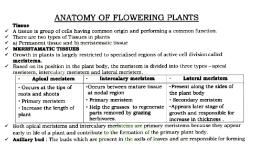
Page 15 :
THE TISSUE SYSTEM, , , , , Tissues vary depending on their location in the, plant body. Their structure and function would, also be dependent on location. On the basis of, their structure and location, there are three types, of tissue systems. These are the epidermal tissue, system, the ground or fundamental tissue system, and the vascular or conducting tissue system., , Vascular tissue system, , , Epidermal tissue system, , , The epidermal tissue system forms the outermost covering of the whole plant body and, comprises epidermal cells, stomata and the, epidermal appendages- the trichomes and hairs., , , , Ground tissue system, , , are present in a central cylinder which is known as, stele. Outside this stele, ground tissues are called, extrastelar ground tissues (cortex) and the ground, tissues within the stele are known as intrastelar, ground tissues (pericycle, medullary rays, pith)., Different components of ground tissue systems, are hypodermis, cortex, endodermis (starch, sheath), pericycle and pith or medulla., , The tissue present between epidermis and, vascular tissue system constitutes ground tissue, system. In the stems and roots, vascular bundles, , Vascular tissue system is composed of a number, of vascular bundles, present in the central, cylinder or column of the axis of root and stem, which is known as stele. The members of vascular, bundle are derived from the procambium of, apical meristem., The vascular bundle is composed of primary, xylem, primary phloem and cambium. Cambium, is not the utmost necessary element of vascular, tissue system. It is present in dicot plants and, absent in monocots., ANATOMY OF DICOT AND MONCOT PLANTS, , 16
Page 16 :
, , -, , For a better understanding of tissue organization, of roots and stems, it is convenient to study the, transverse sections of the mature zones of these, organs., , -, , Dicot stem, , , , , , , , , , , , The primary structure of a dicotyledonous stem, may be well understood by using sunflower as an, example. The transverse section of the young, sunflower stem shows the following structure:, Epidermis - Epidermis is the outermost layer of, the stem. It consists of a single layer of cells. The, epidermis, bears multicellular,, uniseriate, trichomes. A thin layer of cuticle is present on the, epidermis as well as trichomes., , Xylem: The xylem tissue lies below the phloem., This is composed of the vessels, tracheids,, xylem parenchyma and the fibres., Cambium: This is present in between the xylem, and the phloem. It consists of 2- 3 layers of thinwalled, rectangular cells. The cambium, divides, to produce new xylem., Monocot stem, , In the stems of monocotyledonous plants, the, vascular bundles are scattered and no distinct corte or, pith can be delimited. The internal structure of the, young maize (Zea mays) stem shows various layers in, a transverse section., Epidermis - It is single layered with, compact,, rectangular, living cells. It is covered with a thick, cuticle and have a few stomata. There are no, hairs,, , Hypodemns - This is just below the epidermis and, of 3 to 4 layers of collenchymatous cells. The, collenchyma cells contain chloroplasts. This tissue, serves to strengthen the youno stem., Endodermis - It is the innermost layer of the, cortex, and consists of a single layer of barrel, shaped cells., Pericycle - The pericycle is in the form of, semilunar patches of sclerenchyma cells, which, are lignified., Medullary rays - In between the vascular bundles, there are a few layers of parenchymatous cells, that constitute medullary rays., Vascular bundles - The vascular bundles are, situated in a ring on the inside of the pericycle of, the plant. Each vascular bundle is conjoint,, collateral, endarch and open. It is composed of, Xylem, phloem and cambium., - Phloem: This is situated on the outer side of, the vascular bundle. The cells are thin-walled, and polygonal. Phloem is a complex tissue and, is composed of sieve tube elements,, companion cells the phloem parenchyma and, phloem fibres., , , , , 17, , Hypodermis - The cells of hypodermis are, sclerenchymatous. This layer is located just below, the epidermis., Vascular bundles - The vascular bundles are found, scattered throughout the ground tissue the, vascular bundles contain no cambium and, consequently secondary thickening does not, occur. Each vascular bundle is oval and usually, surrounded by a sheath of sclerenchymatous, cells, the bundle sheath. The bundle consists of, two parts, xylem and phloem., The xylem consists of 3-4 distinct vessels, which, are arranged, in the form of a “Y” One or two, smaller vessels lying at the arm base constitute, the protoxylem. The two bigger vessels lying, laterally constitute the metaxylem. The lower, most protoxylem vessels open into a cavity called, water containing cavity. The phloem lies outer-to, the xylem. It consist of sieve tube elements and, companion cells. Phloem parenchyma is absent.
Page 17 :
Table: Differences between dicot and monocot stem, , 1., 2., 3., 4., 5., 6., 7., 8., , Dicot stem, Epidermis is single layered with hair (trichome)., Collenchymatous hypodermis, Cortex is made up of several layers of parenchymatous, tissue., Endodermis is single layered which is usually not well, differentiated., Pericycle is made up of one or more layers of, parenchymatous or sclerenchymatous cells., Medullary ray is found between the vascular bundles., Pith is made up of parenchymatous cells situated hollow, in the centre of stem., Vascular bundles arranged in ring. Conjoint, collateral or, bicollateral, endarch and open. Almost all of them, uniform in size., , , , , , , , , It is absent., It is absent., It is absent (pith cavity is present in stems)., Scattered, throughout the ground tissue. Conjoint,, collateral, exarch and closed. Larger towards centre, and smaller towards outer side., , , Dicot root, , , Monocot stem, It is single layered without hair., It is sclerenchymatous., It is absent but parenchymatous ground tissue present, from hypodermis to centre of stem., It is absent., , The primary structure of a typical dicotyledonous, root can be studied by examining the transverse, section of a young root of sunflower, pea or gram., A transverse section of a young dicotyledonous, root shows three regions, namely, the epiblema,, cortex and stele., Epiblema - Epiblema is also known as the, piliferous layer. It is characteristically single, layered, comprising tubular living components., There is no cuticle on the epidermis. Some cells of, the epidermis show tubular outgrowths known as, root hairs., Cortex - The cortex is uniform and simple. It, consists of several layer of thin walled, parenchyma cells with conspicuous intercellular, spaces. The cells contain storage materials like, starch. The innermost layer of cortex is the, endodermis, which completely surrounds the, stele. It is structurally and physiologically different, from the cells on either side of it. The radial and, transverse walls of the endodermal cells contain a, band of lignin and suberin, known as the, Casparian strip and considered to control the, movement of materials in the roots. Later in the, development of the root, all cells of the, endodermis become thick walled, except at, positions opposite the protoxylem. This thin, walled cells are known as passage cells., Stele - All tissues inside of the endodermis, comprise the stele of the root. These include, pericycle, vascular bundles, and pith., , Vascular bundles are radial, that is xylem and, phloem that occur in separate patches, are, arranged on alternate radii. Xylem consists of, both protoxylem and metaxylem. It is exarch,, since the protoxylem lies towards the epidermis., Based on the number of xylem arch, the roots are, known as the monarch, diarch, triarch, tetrarch,, and so on. The root of the bean plant possesses a, tetrach condition, while that in the sunflower, plant is triarch. In the dicotyledonous plants, the, xylem plates usually extent into the centre to, form a solid centre core without any pith. Phloem, patches are rather small. They form oval groups, under the pericycle. These oval masses are, separated from the xylem by parenchymatous, cells that are known as the conjunctive tissue., , Monocot root, , , 18, , A transverse section of a monocotyledonous root, also shows three regions, the epiblema, cortex, and stele.
Page 18 :
, , , , , , Epiblema or piliferous layer - It is the outermost, layer consisting of a single row of thin-walled cells, without any intercellular spaces. The structure, and fate of this layer is more or less similar to that, of dicot roots. It becomes disintegrated after the, root hairs are shed., Cortex - The cortex consists of several layers of, parenchymatous cells with intercellular spaces., The cortex is smaller than in the dicotyledonous, root. The endodermis or inner boundary of the, cortex is characterized by the presence of, Casparian strip. As the epiblema dies off, a few, outer layers of the cortex become cutinized and, form the exodermis. Certain endodermal cells,, which are present opposite to the xylem bundles,, remain thin walled. These are known as passage, cells. Their functions is to transfer water and, dissolved salts from cortex directly into the xylem., Stele - All tissues inside of the endodermis, comprise the stele. These include pericycle,, vascular bundles and pith., Table: Differences between dicot and monocot root, Dicot root, 1. Cortex is comparatively narrow., 2. Endodermis is less thickened, Casparian strips are more, prominent and no passage cells., 3. Pericycle gives rise to secondary (lateral) roots and, lateral meristem., 4. Vascular bundles are diarch to hexarch (2 to 6)., 5. Conjunctive tissue is parenchymatous., 6. Cambium present (at the time of secondary growth)., 7. Secondary growth takes place., 8. Pith is small or absent., , , TRANSITIONAL EPITHELIUM (=UROTHELIUM), , , , , This epithelium consists of 4 to 6 layers of cells., The cells of deepest (= basal) layer are columnar, or cuboidal. The cells of middle layer are, polyhedral or pear shaped. The cells of the surface, layer are large and globular or umbrella shaped., There is no germinative layer or basement, membrane but shows mitosis. The cells of inner, most (= basal) layer rest on underlying connective, tissue., This epithelium is found in the renal calyces, renal, pelvis, ureters, urinary bladder and part of the, urethra. Because of its distribution, transitional, epithelium is also called urothelium (epithelium, present in the urinary system)., , Monocot root, It is very wide., It is highly thickened, Casparian strip visible only in, young root and passage cells occur., It gives rise to lateral roots only., Polyarch (more than 6)., May be parenchymatous or sclerenchym atous., Absent., Absent., Large and well developed., It permits distention. The transitional epithelium, of the urinary bladder can be stretched, considerably without being damaged., TYPES OF CARTILAGE, , , , , , 19, , The cartilage is of three types –, Hyaline cartilage (hyalos = glass), It contains clear, large amount of transluscent,, slightly elastic matrix with less fibres., It is the most prevalent cartilage. It forms articular, surfaces at the joints of long knees, where it is, called articular cartilage. It also forms part of, larynx and sternum (breast bone), rings of trachea
Page 19 :
, , , and bronchi, sternal parts of ribs (= costal, cartilages), hyoid apparatus and nasal cartilages., Most of the embryonic skeleton consists of, hyaline cartilage., Hyaline cartilage forms the skeleton of, elasmobranch fishes (cartilaginous fishes) and the, embryonic skeleton in bony vertebrates., , Unipolar neuron, , , It is a neuron with a single process. The single, process is generally axon but can also be dendron., Unipolar neuron occurs in invertebrates and in, embryonic state in vertebrates., Bipolar neuron, , Fibrous cartilage, , , , , , It has well developed fibres in the matrix. It is of, two types: white fibrocartilage and yellow elastic, fibrocartilage., This type of cartilage is found in the pinna and, external auditory canal of the ear, Eustachian, tube, epiglottis and tip of the nose., , , , Multipolar neuron, , , Calcified cartilage, , , Sometimes matrix contains granules of calcium, carbonate, then the cartilage is called calcified, cartilage. Calcium carbonate makes the cartilage, hard and inelastic. This cartilage is found in, suprascapula of pectoral girdle of frog and, vertebrae of shark., , , , Osteoprogenitor cells: They are mesenchymal, stem cells which occur in periosteum and, endosteum., , They are elongated and slender processes of the, neurons which are formed by ensheathing of, axons. These are of given types:, Myelinated or Medullated nerve fibres, , , , (ii) Osteoblasts: They are bone forming cells which, occur in monolayers on the forming surface of, the bones., , , , , (iii) Osteocytes: They are bone cells that occur, entrapped inside the matrix. Osteocytes lie inside, fluid-filled spaces or lacunae and their canaliculi., , Myelin or medullary sheath is secreted by, Schwann cells in peripheral nerve fibres and, oligodendrocytes in central nervous system., The area of myelinated nerve fibres devoid of, myelin sheath are called nodes of Ranvier., Area of nerve fibre between two adjacent nodes, of Ranvier is called internode., Nonmyelinated or Nonmedullated nerve fibres, , (iv) Osteoclasts: They are polymorphic cells which, develop by fusion of several monocyte type of, phagocytic cells developed by bone marrow. An, osteoclast may have 15-20 nuclei. Osteoclasts are, specialized to cause resorption of bone., , , , , TYPES OF NEURON, Apolar neuron, , , The neuron possesses more than two processes., Multipolar neurons are the common type of, neurons being present in grey matter of central, nervous system and ganglia of autonomic nervous, system., NERVE FIBRES, , BONE CELLS, (i), , The neuron has two processes, arising from the, two ends of a cell body or cyton., Bipolar neurons occur in olfactory cells, retina and, sensory cells of ear., , The fibres lack myelin sheath. They are covered by, only one sheath, that of neurilemma., Nonmedullated or nonmyelinated nerve fibres, form about 75% of nerve supply to skin and dorsal, spinal roots, 50% of nerve supply to muscles and, majority of nerve fibres of autonomic system., They are greyish in fresh state., Afferent and efferent nerve fibres, , The nerve cell is without any process, neither, dendron nor axon. The condition is found in, neuroblasts or immature embryonic nerve cells., , , 20, , Nerve fibres bringing information from sense, organs to central nervous system are called
Page 20 :
afferent or sensory nerve fibres. Nerve fibres, taking information from central nervous system to, muscles, glands and other effector organs are, called efferent or motor nerve fibres., SYNAPSE, , , , , Synapse is an area of specialized apposition, between two neurons for transmission of impulse., A narrow gap of about 200 A synaptic cleft occurs, in the synapse. One neuron develops a number of, synapses., Chemicals used for neurotransmission are called, neurotransmitters, e.g., acetylcholine (Ach),, adrenaline, noradrenaline, dopamine, serotonin,, histamine, gamma amino butyric acid (GABA),, nitrogen oxide and several types of, neuropeptides., , 21
 Learn better on this topic