Notes of VI th semester, Physiology Hormones U satyanarayana - Study Material
Page 1 :
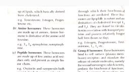
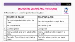
Section 4, , Clinical Biochemistry and Nutrition, , Chapter, , Hormones, , 19, , The hormones speak :, , “We are the chemical messengers of the body!, Diversified in our structure and function;, Act either directly or through messengers;, ‘Growth, health and welfare’ is our motto.”, , T, , he living body possesses a remarkable, communication system to coordinate its, biological functions. This is achieved by two, distinctly organized functional systems., 1. The nervous system coordinates the body, functions through the transmission of electrochemical impulses., , 2. The endocrine system acts through a wide, range of chemical messengers known as, hormones., , Hormones are conventionally defined as, organic substances, produced in small amounts, by specific tissues (endocrine glands), secreted, into the blood stream to control the, metabolic and biological activities in the target, cells. Hormones may be regarded as the, chemical, messengers, involved, in, the, transmission of information from one tissue to, another and from cell to cell. The major, endocrine organs in human body are depicted in, Fig.19.1)., , CLASSIFICATION OF HORMONES, Hormones may be classified in many ways, based on their characteristics and functions. Two, types of classification are discussed here, , I. Based on the chemical nature, The hormones can be categorized into three, groups considering their chemical nature., 1. Protein or peptide hormones e.g. insulin,, glucagon, antidiuretic hormone, oxytocin., 2. Steroid hormones e.g. glucocorticoids,, mineralocorticoids, sex hormones., 3. Amino acid derivatives e.g. epinephrine,, norepinephrine, thyroxine (T4), triiodothyronine, (T3)., , II. Based on the mechanism of, action, Hormones are classified into two broad, groups (I and II) based on the location of the, , 427
Page 2 :
428, , BIOCHEMISTRY, , Pineal gland, Pituitary gland, , Thyroid gland, , Pancreas, Adrenal gland, , Testis (male), , Ovary, (female), , stimulate the release of certain molecules,, namely the second messengers which, in, turn, perform the biochemical functions., Thus, hormones themselves are the first, messengers., Group II hormones are subdivided into three, categories based on the chemical nature of the, second messengers., (a) The second messenger is cAMP e.g., ACTH, FSH, LH, PTH, glucagon,, calcitonin., (b) The second messenger is phosphatidylinositol/calcium e.g. TRH, GnRH, gastrin,, CCK., (c) The second messenger is unknown e.g., growth hormone, insulin, oxytocin,, prolactin., The principal human hormones, their classification based on the mechanism of action, and, major functions are given in Table 19.1., , Mechanism of action of, group I hormones, , Fig. 19.1 : Diagrammatic representation of, major endocrine glands., , receptors to which they bind and the signals, used to mediate their action., 1. Group I hormones : These hormones bind, to intracellular receptors to form receptorhormone, complexes, (the, intracellular, messengers) through which their biochemical, functions are mediated. Group I hormones, are lipophilic in nature and are mostly, derivatives of cholesterol (exception—T3 and, T4). e.g. estrogens, androgens, glucocorticoids,, calcitriol., 2. Group II hormones : These hormones bind, to cell surface (plasma membrane) receptors and, , These hormones are lipophilic in nature and, can easily pass across the plasma membrane., They act through the intracellular receptors, located either in the cytosol or the nucleus., The, hormone-receptor complex binds to, specific regions on the DNA called hormone, responsive element (HRE) and causes increased, expression of specific genes (Fig.19.2). It is, believed that the interaction of hormone, receptor complex with HRE promotes initiation, and, to a lesser extent, elongation and, termination of RNA synthesis (transcription). The, ultimate outcome is the production of specific, proteins (translation) in response to hormonal, action., , Mechanism of action of, group II hormones, These hormones are considered as the first, messengers. They exert their action through, mediatory molecules, collectively called second, messengers.
Page 3 :
429, , Chapter 19 : HORMONES, , TABLE 19.1 Principal human hormones—classification, (by mechanism of action), origin and major functions, , Hormone(s), , Origin, , Major Function(s), , Group I. HORMONES THAT BIND TO INTRACELLULAR RECEPTORS, Estrogens, , Ovaries and adrenal cortex Female sexual characteristics, menstrual cycle., , Progestins, , Ovaries and placenta, , Androgens, , Testes and adrenal cortex Male sexual characteristics, spermatogenesis., , Glucocorticoids, , Adrenal cortex, , Affect metabolisms, suppress immune system., , Mineralocorticoids, , Adrenal cortex, , Maintenance of salt and water balance., , Calcitriol (1, 25–DHCC), , Kidney (final form), , Promotes absorption of Ca2+ from intestine, kidney and bone., , Thyroid hormones (T3, T4), , Thyroid, , Promote general metabolic rate., , Involved in menstrual cycle and maintenance of pregnancy., , Group II. HORMONES THAT BIND TO CELL SURFACE RECEPTORS, A. The second messenger is cAMP, Adrenocorticotropic hormone (ACTH) Anterior pituitary, , Stimulates the release of adrenocorticosteroids., , Follicle stimulating hormone (FSH), , Anterior pituitary, , In females, stimulates ovulation and estrogen synthesis., In males, promotes spermatogenesis., , Luteinizing hormone (LH), , Anterior pituitary, , Stimulates synthesis of estrogens and progesterone and, causes ovulation. Promotes androgen synthesis by testes., , Chorionic gonadotropin (hCG), , Anterior pituitary, , Stimulates progesterone release from placenta., , Thyroid stimulating hormone (TSH), , Anterior pituitary, , Promotes the release of thyroid hormones (T3, T4)., , E-Endorphins and enkephalins, , Anterior pituitary, , Natural endogenous analgesics (pain relievers)., , Antidiuretic hormone (ADH), , Posterior pituitary (stored) Promotes water reabsorption by kidneys., , Glucagon, , Pancreas, , Increases blood glucose level, stimulates glycogenolysis, and lipolysis., , Parathyroid hormone (PTH), , Parathyroid, , Increases serum calcium, promotes Ca2+ release from bone., , Calcitonin, , Thyroid, , Lowers serum calcium. Decreases Ca2+ uptake by bone and kidney., , Epinephrine, , Adrenal medulla, , Increases heart rate and blood pressure. Promotes glycogenolysis in liver and muscle and lipolysis in adipose tissue., , Norepinephrine, , Adrenal medulla, , Stimulates lipolysis in adipose tissue., , B. The second messenger is phosphatidyl inositol/calcium, Thyrotropin-releasing hormone (TRH), , Hypothalamus, , Promotes TSH release., , Gonadotropin-releasing hormone (GnRH), , Hypothalamus, , Stimulates release of FSH and LH., , Gastrin, , Stomach, , Stimulates gastric HCI and pepsinogen secretion., , Cholecystokinin (CCK), , Intestine, , Stimulates contraction of gall bladder and secretion of pancreatic, enzymes., , C. The second messenger is unknown/unsettled, Growth hormone (GH), , Anterior pituitary, , Promotes growth of the body (bones and organs)., , Prolactin (PRL), , Anterior pituitary, , Growth of mammary glands and lactation., , Oxytocin, , Posterior pituitary (stored) Stimulates uterine contraction and milk ejection., , Insulin, , Pancreas, , Lowers blood glucose (hypoglycemic effect), promotes protein, synthesis and lipogenesis., , Somatomedins (insulin-like, growth factors, IGF-I, IGF-II), , Liver, , Growth related functions of GH are mediated., Stimulates growth of cartilage.
Page 4 :
430, , BIOCHEMISTRY, , cAMP—THE SECOND, MESSENGER, , Plasma membrane, H, , Cyclic AMP (cAMP, cyclic, adenosine 3c,5c-monophosphate), is a ubiquitous nucleotide. It, consists of adenine, ribose and a, phosphate (linked by 3c,5c, linkage). cAMP acts as a second, messenger for a majority of, polypeptide hormones., The membrane-bound enzyme, adenylate cyclase converts ATP to, cyclic AMP. cAMP is hydrolysed, by phosphodiesterase to 5c-AMP, (Fig.19.3)., , Adenylate cyclase, system, A series of events occur at the, membrane level that influence, the activity of adenylate cyclase, leading to the synthesis of cAMP., This process is mediated by, G-proteins, so designated due to, their ability to bind to guanine, nucleotides., , Action of cAMP—a, general view, , H, , H, , R, , H, , R, , R, , Cytosol, , Nucleus, , DNA, , Acceptor, Gene, Transcription, mRNA, , mRNA, Translation, Specific protein, , Biochemical, response, , Once, produced,, cAMP, performs its role as a second, Fig. 19.2 : Mechanism of action of steroid hormones (H–Hormone;, R–Receptor; HR–Hormone-receptor complex)., messenger in eliciting biochemical responses (Fig.19.4)., cAMP activates protein kinase A, (A stands for cAMP). This enzyme is a heterote- protein that ultimately causes the biochemical, tramer consisting of 2 regulatory subunits (R) and response., 2 catalytic subunits (C)., It should, however, be remembered that, cAMP binds to inactive protein kinase and cAMP does not act on all protein kinases. For, causes the dissociation of R and C subunits., instance, on protein kinase C (the second, messenger is diacylglycerol)., 4cAMP + R2C2 o� R2(4 cAMP) + 2C, (inactive), (inactive), (active), Dephosphorylation of proteins : A group of, The active subunit (C) catalyses phosphory- enzymes called protein phosphatases hydrolyse, lation of proteins (transfer of phosphate group to and remove the phosphate group added to, serine and threonine residues). It is the phospho- proteins.
Page 5 :
431, , Chapter 19 : HORMONES, , ATP, Adenylate, cyclase, PPi, , Mg, , 2+, , NH2, N, , N, , N, , N, 5c, , O, , O, , CH2, , –, , O P O H, , H, , H, H, , 3c, , O, , OH, , 3c,5c-Cyclic adenosine, monophosphate (cAMP), H2O, , hormonal action. In response to the stimuli of, central nervous system, hypothalamus liberates, certain releasing factors or hormones. These, factors stimulate or inhibit the release of, corresponding tropic hormones from the anterior, pituitary. Tropic hormones stimulate the target, endocrine tissues to secrete the hormones they, synthesize., The, relationship, between, hypothalamus and pituitary with endocrine, glands is illustrated in Fig.19.6. In general, the, hormonal system is under feedback control. For, instance, adrenocorticotropic hormone (ACTH), inhibits the release of corticotropin releasing, hormone (CRH)., , Phosphodiesterase, 5c-AMP, , Fig. 19.3 : Synthesis and degradation of cAMP., , Degradation of cAMP : cAMP undergoes, rapid hydrolysis, catalysed by the enzyme, phosphodiesterase to 5’ AMP which is inactive., Hence, the effect of cAMP will be shortlived, if the hormone stimulating adenylate cyclase, is removed. Caffeine and theophylline, (methylxanthine, derivatives), can, inhibit, phosphodiesterases and increase the intracellular, levels of cAMP., , HYPOTHALAMIC AND, PITUITARY HORMONES, The pituitary gland or hypophysis (weighing, about 1 g) is located below the hypothalamus, of the brain. It consists of two distinct parts—, the anterior pituitary (adenohypophysis) and the, posterior pitutitary (neurohypophysis) connected, by pars intermedia (Fig.19.5). The latter is, almost absent in humans, although found in, lower organisms., , Hypothalamus is a specialized center in the, brain that functions as a master coordinator of, , HYPOTHALAMIC HORMONES, Hypothalamus produces at least six releasing, factors or hormones., 1. Thyrotropin-releasing hormone (TRH) : It, is a tripeptide consisting of glutamate derivative, (pyroglutamate), histidine and proline. TRH, stimulates anterior pituitary to release thyroidstimulating hormone (TSH or thyrotropin) which,, in turn, stimulates the release of thyroid, hormones (T3 and T4)., 2. Corticotropin-releasing hormone (CRH) :, It stimulates anterior pituitary to release, adrenocorticotropic hormone (ACTH) which in, turn, acts on adrenal cortex to liberate, adrenocorticosteroids. CRH contains 41 amino, acids., 3. Gonadotropin-releasing hormone (GnRH) :, It is a decapeptide. GnRH stimulates anterior, pituitary to release gonadotropins, namely, luteinizing hormone (LH) and follicle stimulating, hormone (FSH)., 4. Growth hormone-releasing hormone, (GRH) with 44 amino acids stimulates the, release of growth hormone (GH or somatotropin), which promotes growth., 5. Growth, hormone, release-inhibiting, hormone (GRIH) : It contains 14 amino acids, and is also known as somatostatin. GRIH inhibits, the release of growth hormone from the anterior, pituitary.
Page 6 :
432, , BIOCHEMISTRY, , Hormone, , Receptor, Plasma membrane, , GTP regulatory protein, Adenylate, cyclase, , Cytosol, PPi, , ATP, Phosphodiesterase, AMP, , 4cAMP, ( ), , R, , C, , R, , C, , R2C2, R, C, , C, , R, R 2(4 cAMP), , ATP, , ADP, , Phosphoprotein, , Protein, Phosphatase, , Pi, , Ultimate, biochemical, response, , Fig. 19.4 : Overview of synthesis and action of cAMP (R2C2–cAMP dependent protein kinase A;, R2–Regulatory subunits; C2–Catalytic subunits; C–Active catalytic unit of R2C2)., , 6. Prolactin release-inhibiting hormone, (PRIH) : It is believed to be a dopamine and/or, a small peptide that inhibits the release of, prolactin (PRL) from anterior pituitary., , hormones that influence—either directly or, indirectly—a variety of biochemical processes in, the body. The hormones of adenohypophysis are, broadly classified into three categories., I. The growth hormone-prolactin group., , ANTERIOR PITUITARY HORMONES, Anterior pituitary or adenohypophysis is truly, the master endocrine organ. It produces several, , II. The glycoprotein hormones., III. The pro-opiomelanocortin, family., , peptide
Page 8 :
434, 1. Effects on growth : As is obvious from the, name, GH is essential for the growth. The, growth-related effects of GH are mediated, through insulin like growth factor I (IGF-I) which, is also known as somatomedin C (formerly, sulfation factor), produced by liver., 2. Effects on protein metabolism : Growth, hormone has an anabolic effect on protein, metabolism. It promotes the uptake of amino, acids into the tissues and increases the protein, synthesis. The overall effect of GH is a positive, nitrogen balance that leads to increase in body, weight., 3. Effects on carbohydrate metabolism :, Growth hormone is antagonistic to insulin and, causes hyperglycemia. GH increases gluconeogenesis, decreases glucose utilization, impairs, glycolysis and reduces the tissue uptake of, glucose., 4. Effects on lipid metabolism : Growth, hormone promotes lipolysis in the adipose tissue, and increases the circulatory levels of free fatty, acids and their oxidation. It increases, ketogenesis, particularly in diabetes., 5. Effects on mineral metabolism : Growth, hormone promotes bone mineralization and its, growth, as clearly observed in the growing, children., , Abnormalities of GH production, Deficiency of GH :, Impairment in the, secretion of growth hormone in the growing age, causes dwarfism. The other deficiency metabolic, effects are not that serious in nature., Overproduction of GH : Excessive production, of GH causes gigantism in children and, acromegaly in adults. This usually occurs in the, acidophil tumor of pituitary gland. Gigantism is, characterized by increased growth of long bones, and this is observed before the epiphyseal plates, close. Acromegaly occurs after epiphyseal, closure and is characterized by increase in the, size of hands, facial changes (enlarged nose,, protruding jaw), excessive hair, thickening of, skin etc., , BIOCHEMISTRY, , Prolactin, Prolactin (PRL) is also called lactogenic, hormone, luteotropic hormone, mammotropin or, luteotropin., Biochemical functions of PRL : Prolactin is, primarily concerned with the initiation and, maintenance of lactation in mammals. PRL, increases the levels of several enzymes involved, in carbohydrate and lipid metabolism. PRL, promotes, HMP, shunt,, increases, lipid, biosynthesis and stimulates lactose production in, mammary glands., Prolactin promotes the growth of corpus luteum, (hence also known as luteotropic hormone) and, stimulates the production of progesterone., , II. The glycoprotein hormones, The following four hormones are glycoprotein, in nature and possess certain structural, similarities, despite their functional diversity., 1. Thyroid stimulating hormone (TSH), 2. Follicle stimulating hormone (FSH), 3. Luteinizing hormone (LH), 4. Human chorionic gonadotropin (hCG)., The last three hormones (2-4) are collectively, referred to as gonadotropins due to their, involvement in the function of gonads. The, hormone hCG is produced by human placenta, and not by pituitary. However, due to its, structural resemblance with other hormones, it is, also considered here., 1. Thyroid stimulating hormone (TSH) : TSH, is a dimer (DE) glycoprotein with a molecular, weight of about 30,000., Regulation of TSH production : The release of, TSH from anterior pituitary is controlled by a, feedback mechanism. This involves the hormones, of thyroid gland (T3 and T4) and thyrotropinreleasing hormone (TRH) of hypothalamus., Functions of TSH : The biochemical effects of, TSH on thyroid gland are briefly discussed here., TSH binds with plasma membrane receptors and, stimulates adenylate cyclase with a consequent, increase in cAMP level. TSH, through the, mediation of cAMP, exerts the following effects.
Page 9 :
435, , Chapter 19 : HORMONES, , l, , l, , l, , Promotes the uptake of iodide (iodide pump), from the circulation by thyroid gland., , III. The pro-opiomelanocortin, (POMC) peptide family, , Enhances the conversion of iodide (I–) to, active iodide (I+), a process known as, organification., , This family consists of the hormones—, adrenocorticotropic, hormone, (ACTH),, lipotropin (LPH) and melanocyte stimulating, hormone (MSH) and several (about 24), neuromodulators such as endorphins and, enkephalins., , Increases the proteolysis of thyroglobulin to, release T3 and T4 into the circulation., , TSH increases the synthesis of proteins,, nucleic acids and phospholipids in thyroid, gland., Gonadotropins : The follicle-stimulating, hormone (FSH), luteinizing hormone (LH) and, human chorionic gonadotropin (hCG) are, commonly known as gonadotropins. All three, are glycoproteins., The release of FSH and LH from the anterior, pituitary is controlled by gonadotropin-releasing, hormone (GnRH) of hypothalamus., 2. Biochemical functions of FSH : In females,, FSH stimulates follicular growth, increases the, weight of the ovaries and enhances the, production of estrogens., In males, FSH stimulates testosterone, production, required for spermatogenesis., FSH also promotes growth of seminiferous, tubules., 3. Biochemical functions of LH : Luteinizing, hormone, stimulates, the, production, of, progesterone from corpus luteum cells in females, and testosterone from Leydig cells in males. LH, and FSH are collectively responsible for the, development and maintenance of secondary, sexual characters in males., 4. Human chorionic gonadotropin (hCG) :, hCG is a glycoprotein (mol. wt. 100,000),, produced by syncytiotrophoblast cells of, placenta. The structure of hCG closely resembles, that of LH., The levels of hCG in plasma and urine, increase, almost, immediately, after, the, implantation of fertilized ovum. The detection of, hCG in urine is conveniently used for the early, detection (within a week after missing the, menstrual cycle) of pregnancy., , The synthesis of POMC family. is very, interesting. All the members of POMC are, produced from a single gene of the anterior, and intermediate lobes of pituitary. It is, fascinating that a single polypeptide—proopiomelanocortin—is the precursor (approximately 285 amino acids) that contains multiple, hormones. The name pro-opiomelano-cortin is, derived since it is a prohormone to opioids,, melanocyte-stimulating hormone and corticotropin., Products of POMC : The pituitary, multihormone precursor is synthesized as preproopiomelanocortin from which POMC, is formed. The POMC consists of 3 peptide, groups., 1. ACTH that can give rise to D-MSH and, corticotropin like intermediate lobe peptide, (CLIP)., 2. E-Lipotropin (E-LPH) that can produce, J-LPH, E-MSH and E-endorphin. The latter yields, J- and D-endorphins., 3. An N-terminal peptide that forms J-MSH., The products obtained from POMC are, depicted in Fig.20.7. These products undergo, many modifications such as glycosylation,, acetylation etc., 1. Adrenocorticotropic hormone (ACTH) :, ACTH is a polypeptide with 39 amino acids and, a molecular weight of 4,500. This hormone is, primarily concerned with the growth and, functions of adrenal cortex., Regulation of ACTH production : The release, of ACTH from the anterior pituitary is under the, regulation of hypothalamic hormone, namely, corticotropin releasing hormone (CRH).
Page 10 :
436, Biochemical, ACTH, l, , l, , l, , BIOCHEMISTRY, , functions, , of, , ACTH, promotes, the, conversion of cholesterol to, pregnenolone in the adrenal, cortex., , ACTH, , E-LPH (93 A.As), , 1–39, , 42–134, , D-MSH, 1–13, , J-LPH, 42–101, E-MSH, 84–101, , It enhances RNA and protein, synthesis and thus promotes, adrenocortical growth., ACTH increases lipolysis by, activating lipase of adipose, tissue., , CLIP, 18–39, , E-Endorphin (31 A.As), 104–134, J-Endorphin (15 A.As), 104–118, D-Endorphin (14 A.As), 104–117, Enkephalin, 5, , Fig. 19.7 : The members of the pro-opiomelanocortin (POMC) family, , derived from POMC cleavage. (Numbers in blocks represent amino acids, Overproduction of ACTH :, in sequence; In the brackets are the number of amino acids–AAs;, Cushing’s syndrome is caused, (ACTH–Adrenocorticotropic hormone; LPH–Lipotropin;, by an excessive production of, MSH–Melanocyte-stimulating hormone; CLIP–Corticotropin like, intermediate lobe peptide)., ACTH which may be due, to a tumor. This syndrome, is characterized by hyperpigmentation and increased production of is derived from POMC (Fig.19.7). E-Lipotropin, adrenocorticosteroids. The associated symptoms has 31 amino acids while its modified products, include negative nitrogen balance, impaired D and J-endorphins have 15 and 14 amino acids,, glucose tolerance, hypertension, edema, muscle respectively. Methionine enkephalin (Tyr–Gly–, Gly–Phe–Met) and leucine enkephalin (Tyr-Glyatrophy etc., Gly-Phe-Leu) are the two important pentapeptide, 2. E-Lipotropin (E-LPH) : E-LPH is derived, derivatives of E-endorphin., from POMC and contains 93 carboxy terminal, amino acids. This polypeptide consists of, Biochemical actions : Endorphins and, J-LPH and E-endorphin from which E-MSH enkephalins are peptide neurotransmitters that, and J-endorphin are, respectively, formed. produce opiate-like effects on the central, J-Endorphin can be converted to D-endorphin nervous system, hence they are also known as, and then to enkephalins (Fig.19.7). E-LPH is opioid-peptides. They bind to the same receptors, found only in the pituitary and not in other as the morphine opiates and are believed to, tissues since it is rapidly degraded., control the endogenous pain perception., The biochemical functions of E-LPH, as Endorphins and enkephalins are more potent, such, are limited. It promotes lipolysis and (20-30 times) than morphine in their function as, increases the mobilization of fatty acids. The analgesics., , most important function of E-LPH is its precursor, role for the formation of E-endorphin and, enkephalins., , Endorphins and enkephalins : These are the, natural analgesics that control pain and, emotions. They were discovered after an, unexpected finding of opiate receptors in the, human brain., Synthesis : Endorphins and enkephalins, are produced from E-endorphin which, in turn,, , It is believed that the pain relief through, acupuncture and placebos is mediated through, opioid peptides., 3. Melanocyte-stimulating hormone (MSH) :, Three types of MSH (D, E and J) are present in, the precursor POMC molecule. In humans, J, MSH is important while in some animals D and, E are functional. The activity of J-MSH is, contained in the molecule J-LPH or its precursor, E-LPH (Fig.19.7).
Page 11 :
437, , Chapter 19 : HORMONES, , 1, , 9, , (A) Cys — Tyr — Ile — Gln — Asn — Cys — Pro — Leu — Gly, , S, , S, , 1, , 9, , (B) Cys — Tyr — Phe — Gln — Asn — Cys — Pro — Arg — Gly, , S, , S, , Fig. 19.8 : Structures of (A) Human oxytocin and, (B) Human antidiuretic hormone (ADH)., , The functions of MSH has been clearly, established in some animals. MSH promotes the, synthesis of skin pigment melanin (melanogenesis), and disperses melanin granules that ultimately, leads to darkening of the skin. In humans, MSH, does not appear to play any role in melanin, synthesis., , POSTERIOR PITUITARY HORMONES, Two hormones namely oxytocin and, antidiuretic hormone (ADH, vasopressin) are, produced by the posterior pituitary gland, (neurohypophysis)., Both, of, them, are, nonapeptides (9 amino acids). Their structures, are depicted in Fig.19.8., , Oxytocin, The release of oxytocin from posterior, pituitary gland is caused by the neural impulses, of nipple stimulation. The other stimuli, responsible for oxytocin release include vaginal, and uterine distention., , Biochemical functions, 1. Effect on uterus : Oxytocin causes the, contraction of pregnant uterus (smooth muscles), and induces labor., 2. Effect on milk ejection : In mammals,, oxytocin causes contraction of myoepithelial, cells (look like smooth muscle cells) of breast., This stimulates the squeezing effect, causing milk, ejection from the breast., 3. Oxytocin synthesized in the ovary appears, to inhibit the synthesis of steroids., , Antidiuretic hormone (ADH), The release of ADH (also called vasopressin), is mostly controlled by osmoreceptors (of hypothalamus) and baroreceptors (of heart). Any, increase in the osmolarity of plasma stimulates, ADH secretion., Biochemical functions : ADH is primarily, concerned with the regulation of water balance, in the body. It stimulates kidneys to retain water, and, thus, increases the blood pressure., In the absence of ADH, the urine output, would be around 20 l/day. ADH acts on the, distal convoluted tubules of kidneys and causes, water reabsorption with a result that the urine, output is around 0.5-1.5 l/day., Mechanism of action : ADH stimulates, adenylate cyclase causing production of cAMP., Water reabsorption is promoted by cAMP., Inhibitors of adenylate cyclase (e.g. calcium), inhibit the activity of ADH. This supports the, view that ADH action is mostly mediated, through cAMP., Diabetes insipidus : This disorder is characterized by the excretion of large volumes of, dilute urine (polyuria). It may be due to, insufficient levels of ADH or a defect in the, receptors of target cells., , THYROID HORMONES, Thyroid gland (weighs about 30 g in adults) is, located on either side of the trachea below the, larynx. It produces two principal hormones, (Fig.19.9)—thyroxine (T4; 3,5,3’,5’-tetraiodothyronine) and 3,5,3’-triiodothyronine (T3)—, which regulate the metabolic rate of the body., Thyroid gland also secretes calcitonin, a hormone, concerned with calcium homeostasis (discussed, under calcium metabolism, Chapter 18)., , Biosynthesis of thyroid hormones, Iodine is essential for the synthesis of thyroid, hormones. More than half of the body’s total, iodine content is found in the thyroid gland.
Page 12 :
438, , BIOCHEMISTRY, , I, , I, 3c, , 3, , HO, , CH2 CH COOH, , O, 5c, , I, , 5, , NH 2, I, 3, 5, 3c, 5c-Tetraiodothyronine (thyroxine, T4), , I, , I, , HO, , CH2 CH COOH, , O, , NH 2, , I, 3,5,3c-Triiodothyronine (T3), I, , I, , HO, , O, , CH2 CH COOH, NH 2, , I, , 3, 3c, 5c-Triiodothyronine (reverse T3, rT3), , Fig. 19.9 : Structures of thyroid hormones, (Refer Fig. 15.21 for their biosynthesis)., , Thyroglobulin and synthesis of T3 and T4 :, Thyroglobulin (mol. wt. 660,000) is a, glycoprotein and precursor for the synthesis of, T3 and T4. Thyroglobulin contains about 140, tyrosine residues which can serve as substrates, for iodine for the formation of thyroid hormones., Tyrosine (of thyroglobulin) is first iodinated at, position 3 to form monoiodotyrosine (MIT) and, then at position 5 to form diiodotyrosine (DIT)., Two molecules of DIT couple to form thyroxine, (T4). One molecule of MIT, when coupled with, one molecule of DIT, triiodothyronine (T3) is, produced. The mechanism of coupling is not, well understood. The details of synthesis of T3, and T4 are given under tyrosine metabolism, (Chapter 15). A diagrammatic representation is, depicted in Fig.19.10., As the process of iodination is completed,, each molecule of thyroglobulin contains about, 6-8 molecules of thyroxine (T4). The ratio of T3, to T4 in thyroglobulin is usually around 1 : 10., , Uptake of iodide : The uptake of iodide by, the thyroid gland occurs against a concentration, gradient (about 20 : 1). It is an energy requiring, process and is linked to the ATPase dependent, Na+-K+ pump. Iodide uptake is primarily, controlled by TSH. Antithyroid agents such as, thiocyanate and perchlorate inhibit iodide, transport., , Thyroglobulin containing T4 and T3 can be, stored for several months in the thyroid gland. It, is estimated that the stored thyroid hormones can, meet the body requirement for 1-3 months., , Formation of active iodine : The conversion, of iodide (I–) to active iodine (I+) is an essential, step for its incorporation into thyroid hormones., Thyroid is the only tissue that can oxidize I– to, a higher valence state I+. This reaction requires, H2O2 and is catalysed by the enzyme, thyroperoxidase (mol. wt. 60,000). An NADPH, dependent system supplies H2O2., , Thyroglobulin is digested by lysosomal, proteolytic enzymes in the thyroid gland. The, free, hormones, thyroxine, (90%), and, triiodothyronine (10%) are released into the, blood, a process stimulated by TSH. MIT and, DIT produced in the thyroid gland undergo, deiodination by the enzyme deiodinase and the, iodine thus liberated can be reutilized., , I–, , O2, , H2O2, +, NADP NADPH + H, , +, , Thyroperoxidase, I+, , H2 O, , TSH promotes the oxidation of iodide to, active iodine while the antithyroid drugs, (thiourea, thiouracil, methinazole) inhibit., , Storage and release of, thyroid hormones, , Transport of T4 and T3, Two specific binding proteins—thyroxine, binding globulin (TBG) and thyroxine binding, prealbumin (TBPA)—are responsible for the, transport of thyroid hormones. Both T4 and T3, are more predominantly bound to TBG. A small, fraction of free hormones are biologically active., T4 has a half-life of 4-7 days while T3 has about, one day.
Page 13 :
439, , Chapter 19 : HORMONES, , correlated to thyroid hormones and this, in turn,, with ATP utilization. Obesity in some individuals, is attributed to a decreased energy utilization and, heat production due to diminished Na+-K+, ATPase activity., , Tgb, , I+, , Iodination, DIT, MIT, , Tgb, Thioperoxidase, , Coupling, , I–, , Deiodination, (deiodinase), , Tgb, T4, , T3, , Proteolysis, MIT + T , T + A.As, 3 4, DIT, , To target tissues, , Fig. 19.10 : Biosynthesis of thyroid hormones—, diagrammatic representation [Note : Refer Fig. 15.21 for, synthesis with structures; Tgb–Thyroglobulin; I+–Active, iodine; T3–Triiodothyronine; T4–Thyroxine; MIT–Monoiodotyrosine; DIT–Diiodotyrosine;, A. As–Amino acids]., , Biochemical functions of, thyroid hormones, Triiodothyronine (T3) is about four times more, active in its biological functions than thyroxine, (T4). The following are the biochemical functions, attributed to thyroid hormones (T3 and T4)., 1. Influence on the metabolic rate : Thyroid, hormones stimulate the metabolic activities and, increases the oxygen consumption in most of the, tissues of the body (exception—brain, lungs,, testes and retina)., Na+-K+ ATP pump : This is an energy dependent process which consumes a major share of, cellular ATP. Na+-K+ ATPase activity is directly, , 2. Effect on protein synthesis : Thyroid, hormones act like steroid hormones in promoting, protein synthesis by acting at the transcriptional, level (activate DNA to produce RNA). Thyroid, hormones, thus, function as anabolic hormones, and cause positive nitrogen balance and promote, growth and development., 3. Influence on carbohydrate metabolism :, Thyroid hormones promote intestinal absorption, of glucose and its utilization. These hormones, increase gluconeogenesis and glycogenolysis,, with an overall effect of enhancing blood, glucose level (hyperglycemia)., 4. Effect on lipid metabolism : Lipid turnover, and utilization are stimulated by thyroid, hormones. Hypothyroidism is associated with, elevated plasma cholesterol levels which can be, reversed by thyroid hormone administration., , Regulation of T3 and T4 synthesis, The synthesis of thyroid hormones is, controlled by feedback regulation (Fig.19.11). T3, appears to be more actively involved than T4 in, the regulation process. The production of thyroid, stimulating hormone (TSH) by pituitary, and, thyrotropin releasing hormone (TRH) by, hypothalamus are inhibited by T3 and, to a lesser, degree, by T4. The increased synthesis of TSH, and TRH occurs in response to decreased, circulatory levels of T3 and T4. As already, discussed, the body has sufficient stores of, hormones to last for several weeks. Hence it, takes some months to observe thyroid functional, deficiency., , Metabolic fate of T3 and T4, Thyroid hormones undergo deiodination in, the peripheral tissues. The iodine liberated may, be reutilized by the thyroid. T3 and T4 may get, conjugated with glucuronic acid or sulfate in the, liver and excreted through bile. Thyroid, hormones are also subjected to deamination to
Page 14 :
440, , BIOCHEMISTRY, , produce tetraiodothyroacetic acid, (from T4) and triiodothyroacetic acid, (from T3) which may then undergo, conjugation and excretion., , Hypothalamus, , TRH, , Abnormalities of thyroid, function, Pituitary, , Among the endocrine glands,, thyroid is the most susceptible for, hypo- or hyperfunction., , TSH, , Three abnormalities associated, with thyroid functions are known., Goiter : Any abnormal increase, in the size of the thyroid gland is, known as goiter. Enlargement of, thyroid gland is mostly to, compensate the decreased synthesis, of thyroid hormones and is, associated with elevated TSH., Goiter is primarily due to a failure, in the autoregulation of T3 and T4, synthesis. This may be caused by, deficiency or excess of iodide., , Thyroid, gland, , T3 , T4, , Metabolic, , Protein, , Carbohydrate, , Utilization, , Maintenance, , Goitrogenic, substances, raten, synthesisn metabolismn, of lipidsn of H2O, electrolyte, balance, (goitrogens) : These are the, substances that interfere with the, Fig. 19.11 : Regulation of synthesis and functions of thyroid, hormones—an overview (TRH–Thyrotropin-stimulating hormone;, production of thyroid hormones., TSH–Thyroid stimulating hormone; T3–Triiodothyronine;, These include thiocyanates, nitrates, T4–Thyroxine;, –Promoting effect;, –Inhibitory effect)., and perchlorates and the drugs such, as thiourea, thiouracil, thiocarbamide, etc. Certain plant foods—cabbage, cauliflower and irritability, anxiety, rapid heart rate, loss of, turnip—contain goitrogenic factors (mostly thio- weight despite increased appetite, weakness,, diarrhea, sweating, sensitivity to heat and often, cyanates)., protrusion of eyeballs (exopthalmos)., Simple endemic goiter : This is due to iodine, deficiency in the diet. It is mostly found in the, Hyperthyroidism is caused by Grave’s disease, geographical regions away from sea coast where (particularly in the developed countries) or due, the water and soil are low in iodine content. to increased intake of thyroid hormones. Grave’s, Consumption of iodized salt is advocated to disease is due to elevated thyroid stimulating, overcome the problem of endemic goiter. In IgG also known as long acting thyroid stimulator, certain cases, administration of thyroid hormone (LATS) which activates TSH and, thereby,, is also employed., increases thyroid hormonal production., Hyperthyroidism : This is also known as, thyrotoxicosis, and, is, associated, with, overproduction, of, thyroid, hormones., Hyperthyroidism is characterized by increased, metabolic rate (higher BMR) nervousness,, , Thyrotoxicosis is diagnosed by scanning and/, or estimation of T3, T4 (both elevated) and TSH, (decreased) in plasma. The treatment includes, administration of antithyroid drugs. In severe, cases, thyroid gland is surgically removed.
Page 15 :
441, , Chapter 19 : HORMONES, , Hypothyroidism : This is due to an, impairment in the function of thyroid gland that, often causes decreased circulatory levels of T3, and T4. Disorders of pituitary or hypothalamus, also contribute to hypothyroidism. Women are, more susceptible than men. Hypothyroidism is, characterized by reduced BMR, slow heart rate,, weight gain, sluggish behaviour, constipation,, sensitivity to cold, dry skin etc., Hypothyroidism in children is associated with, physical and mental retardation, collectively, known as cretinism. Early diagnosis and proper, treatment are essential. Hypothyroidism in adult, causes myxoedema, characterized by bagginess, under the eyes, puffiness of face, slowness in, physical and mental activities., Thyroid hormonal administration is employed, to treat hypothyroidism., , Laboratory diagnosis of, thyroid function, Measurement of basal metabolic rate (BMR), was once used to reflect thyroid activity. The, estimation of serum protein bound iodine (PBI),, representing the circulating thyroid hormones,, was employed for a long time to assess thyroid, function. The normal serum PBI concentration is, 3-8 Pg/100 ml., Hypothyroidism is associated with decreased, PBI and hyperthyroidism with increased PBI., In recent years, more sensitive and reliable, tests have been developed to assess thyroid, activity. The concentration of free T3 and T4,, and TSH are measured (by RIA or ELISA) and, their serum normal concentrations are, Free triiodothyronine (T3) — 80–220 ng/dl, Free thyroxine (T4), , — 0.8–2.4 ng/dl, , Total thyroxine (T4), , — 5–12 Pg/dl, , Thyroid stimulating, hormone (TSH), , — <10 PU/ml, , Radioactive iodine uptake (RAIU) and, scanning of thyroid gland are also used for, diagnosis., , Zona glomerulosa, Zona fasciculata, Zona reticularis, Medulla, , Fig. 19.12 : Adrenal gland with zones (3) and medulla., , Thyroid activity and, serum cholesterol, Serum cholesterol level is increased in, hypothyroidism and decreased in hyperthyroidism., Unfortunately, cholesterol estimation will be of no, value in the assessment of thyroid function. This is, due to the fact that serum cholesterol level is, elevated in many other disorders (diabetes,, obstructive jaundice, nephrotic syndrome etc.)., However, cholesterol estimation may be utilized, for monitoring thyroid therapy., , HORMONES OF ADRENAL CORTEX, The adrenal glands are two small organs (each, weighing about 10 g), located above the kidneys., Each adrenal consists of two distinct tissues—an, outer cortex (with 3 zones) and inner medulla, (Fig.19.12)., As many as 50 steroid hormones (namely, adrenocorticosteroids), produced by adrenal, cortex, have been identified. However, only a, few of them possess biological activity., , Adrenocorticosteroids are classified into three, groups according to their dominant biological, action. However, there is some overlap in their, functions., 1. Glucocorticoids : These are 21-carbon, steroids, produced mostly by zona fasciculata., They affect glucose (hence the name), amino, acid and fat metabolism in a manner that is, opposite to the action of insulin. Cortisol (also, known as hydrocortisone) is the most important, glucocorticoid in humans. Corticosterone is, predominantly found in rats.
Page 16 :
442, , BIOCHEMISTRY, , 2. Mineralocorticoids : These are also 21carbon containing steroids produced by zona, glomerulosa. They regulate water and electrolyte, balance. Aldosterone is the most prominent, mineralocorticoid., 3. Androgens and estrogens : The innermost, adrenal cortex zona reticularis produces small, quantities of androgens (19-carbon) and, estrogens (18-carbon). These hormones affecting, sexual development and functions are mostly, produced by gonads. Dehydroepiandrosterone—, a precursor for androgens—is synthesized in, adrenal cortex., , Synthesis of adrenocorticosteroids, Cholesterol, elimination of, pregnenolone., precursor for, hormones., , undergoes cleavage with an, a 6-carbon fragment to form, Pregnenolone is the common, the synthesis of all steroid, , Conversion of cholesterol to pregnenolone is, catalysed by cytochrome P450 side chain cleavage, enzyme. This reaction is promoted by ACTH., The enzymes—hydroxylases, dehydrogenases/, isomerases, and, lyases, associated, with, mitochondria or endoplasmic reticulum—are, responsible for the synthesis of steroid hormones., The metabolic pathway for the formation of major, adrenocorticosteroids is given in Fig.19.13., , Biochemical functions of, adrenocorticosteroids, 1. Glucocorticoid hormones : The important, glucocorticoids are—cortisol, cortisone and, corticosterone. They bring about several, biochemical functions in the body., (a) Effects on carbohydrate metabolism :, Glucocorticoids promote the synthesis of, glucose (gluconeogenesis). This is brought, about by increasing the substrates, (particularly amino acids) and enhancing, the synthesis of phosphoenolpyruvate, carboxykinase, the rate limiting enzyme, in gluconeogenesis., The overall influence of glucocorticoids, on carbohydrate metabolism is to increase, , blood, glucose, concentration., The, biological actions of glucocorticoids, generally oppose that of insulin., (b) Effects on lipid metabolism : Glucocorticoids increase the circulating free fatty, acids. This is caused by two mechanisms., (i) Increased breakdown of storage triacylglycerol (lipolysis) in adipose tissue., (ii) Reduced utilization of plasma free fatty, acids for the synthesis of triacylglycerols., (c) Effects on protein and nucleic acid, metabolism : Glucocortiocoids exhibit, both catabolic and anabolic effects on, protein and nucleic acid metabolism., They, promote, transcription, (RNA, synthesis) and protein biosynthesis in, liver., These, anabolic, effects, of, glucocorticoids are caused by the, stimulation of specific genes., Glucocorticoids (particularly at high, concentration) cause catabolic effects in, extrahepatic tissues (e.g. muscle, adipose, tissue, bone etc.). This results in enhanced, degradation of proteins., (d) Effects on water and electrolyte metabolism : The influence of glucocorticoids, on water metabolism is mediated through, antidiuretic hormone (ADH). Deficiency, of glucocorticoids causes increased, production of ADH. ADH decreases, glomerular filtration rate causing water, retention in the body., (e) Effects on the immune system : Glucocorticoids (particularly cortisol), in high, doses, suppress the host immune, response. The steroid hormones act at, different levels—damaging lymphocytes,, impairment of antibody synthesis,, suppression of inflammatory response etc., (f) Other physiological effects of glucocorticoids : Glucocorticoids are involved in, several physiological functions., (i) Stimulate the fight and flight response, (to face sudden emergencies) of, catecholamines.
Page 18 :
444, , BIOCHEMISTRY, , (ii) Increase the production of gastric HCI, and pepsinogen., (iii) Inhibit the bone formation, hence the, subjects are at a risk for osteoporosis., Mechanism of action of glucocorticoids :, Glucocorticoids bind to specific receptors on the, target cells and bring about the action. These, hormones mostly act at the transcription level, and control the protein synthesis., 2. Mineralocorticoid hormones : The most, active and potent mineralocorticoid is, aldosterone. It promotes Na+ reabsorption at the, distal convoluted tubules of kidney. Na+, retention is accompanied by corresponding, excretion of K+, H+ and NH4+ ions., Regulation of aldosterone synthesis : The, production of aldosterone is regulated by, different mechanisms. These include reninangiotensin, potassium, sodium and ACTH., Mechanism of aldosterone action :, Aldosterone acts like other steroid hormones. It, binds with specific receptors on the target tissue, and promotes transcription and translation., Metabolism of adrenocorticosteroids : The, steroid hormones are metabolized in the liver, and excreted in urine as conjugates of, glucuronides or sulfates., The urine contains mainly two steroids—, 17-hydroxysteroids and 17-ketosteroids—derived, from the metabolism of glucocorticoids and, mineralocorticoids., Androgens, synthesized, by gonads also contribute to the formation of, 17-ketosteroids., Urinary 17-ketosteroids estimated in the, laboratory are expressed in terms of, dehydroepiandrosterone and their normal, excretion is in the range of 0.2–2.0 mg/day., , Abnormalities of adrenocortical, function, Addison’s, disease, adrenocortical function, disease. This disorder, decreased blood glucose, , :, Impairment, in, results in Addison’s, is characterized by, level (hypoglycemia),, , loss of weight, loss of appetite (anorexia), muscle, weakness, impaired cardiac function, low blood, pressure, decreased Na+ and increased K+ level, in serum, increased susceptibility to stress etc., Cushing’s syndrome : Hyperfunction of, adrenal cortex may be due to long term, pharmacological use of steroids or tumor of, adrenal cortex or tumor of pituitary. Cushing’s, syndrome is characterized by hyperglycemia, (due to increased gluconeogenesis), fatigue,, muscle wasting, edema, osteoporosis, negative, nitrogen balance, hypertension, moon-face etc., , Assessment of adrenocortical, function, The adrenocortical function can be assessed, by measuring plasma cortisol (5-15 Pg/dl at 9.00, AM), plasma ACTH, urinary 17-ketosteroids etc., , HORMONES OF ADRENAL MEDULLA, Adrenal medulla is an extension of, sympathetic nervous system. It produces two, important hormones—epinephrine (formerly, adrenaline) and norepinephrine (formerly, noradrenaline). Both these hormones are, catecholamines since they are amine derivatives, of catechol nucleus (dihydroxylated phenyl ring)., Epinephrine is a methyl derivative of, norepinephrine., Dopamine, is, another, catecholamine, produced as an intermediate, during, the, synthesis, of, epinephrine., Norepinephrine and dopamine are important, neurotransmitters in the brain and autonomic, nervous system. The structures of the three, catecholamines are given in Fig.19.14., , Synthesis of catecholamines, The amino acid tyrosine is the precursor for, the synthesis of catecholamines. The pathway is, described under tyrosine metabolism (Chapter, 15, Fig.15.22). Catecholamines are produced in, response to fight, fright and flight. These include, the emergencies like shock, cold, fatigue,, emotional conditions like anger etc.
Page 19 :
445, , Chapter 19 : HORMONES, , Biochemical functions, of catecholamines, Catecholamines, cause, diversified biochemical effects, on the body. The ultimate goal, of their action is to mobilize, energy resources and prepare the, individuals to meet emergencies, (e.g. shock, cold, low blood, glucose etc.)., , HO, , HO, , HO, , HO, , CH2 CH2 NH2, , Catechol, , Dopamine, HO, , HO, HO, , CH CH2 NH2, , HO, , CH2 CH2 NH, OH, , OH, Norepinephrine, , CH3, , Epinephrine, , 1. Effects on carbohydrate, Fig. 19.14 : Catecholamines (dopamine, norepinephrine and, metabolism : Epinephrine and, epinephrine) produced by adrenal medulla, norepinephrine, in, general, (Refer Fig. 15.22 for biosynthesis)., increase the degradation of, glycogen, (glycogenolysis),, synthesis of glucose (gluconeogenesis) and transferase (COMT) and monoamine oxidase, (MAO), found in many tissues act on, decrease glycogen formation (glycogenesis)., The overall effect of catecholamines is to catecholamines. The metabolic products, elevate blood glucose levels and make it metanephrine and vanillylmandelic acid (VMA), available for the brain and other tissues to meet are excreted in urine., the emergencies., 2. Effects on lipid metabolism : Both, epinephrine and norepinephrine enhance the, breakdown of triacylglycerols (lipolysis) in, adipose tissue. This causes increase in the free, fatty acids in the circulation which are effectively, utilized by the heart and muscle as fuel source., The metabolic effects of catecholamines are, mostly related to the increase in adenylate, cyclase activity causing elevation in cyclic AMP, levels (refer carbohydrate and lipid metabolisms, for more details)., 3. Effects on physiological functions : In, general, catecholamines (most predominantly, epinephrine) increase cardiac output, blood, pressure and oxygen consumption. They cause, smooth muscle relaxation in bronchi, gastrointestinal tract and the blood vessels supplying, skeletal muscle. On the other hand,, catecholamines stimulate smooth muscle, contraction of the blood vessels supplying skin, and kidney. Platelet aggregation is inhibited by, catecholamines., , Metabolism of catecholamines, Catecholamines are rapidly inactivated and, metabolized. The enzymes—catechol-O methyl-, , Abnormalities of, catecholamine production, Pheochromocytomas : These are the tumors, of adrenal medulla. The diagnosis of, pheochromocytoma is possible only when there, is an excessive production of epinephrine and, norepinephrine that causes severe hypertension., In the individuals affected by this disorder, the, ratio of norepinephrine, to epinephrine is, increased. The measurement of urinary VMA, (normal <8 mg/day) is helpful in the diagnosis of, pheochromocytomas., , HORMONES OF GONADS, The gonads (testes in males, ovaries in, females) perform closely related dual functions., 1. Synthesize sex hormones;, 2. Produce germ cells., The steroid sex hormones are responsible for, growth,, development,, maintenance, and, regulation of reproductive system. Sex hormones, are essentially required for the development of, germ cells.
Page 20 :
446, The sex hormones are categorized into three, groups, 1. Androgens or male sex hormones which, are C-19 steroids., 2. Estrogens or female sex hormones which, are C-18 steroids. Ring A of steroid nucleus is, phenolic in nature and is devoid of C-19 methyl, group., 3. Progesterone is a C-21 steroid produced, during the luteal phase of menstrual cycle and, also during pregnancy., , BIOCHEMISTRY, , l, , Spermatogenesis., , l, , Male pattern of aggressive behavior., , 2. Biochemical functions : Many specific, biochemical effects of androgens that ultimately, influence the physiological functions stated, above are identified. Androgens are anabolic in, nature., l, , ANDROGENS, The male sex hormones or androgens are, produced by the Leydig cells of the testes and to, a minor extent by the adrenal glands in both the, sexes. Ovaries also produce small amounts of, androgens., , l, , l, , Biosynthesis of androgens, Cholesterol is the precursor for the synthesis, of androgens. It is first converted to, pregnenolone which then forms androstenedione, by two pathways—either through progesterone, or through 17-hydroxypregnenolone (Fig.19.15)., Testosterone is produced from androstenedione., The production of androgens is under the control, of LH and FSH., Active form of androgen : The primary, product of testes is testosterone. However, the, active hormone in many tissues is not, testosterone but its metabolite dihydrotestosterone (DHT). Testosterone, on reduction, by the enzyme 5 D-reductase, forms DHT. This, conversion mostly occurs in the peripheral, tissues. Some workers consider testosterone as a, prohormone and dihydrotestosterone, the more, potent form as the hormone., , Physiological and biochemical, functions of androgens, 1. Sex-related physiological functions : The, androgens, primarily DHT and testosterone,, influence :, l, , l, , Effects on protein metabolism : Androgens, promote RNA synthesis (transcription) and, protein synthesis (translation). Androgens, cause positive nitrogen balance and increase, the muscle mass., Effects on carbohydrate and fat metabolisms :, Androgens increase glycolysis fatty acid, synthesis and citric acid cycle., Effects on mineral metabolism : Androgens, promote mineral deposition and bone growth, before the closure of epiphyseal cartilage., , ESTROGENS, Estrogens, are, predominantly, ovarian, hormones, systhesized by the follicles and, corpus luteum of ovary. These hormones are, responsible for maintenance of menstrual cycle, and reproductive process in women., , Synthesis of estrogens, Estrogen synthesis occurs from the precursor, cholesterol (Fig.19.15). Estrogens are produced, by aromatization (formation of aromatic ring) of, androgens. The ovary produces estradiol (E2) and, estrone (E1) while the placenta synthesizes these, two steroid hormones and estriol (E3). The, synthesis of estrogens is under the control of LH, and FSH., , Physiological and biochemical, functions of estrogens, 1. Sex-related physiological functions : The, estrogens are primarily concerned with, Growth, development and maintenance of, female reproductive organs., , Growth, development and maintenance of, male reproductive organs., , l, , Sexual differentiation and secondary sexual, characteristics., , l, , Maintenance of menstrual cycles., , l, , Development of female sexual characteristics.
Page 21 :
447, , Chapter 19 : HORMONES, , Cholesterol, , Pregnenolone, , Progesterone, 17-D-Hydroxyprogesterone, , 17-D-Hydroxyprogesterone, Dehydroepiandrosterone, , Androstenedione, Aromatase, , Dehydrogenase/, Isomerase, , O, , OH, , 5 D-Reductase, , A, , A, O, , HO, , OH, , O, , Estrone (E1), , H, 5 D-Dihydrotestosterone, (DHT), , Testosterone, , 16 D-Hydroxylase, , Aromatase, , OH, , OH, OH, , A, HO, , A, HO, , Estriol (E3), , 17 E-Estradiol (E2), , Fig. 19.15 : Biosynthesis of steroid sex hormones from cholesterol, (Note : Male and female sex hormones are given together)., , 2. Biochemical functions : Estrogens are, involved in many metabolic functions., l, , Lipogenic effect : Estrogens increase lipogenesis in adipose tissue and, for this reason,, women have relatively more fat (about 5%), than men., , l, , Hypocholesterolemic effect : Estrogens lower, the plasma total cholesterol. The LDL fraction, of lipoproteins is decreased while the HDL, fraction is increased. This explains the, low incidence of atherosclerosis and coronary, heart diseases in the women during reproductive age.
Page 22 :
448, , l, , l, , l, , BIOCHEMISTRY, , Anabolic effect : Estrogens in general promote, transcription and translation. The synthesis of, many proteins in liver is elevated e.g., transferrin, ceruloplasmin., Effect on bone growth : Estrogens like, androgens promote calcification and bone, growth. It is believed that decalcification, of bone in the postmenopausal women, leading to osteoporosis is due to lack of, estrogens., Effect on transhydrogenase : Transhydrogenase is an enzyme activated by estrogen. It, is capable of transferring reducing equivalents, from NADPH to NAD+. The NADH so formed, can be oxidized. It is explained that in the, women after menopause, due to deficiency of, , estrogens, the transhydrogenase activity is low., This results in the diversion of NADPH, towards lipogenesis—causing obesity., , PROGESTERONE, Progesterone is synthesized and secreted by, corpus luteum and placenta. Progesterone, as, such, is an intermediate in the formation, of steroid hormones from cholesterol (See, Fig.20.13). LH controls the production of, progesterone., , Biochemical functions of, progesterone, 1. Progesterone is essentially required for the, implantation of fertilized ovum and maintenance, of pregnancy., , + Growth hormone deficiency causes dwarfism while its excessive production results in, gigantism (in children) or acromegaly (in adults)., , + Identification of hCG in urine is employed for the early detection of pregnancy., + Cushing’s syndrome is due to overproduction of ACTH that results in the increased, synthesis of adrenocorticosteroids. The symptoms of this syndrome include hypertension,, edema and negative nitrogen balance., , + Endorphins and enkephalins are the natural pain-killers in the brain. It is believed that, the pain relief through acupuncture and placebos is mediated through these compounds., , + Deficiency of ADH causes diabetes insipidus, a disorder characterized by excretion of, large volumes of dilute urine (polyuria)., , + Thyroid hormones directly influence Na+ – K+ ATP pump which consumes a major share, of cellular ATP. Obesity in some individuals is attributed to decreased energy utilization, (heat production) due to diminished Na+ – K+ ATPase activity., , + Catecholamines are produced in response to fight, fright and flight. The ultimate goal, of catecholamine function is to mobilize energy resources and prepare the individual to, meet emergencies such as shock, cold, fatigue, anger etc., , + Pheochromocytomas are the tumors of adrenal medulla, characterized by excessive, production of epinephrine and norepinephrine, associated with severe hypertension., , + Sex hormones are primarily responsible for growth, development, maintenance and, regulation of reproductive system., , + The low incidence of atherosclerosis and coronary heart disease in the women during, reproductive age is due to estrogens.
Page 23 :
449, , Hormone level, , Chapter 19 : HORMONES, , 2. Luteal phase : After the ovulation occurs,, the ruptured follicles form corpus luteum and, start producing progesterone and estradiol. The, predominant hormone of luteal phase is progesterone which prepares the endometrium of, uterus for implantation of the fertilized ovum., LH maintains the corpus for a few days. In the, absence of implantation, the corpus luteum, regresses and sheds endometrium causing, menstruation. And another new cycle begins., , LH surge, Follicular, phase, , Luteal, phase, Progesterone, , Estradiol, , LH, , FSH, , 0, , 4, , 8, , 12 14 16, , Menstruation, , 20, 24, Days, , 28, , Ovulation, , Fig. 19.16 : Hormonal pattern in women during, mestrual cycle (FSH–Follicle stimulating hormone;, LH–Luteinizing hormone)., , 2. It promotes the growth of glandular tissue, in uterus and mammary gland., 3. Progesterone increases the body temperature by 0.5–1.5 F°. The exact mechanism of this, thermogenic effect is not clearly known. The, measurement of temperature was used as an, indicator for ovulation., , THE MENSTRUAL CYCLE, The occurrence of menstrual cycle is a good, example of coordination among the hormonal, functions. In humans, the menstrual cycle is, under the control of FSH, LH, estrogens and, progesterone. The cycle normally varies between, 25 and 35 days in length, with a mean of 28, days. The menstrual cycle can be divided into, two phases—follicular phase and luteal phase, (Fig.19.16)., 1. Follicular phase : Follicular stimulating, hormone (FSH) causes the development and, maturation of ovarian follicles. As the follicle, enlarges, estradiol progessively rises and reaches, its peak value 24 hours before LH and FSH attain, their respective maximum levels. LH surge or, peak initiates ovulation-release of ovum from the, ruptured follicles. The levels of progesterone are, low during follicular phase, , The luteal phase is always fixed, with 14 ± 2, days in length. The observed variations in the, length of menstrual cycle are due to changes in, the follicular phase. In case of implantation of, the, fertilized, ovum,, human, chorionic, gonadotropin (hCG) is produced by the cells of, implanted early embryo. hCG stimulates corpus, luteum to synthesize progesterone. This, continues till the plancenta starts making high, quantities of progesterone., , Menopause, The menstrual cycles which begin in the, women after puberty, continue till the age of, 45-50 years. The cycles cease around this age, which coincides with the loss of ovarian, function. The progesterone and estrogen levels, are very low in these women. However, the, concentration of LH and FSH are elevated due to, lack of feedback inhibition by estrogens., Post-menopausal women are susceptible to, two complications associated with insufficient, levels of sex hormones., 1. Atrophy of secondary sex tissues : Mainly, the epithelial tissue of vagina and lower urinary, tract., 2. Osteoporosis : Decreased density of bones, and increased susceptibility to fractures., , GASTROINTESTINAL, (OR GUT) HORMONES, The digestion and absorption of nutrients, (Chapter 8) is a complicated process which is, regulated by the autonomic nervous system. This, occurs in association with peptide hormones of, gastrointestinal tract (GIT).
Page 24 :
450, The specialized cells lining the GIT are, responsible for the production of GIT hormones., Hence GIT may be considered as the largest, mass of cells that secrete hormones. A large, number of GIT hormones have been identified., However, only four GIT hormones have been, well characterised., 1. Gastrin : This hormone contains 17 amino, acids and is produced by gastric mucosa. It, stimulates the secretion of gastric HCI and, pepsinogen (proenzyme of pepsin). The release, of gastrin is stimulated by vagus nerve of, stomach and partially digested proteins. HCI and, certain other hormones inhibit gastrin release., 2. Secretin : It is a 27-amino acid containing, polypeptide and resembles glucagon in many, ways. Secretin is synthesized by the mucosa of, the upper small intestine. It is released in, response to the presence of HCI in chyme in the, duodenum which is passed on from the stomach., Secretin stimulates pancreatic cells to produce, bicarbonate (HCO–3) in order to neutralize HCI., 3. Cholecystokinin (CCK) : It contains 33, amino acids and is produced by the upper part, of small intestine. The secretion of CCK is, stimulated by the products of protein and lipid, digestion, namely peptides, amino acids, monoor diacylglycerols, fatty acids and glycerol., Cholecystokinin stimulates the contraction of, gall bladder and increases the flow of bile into, duodenum. It also promotes the secretion of, digestive enzymes and HCO–3 from pancreas., 4. Gastric inhibitory peptide (GIP) : It, contains 43 amino acids and is produced by, duodenal mucosa. The release of GIP is, stimulated by the presence of glucose in the gut., The most important function of GIP is to, stimulate the release of insulin from pancreas., This is evident from the fact that the plasma, insulin level is elevated much before the increase, in blood glucose. GIP also inhibits gastric HCI, secretion, gastric motility and its emptying., , BIOCHEMISTRY, , GIT hormones show certain structural, relations and may be considered under two, families., (i) Gastrin family : Some of the C-terminal, amino acids are identical. This family, includes gastrin and CCK., (ii) Secretin family : Secretin, GIP and, glucagon are structurally related, hence, may be considered under this family., Besides the hormones described above,, several other hormones (in hundreds!) from the, GIT have been identified. These hormones are, often known as candidate hormones, since their, biological functions are yet to be precisely, identified. The candidate hormones include, vasoactive intestinal peptide (VIP), motilin,, enteroglucagon, substance P, neurotensin,, somatostatin and enkephalins., , Mechanism of action, of GIT hormones, Many of the GIT hormones have receptor sites, specific for their action. At least two distinct, mechanisms have been identified through which, these hormones act., 1. Production of cAMP through the activation, of adenylate cyclase e.g. secretin, VIP etc., 2. Stimulation of intracellular Ca2+ usually, mediated, through, the, metabolism, of, phosphatidylinositol e.g. gastrin , CCK., Both these mechanisms ultimately influence, the enzyme secretions/other biological effects., , Other hormones, Besides the hormones discussed above, there, are a few other important hormones which are, not referred to in this chapter. Insulin and, glucagon are described under diabetes mellitus, (Chapter 36) while parathyroid hormone and, calcitonin are discussed under calcium, metabolism (Chapter 18) These hormones are, not given here to avoid repetition.
Page 25 :
Chapter 19 : HORMONES, , 1. Hormones are the organic substances, produced in minute quantities by specific tissues, (endocrine glands) and secreted into the blood stream to control the biological activities, in the target cells. They may be regarded as the chemical massengers involved in the, regulation and coordination of body functions., 2. Hormones are classified based on their chemical nature or mechanism of action., Chemically, they may be proteins or peptides (insulin, oxytocin), steroids, (glucocorticoids, sex hormones) and amino acid derivatives (epinephrine, thyroxine). By, virtue of the function, group I hormones bind to the intracellular receptors (estrogens,, calcitriol), while group II hormones (ACTH, LH) bind to the cell surface receptors and, act through the second messengers., 3. Cyclic AMP (cAMP) is an intracellular second messenger for a majority of polypeptide, hormones. Membrane bound adenylate cyclase enzyme, through the mediation of G, proteins, is responsible for the synthesis of cAMP. cAMP acts through protein kinases, that phosphorylate specific proteins which, in turn, cause the ultimate biochemical, response. Phosphatidylinositol/calcium system also functions as a second messenger for, certain hormones (TRH, gastrin)., 4. Hypothalamus is the master coordinator of hormonal action as it liberates certain, releasing factors or hormones (TRH, CRH, GRH, GRIH) that stimulate or inhibit the, corresponding trophic hormones from the anterior pituitary., 5. Anterior pituitary gland is the master endocrine organ that produces several hormones, which influence either directly or indirectly (through the mediation of other endocrine, organs) a variety of biochemical processes in the body. For instance, growth hormone, is directly involved in growth promoting process while TSH, FSH and ACTH,, respectively influence thyroid gland, gonads and adrenal cortex to synthesize hormones., 6. Thyroid gland produces two principal hormones—thyroxine (T4) and triiodothyronine, (T3)—which are primarily concerned with the regulation of the metabolic activity of the, body. Goiter is a disorder caused by enlargement of thyroid gland and is mainly due, to iodine deficiency in the diet., 7. Adrenal cortex synthesizes glucocorticoids (e.g. cortisol) that influence glucose, amino, acid and fat metabolism, and mineralocorticoids (e.g. aldosterone) that regulate water, and electrolyte balance. Androgens and estrogens (sex hormones) in small quantities, are also synthesized by the adrenal cortex., 8. Adrenal medulla produces two important hormones—epinephrine and norepinephrine, (catecholamines). They influence diversified biochemical functions with an ultimate goal, to mobilize energy resources and prepare the individual to meet emergencies (shock,, anger, fatigue etc.), 9. The steroid sex hormones, primarily androgens in males and estrogens in females, are, respectively synthesized by the testes and ovaries. These hormones are responsible for, growth, development, maintenance and regulation of reproductive system in either sex., 10. Several gastrointestinal hormones (e.g. gastrin, secretin) have been identified that are, closely involved in the regulation of digestion and absorption of foodstuffs., , 451
Page 26 :
452, , BIOCHEMISTRY, , I. Essay questions, 1. Describe the role of second messengers in hormonal action., 2. Write an account of the anterior pituitary hormones., 3. Discuss in detail the synthesis and biochemical functions of thyroid hormones., 4. Describe the hormones of adrenal cortex with special reference to glucocorticoids., 5. Write briefly on the synthesis and biochemical functions of sex hormones., , II. Short notes, (a) ‘G’-Proteins, (b) Inositol triphosphate, (c) Hypothalamic hormones, (d) ACTH, (e) Goiter,, (f) Epinephrine, (g) Cortisol, (h) Gastrin, (i) ADH, (j) Aldosterone., , III. Fill in the blanks, 1. The enzyme that catalyses the formation of cAMP from ATP is _______________., 2. The inorganic ion that can act as a second messenger for certain hormones is _______________., 3. The endocrine organ responsible for the synthesis of trophic hormones is _______________., 4. The compounds that produce opiate-like effects on the central nervous system are, _________________., 5. The enzyme that converts iodide (I–) to active iodine (I+) _______________., 6. The most predominant mineralocorticoid synthesized by adrenal cortex _______________., 7. The major urinary excretory product of catecholamines _______________., 8. The male sex hormone, testosterone, is converted to a more active form, namely, _______________., 9. The precursor for the synthesis of steroid hormones _______________., 10. The gastrointestinal hormone that increases the flow of bile from the gall bladder, _______________., , IV. Multiple choice questions, 11. Impairment in the synthesis of dopamine by the brain is a major causative factor for the disorder, (a) Parkinson’s disease (b) Addison’s disease (c) Cushing’s syndrome (d) Goiter., 12. One of the following hormones is an amino acid derivative, (a) Epinephrine (b) Norepinephrine (c) Thyroxine (d) All of them., 13. The most active mineralocorticoid hormone is, (a) Cortisol (b) Aldosterone (c) 11-Deoxycorticosterone (d) Corticosterone., 14. Name the hormone, predominantly produced in response to fight, fright and flight, (a) Thyroxine (b) Aldosterone (c) Epinephrine (d) ADH., 15. The hormone essentially required for the implantation of fertilized ovum and maintenance of, pregnancy, (a) Progesterone (b) Estrogen (c) Cortisol (d) Prolactin.
 Learn better on this topic