Page 1 :
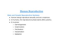
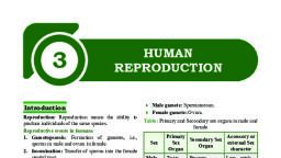
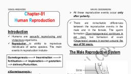
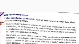
IUHSS, PARAPPUR, Malappuram, , 3. HUMAN REPRODUCTION, , ZLGY-MM: XII, , Humans are sexually reproducing and viviparous., Reproductive Events In Human, In Male, Testes, , In Female, Ovary, (Gametogenesis), , Sperm, , Ovum, (Insemination), Zygote, (Fertilisation), Blastocyst (Implantation), (Embryogenesis), Embryo, (Gestation), Foetus, (Parturition), , THE MALE REPRODUCTIVE SYSTEM, It consists of paired testes, accessory ducts, glands and the, external genitalia., , Accessory Glands, Include, 1. Paired seminal vesicles, 2. A prostate, Secretions= Seminal plasma, 3. Paired bulbourethral glands., , External genitalia (Penis), It is the copulatory organ made up of erectile tissue to facilitate, insemination., The enlarged end of penis called the glans penis is covered by a, loose fold of skin called foreskin., , THE FEMALE REPRODUCTIVE SYSTEM, It consists a pair of Ovaries, Accessory ducts (constitute oviducts,, uterus & vagina) and the external genitalia., , Testes (10 sex organ), Situated in scrotum (helps in maintaining the low temperature), Oval in shape, length: 4-5 cm, width: 2-3 cm., Each testis is subdivided into 250 testicular lobules, Each lobule contains 1-3 highly coiled seminiferous tubules., Each seminiferous tubule is lined by Male germ cells (Spermatogonia) undergo meiotic divisions, sperm, Sertoli cells provide nutrition to the germ cells., The regions outside the seminiferous tubules (interstitial, spaces), contain o Small blood vessels, o Interstitial cells or Leydig cells (secrete androgens), Germ cells, Sertoli cells, , Ovaries (10 sex organs), Located one on each side of the lower abdomen., Almond shaped; Length: 2- 4 cm., Each ovary is covered by a thin epithelium which encloses the, ovarian stroma. The stroma is divided into –, 1. An outer cortex, 2. An inner medulla- contains many ovarian follicles at, various stages of development., Oviducts(fallopian tubes), Muscular tubes of 10-12 cm long which carry the ovum from, ovary to the uterus., It is divided into 3:(i) Infundibulum- Funnel-shaped opening possessing finger-like, fimbriae, which help in collection of the ovum after ovulation., (ii) Ampulla- a wider part., (iii) Isthmus has a narrow lumen and it joins the uterus., , Uterus (womb), Accessory ducts, Seminiferous tubules →Rete testis →Vasa efferentia, →Epididymis→Vas deferens →Ejaculatory duct →Urethra, →Urethral meatus, , for HSS LiVE.IN, prepared by: Minhad. M. Muhiyudeen, #- 9846 29 22 27, , It is a large, inverted pear-shaped, supported by ligaments, attached to the pelvic wall., The wall of the uterus has 3 layers 1. External Perimetrium (thin membrane), 2. Middle Myometrium (thick layer of smooth muscle-exhibits, contraction during delivery of the baby)., 3. Inner Endometrium (glandular layer- undergoes cyclical changes, during menstrual cycle, zone of implantaion)., 13
Page 2 :
Uterus opens into vagina through a narrow cervix., , Vagina, It is a large, elastic, muscular tube about 7.5cm long., Cervical canal +vagina = Birth canal, , External genitalia (Vulva), Consists of mons pubis, labia majora, labia minora, hymen and, clitoris, a) Mons pubis is a cushion of fatty tissue covered pubic hair., b) Labia majora are fleshy folds of tissue surrounding the vaginal, opening., c) Labia minora are paired inner folds of tissue., d) Clitoris is a sensitive structure above the urethral opening., e) Hymen- The membrane covering the opening of the vagina., , After spermiogenesis, sperm heads become embedded in the, Sertoli cells, and are released from the seminiferous tubules by, the process called spermiation., , Hormonal action on spermetogenesis, , Structure of a sperm, , MAMMARY GLAND (Breasts- aids in child care), , The mammary glands are paired structures that contain, glandular and fatty tissue., , A plasma membrane envelops the whole body of sperm., Sperm consists ofa. Head: Oval shaped. Containing nucleus and capped by acrosome, (contains lytic enzymes- helps in penetrating ovum)., b. Middle piece: Contain mitochondria which produce energy for the, sperm motility., c. Tail: It helps the sperm moves in fluid medium., , The human male ejaculates about 200 to 300 million sperms during, coitus., Semen= sperm (10%) + seminal plasma, Each glandular tissue is divided into 15-20 mammary lobes, containing clusters of cells called alveoli. The cells of alveoli, secrete milk, which is stored in the cavities (lumens)., , Oogenesis, , The alveoli open into mammary tubules→mammary duct →, mammary ampulla →lactiferous duct →nipple through which, milk is sucked out., , GAMETOGENESIS, The primary sex organs – the testis produce male gamete, sperms, and the ovaries produce females gametes, ovum., , Spermatogenesis, , for HSS LiVE.IN, prepared by: Minhad. M. Muhiyudeen, #- 9846 29 22 27, , 14
Page 3 :
Comparison between spermatogenesis and oogenesis, Spermatogenesis, Oogenesis, i. It occurs in the testis, i. In Ovary, ii. Gamete is called sperm ii. Ovum, iii. Continuous process, iii. Discontinuous, 0, iv. Each 1 spermatocyte, iv. Each 10 oocyte gives only 1 ovum, gives 4 sperms, and 3 polar bodies, v. It begins at puberty and v. It begins at embryonic stage and, extends up to senility, suspended at the time of birth. The, remaining part takes place only, after puberty., , MENSTRUAL CYCLE, It is the cyclic changes in the activity of ovaries and accessory, ducts as well as hormones during the reproductive phase in a, female body, with bleeding (menstruation), repeated at an, average interval of about 28/29 days, The first menstruation begins at puberty and is called menarche., In human beings, menstrual cycles stop around 50 years of age;, that is termed as menopause., Menstrual cycle has the following phases:Menstrual / Bleeding Phase: 1-5th day, The cycle starts with menstrual flow and it lasts for 3-5 days., It is due to breakdown of endometrial lining of the uterus and its, blood vessels that comes out through vagina., Menstruation only occurs if the released ovum is not fertilised., Lack of menstruation may be indicative of pregnancy. It may, also be caused due to stress, poor health etc., Proliferative / Follicular Phase: 5-13th day, In this phase, the secretion of gonadotropins (LH & FSH), increases gradually from pituitary. They stimulates:o Development of 10 follicles into Graafian follicles. It, secretes estrogens., Estrogens stimulateso Regeneration of ruptured uterine endometrium., Ovulatory Phase: 14 day, Both LH and FSH attain a peak level., Rapid secretion of LH (LH surge) induces rupture of Graafian, follicle and thereby ovulation., th, , Secretory / Luteal Phase: 15-28th day, After ovulation, Graafian follicle is transformed into a yellow, glandular mass called Corpus luteum., - It secretes large amount of progesterone which is essential for, maintenance of the endometrium., Thus the uterus gets ready for implantation (of the fertilised, ovum) and other events of pregnancy., , - During pregnancy all events of menstrual cycle stop and there is, no menstruation., - If fertilization does not occur, corpus luteum degenerates. This, causes disintegration of the endometrium leading to, menstruation, marking a new cycle., for HSS LiVE.IN, prepared by: Minhad. M. Muhiyudeen, #- 9846 29 22 27, , FERTILISATION AND IMPLANTATION, During copulation (coitus), semen is released by the penis into, the vagina (insemination)., Fertilisation (sperm + ovum → zygote) can only occur if the, ovum and sperms are transported simultaneously to the, ampullary-isthmic junction., During fertilisation, a sperm comes in contact with the zona, pellucida layer of the ovum and induces changes in the, membrane that block the entry of additional sperms., Thus, it ensures that only one sperm can fertilise an ovum., The secretions of the acrosome help the sperm enter into the, cytoplasm of the ovum through the zona pellucida and the, plasma membrane., , This induces the completion of the meiotic division of the, secondary oocyte. The second meiotic division is also unequal, and results in the formation of a second polar body and a haploid, ovum (ootid)., Soon the haploid nucleus of the sperms and that of the ovum fuse, together to form a diploid zygote., The mitotic division (cleavage) starts as the zygote moves, through the isthmus towards the uterus and forms 2, 4, 8, 16, daughter cells called blastomeres., The embryo with 8 to 16 blastomeres is called a morula., The morula continues to divide and transforms into blastocyst., The blastomeres in the blastocyst are arranged into an outer layer, (trophoblast) and an inner group of cells (inner cell mass), attached to trophoblast., The trophoblast layer then gets attached to the endometrium and, the inner cell mass gets differentiated as the embryo., , , , After attachment, the uterine cells divide rapidly and cover the, blastocyst. As a result, the blastocyst becomes embedded in the, endometrium of the uterus (implantation) and it leads to, pregnancy., , 15
Page 4 :
PREGNANCY & EMBRYONIC, DEVELOPMENT, After implantation, finger-like projections (chorionic villi), appear on the trophoblast which is surrounded by the uterine, tissue and maternal blood., The chorionic villi and uterine tissue become inter digitated, with each other and jointly form a structural and functional unit, between developing embryo (foetus) and maternal body called, placenta., o Placenta is connected to the embryo by an umbilical cord. It, transports substances to and from the embryo., , The mammary glands of the female undergo differentiation, during pregnancy and starts producing milk towards the end of, pregnancy by the process called lactation., o The milk produced during the initial few days of lactation, (colostrum) which contains several antibodies absolutely, essential to develop resistance for the new-born babies., , Functions of placenta:, Supply O2 and nutrients to the embryo, Remove CO2 and waste materials produced by the embryo., Acts as an endocrine gland by secreting human chorionic, gonadotropin (hCG), human placental lactogen (hPL),, oestrogens, progesterone., In the later phase of pregnancy, a hormone called relaxin is also, secreted by the ovary., In addition to hCG, hPL and relaxin, during pregnancy the, levels of other hormones like estrogens, progestogens, cortisol,, prolactin, thyroxine, etc., are increased several folds in the, maternal blood. They support:o The fetal growth, o Metabolic changes in the mother, o Maintenance of pregnancy., Immediately after implantation, the inner cell mass (embryo), differentiates into an outer ectoderm, middle mesoderm and, an inner endoderm. These 3 layers give rise to all tissues, (organs) in adults., Developmental stages of human embryo during, pregnancy, The human pregnancy lasts 9 months (gestation period)., st, 1 month - Heart is formed., 2nd month - Limbs and digits develops., 3rd month - Limbs and external genital organs well develops., 5th month- Shows movements and appear hair on the head., 6th month- The body is covered with fine hair, eye-lids separate, and, eyelashes are formed., th, 9 month- The foetus is fully developed and is ready for delivery., , PARTURITION & LACTATION, Vigorous contraction of the uterus at the end of pregnancy, causes delivery of the foetus (parturition)., Neuroendocrine mechanism of parturition, a. The signals from the fully developed foetus and the placenta, which induce mild uterine contractions (foetal ejection reflex)., b. This induce release of oxytocin from the maternal pituitary., c. Oxytocin acts on the uterine muscle and causes stronger uterine, contractions, which in turn stimulates further secretion of, oxytocin., d. Contraction get stronger, which leads to expulsion of the baby, out of the uterus through the birth canal., Soon after the infant is delivered, the placenta is also expelled out, of the uterus., , for HSS LiVE.IN, prepared by: Minhad. M. Muhiyudeen, #- 9846 29 22 27, , 16