Page 1 :
SURYA CLASSES, , LECTURE NOTES BIOLOGY [PAPER-II], , XII SCIENCE, , CHAPTER NO, NO:: 08, CHAPTER, 8, , 1, , RESPIRATION &, CIRCULATION, , ❖ Circulation: The process of transport of various substances like, oxygen, nutrients ,hormones and metabolic wastes through circulatory, system is called as circulation, , ❖ Circulatory System in Human: The human circulatory system, composed of blood vascular system and lymphatic system., The interconnected blood vascular and lymphatic system is, known as circulatory system, , ❖ Blood vascular, , System/Cardiovascular system:, , o The system in which blood is circulated throughout the body is called, as blood vascular system., o The heart together with blood vessels helps in transport of blood, around the body to supply nutrients and oxygen to the tissue and, removal of waste products is known as blood vascular system ., o Blood vascular system consist of blood, heart and blood vessels and, helps in transport, homeostasis and protection, o Types of Circulatory system :, 1. Open circulatory system: In this system, blood does not confined to, blood vessels., • Example: Leech, Crab, Spider, Snail, Lobster, Bivalve, hardmania ,, Pila., 2. Closed circulatory system: In this system, blood confined to blood, vessels., • Example: Higher animals, ., , William Harvey: Discovered closed circulatory system, , ❖Hematology: The science which deals with the study of blood., ❖Blood: Blood is fluid connective tissue. It is main circulatory fluid in, human body. Blood derived from mesoderm., 1. Color: Bright Red, 2. Nature: Slightly alkaline(pH : 7.4), 3. Consistency: Viscous fluid and salty [5times thicker than water], 4. Specific gravity: Higher than water & Heavier than water, 5. Quantity: 4-6 Liters of blood [ Adult person], 6. Constitution: About 8 % of the total body weight of adult person, 7. Composition of Blood: A) Plasma (55%) B) Blood Corpuscles (45%), 1, , RESPIRATION & CIRCULATION, , | SIGN OUT BY DR. VIRESH SHELKE
Page 2 :
SURYA CLASSES, , LECTURE NOTES BIOLOGY [PAPER-II], , XII SCIENCE, , BLOOD, , PLASMA [55%], , Water [90-92%], , BLOOD C0RPUSCLES [45 %], , Dissolved Substances [8-10 %], , Blood, protein, , Digested, Nutrient, s, , Regulatory, Substances, , Nitrogenous, Waste, , Gases, , Albumin, , Glucose, , Hormones, , Ammonia, , Oxygen, , Globulin, , Amino acid, , Enzymes, , Urea, , Antibodies, , Uric acid, , acid, , Heparin, , Fatty acid, , Prothrombin, , Creatinin, , Glycerol, , Fibrinogen, , Nitrogen, , Inorganic substanaces, , Red Blood Cells, , White blood cells, , Granulocytes, , Neutrophills, , Carbon, dioxide, , Acidophills, , Platelets/Thrombocytes, Agranulocytes, , Basophills, , Lymphocyte, s, , Monocytes, , A. Plasma: Plasma is straw colored fluid, slightly alkaline and viscous, fluid. Plasma contains : a) Water: 90-92 %, b) Solutes/Dissolved substances: 8-10 %, 1. Proteins(7-8 %) : Serum albumin, serum globulin, Heparin, [Anticoagulant], Properdin , Fibrinogen and Prothrombin, 2. Others : 1-2 %, a. Digested nutrients: Glucose, Amino acids, fatty acids and glycerol, b. Regulatory substances: Hormones, Enzymes,Vitamins, c. Nitrogenous wastes: Urea ,Uric acid, Ammonia, Creatinine, 2, , RESPIRATION & CIRCULATION, , | SIGN OUT BY DR. VIRESH SHELKE
Page 3 :
SURYA CLASSES, , LECTURE NOTES BIOLOGY [PAPER-II], , XII SCIENCE, , d. Inorganic substances/Salts : Bicarbonates[ HCO-3 ] ,chlorides[Cl-],, phosphates[ PO3-4 ] , sulphates[SO4] of Na, K, Ca ,Mg , Fe & Mn, e. Gases: CO2,O2, N2, f. Cholesterol and Antibodies, B. Formed elements / Blood Corpuscles/ Blood cells: Blood corpuscles, that produced in body are collectively called as formed elements and, they are suspended in plasma., o Types of Blood corpuscles:, A. Erythrocytes/Red blood cells(RBC’s), B. Leucocytes/White blood cells(WBC’s), C. Platelets/Thrombocytes, , A. Erythrocytes/Red blood cells(RBC’s):, 1., 2., 3., 4., , Shape: Circular and biconcave [Disc shaped with thicker edges], Nature: Non nucleated [nucleated & oval in camel and lamas), Size: 7µm in diameter and 2.5 µm in thickness, Total RBC count: a) Male: 5.1-5.8 million/cubic mm[per μL], b) Female: 4.3-5.2 million/cubic mm [ per μL], 5. Life span: 120days, 6. Erythropoiesis: Formation of RBC’s ., 7. Erythropietic cells: Haemocytoblasts /reticulocytes., 8. Site of erythropoiesis: a) Foetus: liver & spleen, b) Adults: Red bone marrow., Vitamin Cyanocobalamine [ B12], Folic acid and heme protein are, required for production of RBC’s., 9. Erythropoietin hormone : produced by the kidney cells stimulate, bone marrow for production of RBC’s], 10. Destruction of RBC’s: In liver and Spleen[ Graveyard of RBC’s], 11. Polycythemia : Increase in number of RBC’s, 12. Erythrocytopenia: Decrease in number of RBC’s, 13. Nature of mature RBC’s : It is devoid of nucleus, mitochondria, and other membrane bound cell organelles. Its cytoplasm[stroma], is rich in haemoglobin andO2 carrying proteinaceous pigment, which provide red color . It also contains an enzyme carbonic, anhydrase., 14. Haemocrit is ratio of volume of RBC’s to total blood volume., 15. Hemoglobin: Cytoplasm of RBC’s contains haemoglobin., Haemoglobin is respiratory pigment helps in transport of oxygen, and carbon dioxide ., o Normal haemoglobin content:, a) Male:14-17 gram/100 ml of blood or gram % of blood, b) Female: 13-15 gram /100 ml or gram % of blood, o Anaemia: Deficiency disorder in which Hb level in blood decreases., 3, , RESPIRATION & CIRCULATION, , | SIGN OUT BY DR. VIRESH SHELKE
Page 4 :
SURYA CLASSES, , LECTURE NOTES BIOLOGY [PAPER-II], , XII SCIENCE, , o Composition of Haemoglobin: Hb is protein iron complex, [conjugated protein ] made up of 4 polypeptide chain of globin i.e. 2, alpha and 2 beta chain., An iron – porphyrin (haem) group is attached to, each chain and all four chains are bound together. Each haem, group can carry one O2 molecule and thus one haemoglobin, molecule can carry four O2 forming oxyhaemoglobin. CO2, interacts with amino acid residues of globin chains and forms, carbaminohaemoglobin., After haemolysis, haemoglobin is broken down., Its globin part is broken to recycle the amino acids. Iron of heme, group is stored as ferritin in the liver and porphyrin group of, heme is converted into green pigment biliverdin and then into, red-orange coloured bilirubin. These pigments (mainly bilirubin), are added to bile and finally removed out of body along with, 16. Function:, 1. Transport of oxygen from, lungs to tissue., 2. Transport of carbon, dioxide from tissues to, lungs, 3. Haemoglobin acts as, buffer and maintains, blood pH, 4. Maintains viscosity of, blood, , B. Leucocytes/White blood cells(WBC’s):, 1. Shape: Amoeboid, 2. Diapedesis: Due to amoeboid nature ,WBC’s can squeeze/move out of, blood capillaries known as diapedesis, 3. Nature: nucleated, colorless and phagocytic cells, 4. Size: 8-15 µm [larger than RBC’s], 5. Total WBC count: 5000-11000/cubic mm of blood, 6. Life span: 3-4 days, 7. Leucopoiesis: Formation of WBC’s, 8. Site of leucopoiesis: Red bone marrow, spleen, lymph nodes, tonsils,, thymus and Payer’s patches, 9. Destruction of WBC’s: By liver and lymph nodes, 10. Leucocytosis: Temporary Increase in number of WBC’s, It occurs during infection, pregnancy period and in newborns, 11. Leucopenia: Decrease in number of WBC’s [less than 4000], 4, , RESPIRATION & CIRCULATION, , | SIGN OUT BY DR. VIRESH SHELKE
Page 5 :
SURYA CLASSES, , LECTURE NOTES BIOLOGY [PAPER-II], , XII SCIENCE, , Leucopenia is common in HIV, AIDS, TB and persons exposed, to radiations and shock., 12. Leukemia/Blood cancer: Pathological uncontrolled increase in, number of WBC’s, 13. Function: Concerned with defense mechanism i.e. protection, [ provide cell mediated and humoral immunity] ., 14. Types: Colorless, irregular nucleated cells which show polymorphism, [ exist in variable forms.], A] Granulocytes: a] Neutrophills, b] Eosinophills/Acidophills, c] Basophills/Cyanophils, B] Agranulocytes: a] Lymphocytes b] Monocytes, , Leucocytes, o Granulocytes: Produced by bone, marrow, , o Agranulocytes: Produced, by lymph nodes, tonsils, and spleen, ✓ Shows granular cytoplasm and lobed, ✓ Granules are absent and, nucleus., unlobed/ non lobulated, ✓ Also known as polymorpho nuclear, nucleus., leucocytes [ PMN], ✓ Nucleus is large in size ., ✓ Formed from myeloid stem cells and, ✓ Formed from lymphoid, unable to divide., stem cells and able to, ✓ It constitutes about 72 % of total WBC’s., divide, ✓ Acts as secretary vesicles and secrete, ✓ It constitutes about 28 %, various secretions, enzymes., of total WBC’s., Neutrophills, Basophils, Eosinophils Lymphocytes, Monocytes, Polymorphs, , Cyanophills, , Acidophills, , Fine granules, of large size., , Few Coarse, granules of, large size, , Lysosomal, granules], , Stained with, neutral dyes, , Stained with, basic dyes [, Methylene, blue], , stained to, red color, with acidic, dyes[Eosin, , 3-7 lobed, , Twisted/ S, , Bilobed /, , 5, , RESPIRATION & CIRCULATION, , Immune cells, , Scavengers, , Smallest, WBC’s, , Largest, WBC's, , Large and, , Large bean/, , | SIGN OUT BY DR. VIRESH SHELKE
Page 6 :
SURYA CLASSES, , LECTURE NOTES BIOLOGY [PAPER-II], , XII SCIENCE, , nucleus and, spherical, cells., , shaped, nucleus and, spherical cell, , Spectacle, shaped, nucleus, , round, /spherical, nucleus, , Kidney, shaped, nucleus, , 10-12 µm, , 8-10 µm, , 10-12 µm, , 8 µm, , 12-15 µm, , Constitute 70, % of total, WBC’s, Phagocytic &, amoeboid, cells, , Constitute 0.5, -1 % of total, WBC’s, Non, phagocytic, cells, , Constitute, 1-3% of, total WBC’s, Antihistaminic, cells, , Constitute, 25- 30 % of, total WBC’s, Antibody, producing, cells, , Constitute, 3-5% of, total WBC’s, Phagocytic, cells, , Helps in, engulfing, microbes and, able to, destroy, pathogens by, Phagocytosis., , Release, heparin, [Anticoagulant],, serotonin &, Histamine, [inflammatory, & allergic sub], , They destroy, antigenantibody, complex by, Phagocytosis, , Provide, immune, response to, body., Lymhocytes, are of 2 types, , Actively, motile cells., , Pus is, mixture of, dead, Neutrophills ,, damaged, tissues and, dead, microbes, , Present in, infected and, allergic, conditions, only., , Produces, antitoxins to, Helpsin, , A] Blymhocytes:, mature in, detoxificatioin bone marrow, & provide, Increase in, humoral, numbers, immunity [, during, antibody, allergic, prodn], condition, B] T[Eosinophilia], lymphocytes:, mature in, thymus and, provide cell, mediated, immunity, , Monocytes, enlarge and, differentiated, , into, macrophages, , which, engulf, microbes, and, removes, dead/dama, ged tissue, by, phagocytosis, , cell debris, , C. Platelets/Thrombocytes :, 1. Shape: Round, biconvex, irregular and smallest cells and present in, the form of fragments., 2. Nature: Non nucleated, 3. Size:2.5-5 µm in diameter, 4. Total platelet count: 2.5 -4.5 lakh /mm3 [cubic mm of blood], 5. Life span: 5-10 days, 6. Thrombopoiesis: formation of platlets, 7. Site of Thrombopoiesis: large Megakaryocytes cells of bone marrow, 8. Thrombocytosis: Increase in number of platelets, 6, , RESPIRATION & CIRCULATION, , | SIGN OUT BY DR. VIRESH SHELKE
Page 7 :
SURYA CLASSES, , LECTURE NOTES BIOLOGY [PAPER-II], , XII SCIENCE, , 9. Thrombocytopenia: Decrease in number of platelets. It causes, internal bleeding [haemorrhage]., 10., Function:, 1. Platelets secrete platelet, factor which is essential in, blood clotting., 2. Helps to seal the ruptured, blood vessel by formation of, platelet plug/thrombus at, site of injury [Aggregate at the, site of injury and forms clot], 3. Release of thromboplastin, 4. Secrete serotonin, [ vasoconstrictor], , ❖ Mechanism of Blood clotting / Coagulation of blood:, 1. Blood coagulation: Conversion of liquid blood into semisolid jelly or, solid form is called as clotting, 2. In Intact blood vessels: Blood does not coagulate due to, anticoagulants like heparin and antithrombins., 3. During rupture of blood vessels: Clotting process starts by contact, of blood with any foreign surface [ intrinsic process or with damaged, tissue [extrinsic process] . Intrinsic and extrinsic process involves, interactions of various substances known as clotting factors by a, stepwise/ cascade mechanism. All twelve Clotting factors [ I to XII, with inactive VI factor ] responsible for clotting. [ Interaction of, clotting factor in a cascade manner leads to formation of enzyme, thrombin], 4. Role of thromboplastin: . During rupture of blood vessels , platelets, and injured tissues release Thromboplastin. Thromboplastin initiates, formation of enzyme prothrombinase., 5. Role of prothrombinase: In the presence of calcium ions, prothrombinase converts inactive plasma protein prothrombin into, active thrombin as well as inactivates activity of heparin., 6. Role of thrombin: Thrombin converts soluble fibrinogen into, insoluble fibrin, 7. Role of fibrin: Fibrin forms a mesh in which platelets and other, blood cells are trapped to form the clot., 8. Clotting time: 2-8 minutes, , 7, , RESPIRATION & CIRCULATION, , | SIGN OUT BY DR. VIRESH SHELKE
Page 8 :
SURYA CLASSES, , LECTURE NOTES BIOLOGY [PAPER-II], , XII SCIENCE, , Prothrombinas, e, Ca++, , ❖ Functions of Blood :, 1. Distribution of digested and absorbed food/nutrients: Blood helps, in distribution of digested and absorbed nutrients from alimentary, canal to all parts of body., 2. Transport of hormones and enzymes: Blood helps in transport of, hormones[Endocrine secretions] and enzymes [Exocrine secretions], 3. Transport of respiratory gases: Blood plays important role in, transport of respiratory gases from lungs to cells and vice versa., 4. Collection and transportation of nitrogenous waste: Blood collects, nitrogenous waste from liver and transport to the kidneys for excretion., 5. Distribution of heat or Thermoregulation: Blood distributes heat and, helps in maintaining body temperature., 6. Prevent dehydration by distributing adequate quantity of water:, Blood distributes adequate quantity of water to all cells and prevents, dehydration., 7. Antibody production: Blood contains WBC’s [lymphocytes] which, helps in defense mechanism., 8. Protection : Neutrophills and Monocytes acts as phagocytic cells and, provides protection from pathogens, 9. Regulation of osmotic balance: The proteins and minerals exert, osmotic pressure and regulate blood pressure., 10. Homeostasis: The blood keeps the internal environment of the body, constant., 11. Prevents loss of blood: Platelets in blood prevents loss of blood by, formation of blood clot., , 8, , RESPIRATION & CIRCULATION, , | SIGN OUT BY DR. VIRESH SHELKE
Page 9 :
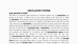
SURYA CLASSES, , ❖ Heart:, , LECTURE NOTES BIOLOGY [PAPER-II], , XII SCIENCE, , Heart is pumping organ helps in transport of blood to the, , cells of body., 1. Location: Mediastenum ( in the middle of thoracic cavity in between two, lungs), 2. Nature: Hollow ,muscular conical organ, 3. Derived from : Mesoderm, 4. Shape: Fist shaped with broad base [upper side] and narrow apex, [lower side], 5. Position : Conical end of heart is tilted towards left side and rests, above the diaphragm., 6. Size : Length-12cm and Breadth:9 cm, 7. Weight: 250grams [ female] - 300 grams [male] gram, 8. Pericardium: Heart enclosed double layered peritoneum[membranous, sac] filled with pericardial fluid is pericardium., The two main layers of pericardium is outer fibrous and inner serous, layer., 1. Fibrous pericardium: It is outer tough, inelastic, loosely fitted layer. It, is made up of fibrous connective tissue., 2. Serous pericardium: It is inner smooth, moist, elastic and thin layer., It is contains outer parietal and inner visceral layer., a] Parietal layer: Forms inner linings of fibrous pericardium, b] Visceral layer: it encloses the heart and forms outer covering, of heart i.e. Epicardium, o Pericardial space: The space present between visceral and parietal, layer of serous pericardium is known as pericardial, o Pericardial fluid: It is present in pericardial space[about 50ml]., Pericardial fluid acts as shock absorber and lubricant. It protects, heart from mechanical injury and desiccation i.e. keeps heart moist., 9. Heart Wall: Heart is mesodermal in origin and contains 3 layers., A. Epicardium: The visceral layer of serous, pericardium forms epicardium. It is thin, outer wall of heart. It is made up of single, layer of flat epithelial cells [mesothelium], resting on basement membrane. Function :, Protection, B. Myocardium: It is middle thick layer of heart, made up of cardiac muscles. Cardiac muscle, helps in contraction and relaxation of heart, C. Endocardium: It is inner thin wall of heart., It is made up of single layer of flat epithelial, cells [endothelium]. function: Protection, , 9, , RESPIRATION & CIRCULATION, , | SIGN OUT BY DR. VIRESH SHELKE
Page 10 :
SURYA CLASSES, , ❖, , LECTURE NOTES BIOLOGY [PAPER-II], , External structure of Heart:, , XII SCIENCE, , Human heart is four, , chambered i.e. 2 atria/auricles [superior] and 2 ventricles[inferior], , A] Atria/Auricles: Atria are superior, small, thin walled receiving, chambers. Atria are 2 in numbers., 1. Right atrium:, o Right atrium is larger in size than left atrium., o Right atrium receives deoxygenated blood from all over the body, through superior and inferior vena cava., o Coronary sinus [atrio- ventricular groove] also opens into the right, atrium.(Coronary veins joins to form coronary sinus), 2. Left atrium:, o Left atrium is smaller in size than right atrium., o Left atrium receives blood from pulmonary veins along dorsal, surface of heart., • Interatrial groove/Sulcus [Anterior and Posterior]: separates right, and left auricle., B] Ventricles: Ventricles also 2 in numbers. Ventricles are inferior,, large , thick walled ,distributing chambers, 1. Right ventricle:, o Right ventricle is smaller and thinner than left ventricle., o Right ventricle receives deoxygenated blood from right atrium., o Pulmonary trunk arises from right ventricle present on anterior, surface of heart. It passes deoxygenated blood to lungs via right & left, pulmonary arteries., 10, , RESPIRATION & CIRCULATION, , | SIGN OUT BY DR. VIRESH SHELKE
Page 11 :
SURYA CLASSES, , LECTURE NOTES BIOLOGY [PAPER-II], , XII SCIENCE, , 2., o, o, o, , Left ventricle:, Left ventricle is bigger and thicker than right ventricle., Left ventricle receives oxygenated blood from left atrium, Systemic aorta arises from left ventricle present on anterior surface of, heart., o Systemic aorta/Aortic arch is divisible into three regions as, ascending aorta, systemic arch/aortic arch and descending aorta., o Systemic aorta passes oxygenated blood to the all body parts., • Interventricular groove/Sulcus [Anterior and Posterior] : It, separates right and left ventricle or present in between right and left, ventricles., C] Ligamentum arteriosum:, o Pulmonary trunk and systemic aorta are connected by ligamentum, arteriosum., o It also represent remnant of an embryonic duct called ductus, arteriosus of foetus., o Aortic arch gives out gives out three arteries viz. brachiocephalic, (innominate) artery, left common carotid and left sub clavian ., D] Atrio-ventricular groove/Coronary Sulcus:, o The transverse groove that separates auricle and ventricle is atrioventricular groove i.e. a] Right atrio-ventricular groove, b] Left atrio –ventricular groove, o Coronary arteries and coronary veins situated in coronary sulcus., E] Coronary artery:, o It arises from systemic aorta, o It supplies oxygenated blood to the walls of heart, F] Coronary vein:, o It receives deoxygenated blood from walls of heart .It joins and, forms coronary sinus which opens in right, , 11, , RESPIRATION & CIRCULATION, , | SIGN OUT BY DR. VIRESH SHELKE
Page 12 :
SURYA CLASSES, , LECTURE NOTES BIOLOGY [PAPER-II], , XII SCIENCE, , ❖Internal structure of heart:, A., , Atria: Atria are two thin walled upper /superior receiving chambers., Inter-atrial/Inter-auricular septum divides atria into right and left, atrium., • Interatrial septum: The partition or dividing wall which, separates two auricles known as inter-auricular septum., o Fossa ovalis: An oval area/depression/marking present on the, right side of Interatrial septum is fossa ovalis. Fossa ovalis, represents ramnant of foramen ovale., o Foramen ovale: An oval opening on interatrial septum is, foramen ovale. Foramen ovale is opening between right and left, auricle in the foetus which closes at the time of birth., , a. Right auricle: Right atrium is larger in size than left atrium., Right atrium receives deoxygenated blood from all over the body, through superior vena cava, inferior vena cava and coronary sinus., o Superior vena cava[ precaval] : It receives blood from upper parts of, body. The opening of superior vena cava is not guarded by any valve., o Inferior vena cava[ postcaval] : It receives blood from lower parts of, body. It is guarded by Eustachian valve. It prevents backward flow, of blood during auricular systole., o Coronary sinus: It receives blood from the walls of heart and, guarded by thebesian valve., o, , b. Left auricle: Left atrium is smaller in size than right atrium., o Four pulmonary veins open into the left atrium and carries, oxygenated blood from lungs., , Atrio-ventricular aperture/Auriculo-ventricular, opening: Each atrium opens into the ventricles through atrioventricular aperture. Atrioventricular aperture is guarded by, valves. Valves made up of C.T., o Atrio-ventricular apertures are : Right atrioventricular aperture, and Left atrioventricular aperture, o Right atrio-ventricular aperture : It is guarded by right, atrioventricular valve. It has 3 flaps of connectives so called as, tricuspid valves., o Left atrio-ventricular aperture: It is guarded by left, atrioventricular valve. It has two flaps of connectives so also, known as bicuspid /mitral valve. It prevents backward flow of, blood during ventricular systole., 12, , RESPIRATION & CIRCULATION, , | SIGN OUT BY DR. VIRESH SHELKE
Page 13 :
SURYA CLASSES, , B., , LECTURE NOTES BIOLOGY [PAPER-II], , XII SCIENCE, , Ventricles: Ventricles are two thick walled distributing, chambers.Each ventricle separated by inter ventricular septum., , Interventricular septum: The partition or dividing wall which, separates two ventricles known as Interatrial/interauricular, septum., , a. Right Ventricle: Right ventricle is smaller and thinner than left, o, o, o, o, o, , ventricle., Right ventricle receives deoxygenated blood from right atrium., Pulmonary trunk/aorta arises from right ventricle., It passes deoxygenated blood to lungs via pulmonary arteries for, oxygenation., The three pocket like valves present at the base of pulmonary aorta is, semilunar valve., Semilunar valve prevents backward flow of blood during ventricular, diastole., , b. Left ventricle: Left ventricle is bigger and thicker [3times thicker], o, o, o, o, o, , than right ventricle., Left ventricles requires more force to push blood along a great, distance so thicker in nature., Left ventricle receives oxygenated blood from left atrium, Systemic aorta arises from left ventricle., Systemic aorta passes oxygenated blood to the all body parts., The three pocket like valves present at the base of systemic aorta is, semilunar valve., , 13, , RESPIRATION & CIRCULATION, , | SIGN OUT BY DR. VIRESH SHELKE
Page 14 :
SURYA CLASSES, , LECTURE NOTES BIOLOGY [PAPER-II], , XII SCIENCE, , o Chordae tendinae: The special inelastic fibrous/ fibers cords, attached to the flaps of bicuspid and tricuspid valves [to the, ventricular wall i.e papillary muscles] is known as chordate, tendinae. It prevents valves from turning back into the atria during, ventricular systole.Also regulates opening and closing of valves., o Papillary muscles: Chordae tendinae attached to the ventricular, walls of heart with special muscles known as papillary muscles., o Columnae carnae/Trabaeculae carnae: A series of irregular, muscular ridges present at the inner surface of the ventricles., o Fissures: Columnae carnae divides cavity / lumen of ventricles, into small spaces/pockets known as fissures., , ❖Pumping action of Heart :, ❖Cardiac out put: Heart beat X Stroke volume, 72, , X 70, , Cardiac output = 5040 ml, , o Heart beat: The rhythmic contraction and relaxation of heart by the, action of cardiac muscles is called as heart beat., The normal heart beat = 70-72 times/ minute, //times/minute, , o Stroke volume: The ventricles pumps blood during ventricular, systole is called as stroke volume., Stroke volume =70 ml blood, , ❖Conducting system of heart: The heart acts as pumping, organ due to myogenic nature. The conducting system of heart consist, of :- a] Sinoatrial node/SA node, b] Atrioventricular node/AV node, c] Bundle of His, d] Purkinje fibers, , A. Sino atrial node/SA node:, o The special node of modified cardiac muscles which generates heart, beat is known as SA node., o SA node present in the wall of right atrium near the opening of, superior vena cava., o SA node has power of generation of wave of contraction so also known, as pace maker. It initiates and maintains rhythmicity of heart., 14, , RESPIRATION & CIRCULATION, , | SIGN OUT BY DR. VIRESH SHELKE
Page 15 :
SURYA CLASSES, , LECTURE NOTES BIOLOGY [PAPER-II], , XII SCIENCE, , o SA node spreads wave of contraction at the rate of 1 meter/seconds, o SA node responsible for auricular systole/Atrial systole., o The wave of contraction or cardiac impulse generated by SA node is, conducted by cardiac muscle fibers of both atria causes their, contraction, , B. Atrio ventricular node /AV node:, o Second special node of modified cardiac muscles is known as AV node., o AV node present in the wall of right atrium near the opening of, coronary sinus. Or AV node present in the interauricular septum near, the right auriculoventricular groove., o AV node receives wave of contraction generated by SA node through, internodal pathway., o AV node acts as pacesetter of heart., o AV node gives rise to bundle of His, , C. Bundle of His :, o The muscular bridge arises from AV node which conducts stimulation, from auricles to ventricles are known as Bundle of His., o Bundle of His present in interventricular septum., o The bundle of His bifurcates into 2 branches and going to right and, left ventricles., o It spreads wave of contraction from AV node to purkinje fibers., o The speed of wave of contraction of bundle of His is 5 meter /second., , D. Purkinje fibers :, o The fine thread like branches arises from bundle of His is purkinje, fibers or the terminal branches of bundle of his forms network of, fibers known as purkinje fibers., o It penetrates into myocardium of ventricles, 15, , RESPIRATION & CIRCULATION, , | SIGN OUT BY DR. VIRESH SHELKE
Page 16 :
SURYA CLASSES, , LECTURE NOTES BIOLOGY [PAPER-II], , XII SCIENCE, , o It present all over the walls of ventricles, o It is responsible for contraction of right and left ventricle (ventricular, systole), o Bundle of His along with purkinje fibers conducts wave of, contraction from AV node to myocardium of ventricles., , ❖Cardiac cycle/Working of heart: The sequence of events, between one heart beat to next heartbeat is called as cardiac cycle., o Heart beat: The rhythmic contractions and relaxations of heart by the, action of cardiac muscles is known as heart beat., o Normal heart beat : 70-72 time/ minute, o Duration of heart beat : 0.83sec, o Systole: Contraction of the heart Diastole: Relaxation of the heart, , ❖Events /Sequence /phases of cardiac cycle:, 1. Auricular systole, 2. Auricular diastole, 3. Ventricular systole, 4.Ventricular diastole, 5. Complete/Joint diastole, , 1. Auricular systole:, 1. SA node generates wave of contraction which causes contraction of, auricles., 2. During auricular systole, blood flows in respective ventricles., 3. Duration: 0.1 second, , 2. Auricular diastole:, 1. During relaxation of auricles, blood is received from superior vena, cava, inferior vena cava and coronary sinus in right atrium and from, lungs in left atrium., 2. Duration: 0.7 seconds, , 3. Ventricular systole:, 1. During ventricular systole, bundle of His receives impulse from AV, node and passes to purkinje fibers., 2. Purkinje fibers cause contraction of ventricles., 3. During ventricular contraction, deoxygenated blood from right, ventricle pushed towards pulmonary trunk and oxygenated blood, from left ventricle pushed into systemic aorta., 4. During ventricular systole bicuspid and tricuspid valves closed which, prevent backward flow of blood in auricles., 5. During ventricular systole, semilunar valves open, 6. Lub sound: The first heart beat produced due to closing of bicuspid, and tricuspid valves known as lubb sound.Lubb sound is systolic, sound. It is louder and lasts for longer period., 7. Duration: 0.3 seconds, 16, , RESPIRATION & CIRCULATION, , | SIGN OUT BY DR. VIRESH SHELKE
Page 17 :
SURYA CLASSES, , LECTURE NOTES BIOLOGY [PAPER-II], , XII SCIENCE, , 4. Ventricular diastole:, 1. During ventricular diastole blood passes from auricles to ventricles., 2. During ventricular diastole, bicuspid and tricuspid valves open and, semilunar valves closed sharply to prevent blood flow., 3. Dub sound: The second heart sound produced due to closing of, semilunar valves is dub sound. Dub sound is diastolic sound. It is, less loud than dub sound, 4. Duration: 0.5 seconds, 5. Heart murmur: Hissing sound produced due to leakage of blood from, defective valves is heart murmur. The murmur sound observed in, heart patients, , 5. Complete/Joint diastole:, 1. The phase in which both auricles and ventricles are relaxed abd, blood from great veins flows in auricles and new cardiac cycle begins, known as complete diastole., 2. Duration: 0.4 seconds, , ❖Regulation of Cardiac activity:, 1. Cardiac muscles: Rhythmic contraction and relaxation of heart is, regulated by cardiac muscles which are auto regulated so heart is also, called as myogenic. Heart shows autorhymicity [self excitability ] due to, presence of cardiac muscles. Cardiac muscle fibres also known as, autorhythmic fibres because it acts as pace maker, setting rhythm for, heart and forms conducting system., 2. Medulla ablongata: The cardio vascular centre present in the medulla, oblongata of brain, 3. SA node: SA node is pacemaker ,it generates wave of contraction, o SA node receives sympathetic and parasympathetic nerves, o Sympathetic nerves secrete neurotransmitter adrenalin which is stress, hormone. Adrenalin responsible for increasing heart beat [tachycardia]., Sympathetic nervous system works during emotional stress [fear,, anger, excitement] and during excrcise., o Parasympathetic nerves secretes neurotransmitter acetylcholine causes, decrease in heart beat (Bradycardia), o Tachycardia: Increase in heart rate above the normal is known as, tachycardia(100 beats/minutes), o Bradycardia: Decrease in heart rate below the normal is known as, bradycardia(60 beats/minute, o Sinus arrhythmias: Variation in normal rhythm of heart beat during, inspiration[increase in heart beat ] & expiration[decrease in heart beat], is known as sinus arrhythmias., 17, , RESPIRATION & CIRCULATION, , | SIGN OUT BY DR. VIRESH SHELKE
Page 18 :
SURYA CLASSES, , LECTURE NOTES BIOLOGY [PAPER-II], , XII SCIENCE, , o Pulse: Pressure wave that travels through the arteries after each, ventricular systole. The pulse rate per minute indicates heart rate, [72/minute] .The pulse rate determined at wrist region in radial artery., Each heart beat generates one pulse. High pulse rate present in, childrens, female and in standing position where as it is lower in adult, male and in lying position., , ❖Double circulation:, o The circulation in which blood flows in two different circuits without, o, o, , mixing of oxygenated and deoxygenated blood is called as double, circulation., The starting from body and before reaching it again, blood passes, twice through the heart is called as double circulation, Pathways:, 1. Systemic circulation 2. Pulmonary circulation, , Types, Definition, , Coarse of, blood, , 18, , Systemic, circulation, The circulation, between heart and, body organs, Left ventricle to, right atrium, through body, organs, , RESPIRATION & CIRCULATION, , Pulmonary, circulation, The circulation, between heart, and lungs, , Coronary, circulation, The circulation, between heart, and heart, walls, Right ventricle to Left ventricle to, left auricle, right atrium, through lungs, through heart, walls, , | SIGN OUT BY DR. VIRESH SHELKE
Page 19 :
SURYA CLASSES, , Path, , LECTURE NOTES BIOLOGY [PAPER-II], , XII SCIENCE, , Left ventricle, , Right ventricle, , Systemic aorta, , Pulmonary trunk Systemic arota, Coronary artery, Pulmonary, arteries(Rt& Lt), Heart walls, , Arteries, , Left ventricle, , Arterioles, Arterioles, , Coronary veins, , Arteries, , Coronary sinus, , Capillaries, , Right atrium, , Capillaries, Body organs, ( tissues/cells), Venules, Veins, Vena Cava(, Superior and, inferior), , Alveoli/Air sac of, lungs, Venules, Pulmonary veins, Left ventricles, , Right auricle, , ❖Angiology:, , The science which deals with study of blood vessels is, , known as angiology. In closed blood vascular system, blood always flows, through blood vessels and never comes out., , ❖Blood vessels:, , 1.Arteries 2. Veins, , 3.Capillaries, , Points, , Arteries, , Veins, , Nature of, blood carries, , Oxygenated blood from, heart to body parts, , Deoxygenated blood from, body parts to heart., , Exception, , Pulmonary arteries, carries deoxygenated, blood, , Pulmonary veins carries, oxygenated blood, , Nature, , Thick walled, , Thin walled, , 19, , RESPIRATION & CIRCULATION, , | SIGN OUT BY DR. VIRESH SHELKE
Page 20 :
SURYA CLASSES, , LECTURE NOTES BIOLOGY [PAPER-II], , XII SCIENCE, , Elasticity, , Highly elastic, , Less elastic, , Valves, , absent, , Present[Semilunar], , Location, , Deeply situated in the, body, , On the surface of body or, situated superficially, , Division, , Arterioles [arteries divides, and redivides into, arterioles], , Venules[ Capillaries, unites and forms venules], , Lumen, , Small, , Large, , Walls/, layers/, Histology, Tunica, Externa, , 1) Tunica externa, 2) Tunica media, 3) Tunica interna, Thick outer layer made, up of collagen fibers, , 1) Tunica externa, 2) Tunica media, 3) Tunica interna, Thin outer layer made up, of fibrous C.T., , Tunica media, , Thick and muscular, middle layer made up of, smooth muscle fibers and, elastic tissue, , Thick middle layer made, up of smooth muscle, fibers, , Tunica, Inner layer made up of, interna/intima single layer of squmous, epithelium (Endothelium, , Inner layer made up of, single layer of squmous, epithelium (Endothelium), , Capillaries, , Veins arises from, capillaries, , Arteries ends in, capillaries, , Blood pressure High, , Low, , Flow of blood, , Slow, , Rapid, , 3. Capillaries: The tiny vessels made up of squmous epithelium, [endothelium] called as capillaries or thinnest blood, vessels formed by division and redivison of arteriole c/a capillaries., o Capillaries are permeable to water and dissolved substances(Except, protein), o Arterioles ends in capillaries and capillaries unite to form venules, o Functions: Exchange of respiratory gases nutrients and excretory, products between blood and tissues., , ❖ Arterial Blood pressure:, o The lateral pressure /force exerted by blood on the walls of artery is, o, , called as blood pressure, The normal blood pressure =120/80 mmHg[millimeters of mercury], 20, , RESPIRATION & CIRCULATION, , | SIGN OUT BY DR. VIRESH SHELKE
Page 21 :
SURYA CLASSES, , LECTURE NOTES BIOLOGY [PAPER-II], , XII SCIENCE, , Ventricular systolic pressure, o ABP = ----------------------------------------Ventricular diastolic pressure, o Sphygmomanometer: Instrument used for measuring blood pressure, (Sphygmo= pulse), o Types of Blood pressure :, 1. Systolic blood pressure: The pressure of blood during ventricular, systole is known as systolic blood pressure. It is maximum and, responsible for flow of blood in arteries., Systolic blood pressure= 120 mmHg [100-140mmHg], 2. Diastolic blood pressure: The pressure of blood during ventricular, diastole is known as diastolic pressure. It is minimum., Diastolic blood pressure: 80 mmHg[60-90 mmHg], o Pulse pressure: The difference between systolic and diastolic, pressure is pulse pressure., Pulse pressure = Systolic pressure – Diastolic pressure, = 120-80, Pulse pressure = 40 mmHg, , o Persistant systolic blood pressure = 140/90 mmHg, , ❖ Blood related Disorders:, 1. Hypertension:, o Elevation of arterial blood pressure above the normal range is known as, hypertension, o Hypertension is High blood pressure [140-90 mmHg], o During hypertension ,systolic blood pressure is more than 140mmHg, and diastolic blood pressure is more than 90 mmHg, o The excessive blood pressure [220/120 mmHg] causes harmful effect, i.e. Rupturing of blood vessels of eye [blindness], Nephritis, [Inflammation of nephron] and Brain stroke or paralysis., o Hypertension causes damage to other system slowly without showing, any distinct symptoms so called as silent killer., o Causes of Hyper tension:, 1. Cholesterol rich diet, 2. Physical /Emotional stress, 3. Obesity, 4. Smoking and alcoholism, 5. Arteriosclerosis [ Deposition of calcium salts in the lumen of arteries, causes narrowing and hardening of blood vessels], 6. Atheleriosclerosis [Deposition of cholesterol in the linings of arteries, results in formation of plaques /scar formation in the blood vessels], 21, , RESPIRATION & CIRCULATION, , | SIGN OUT BY DR. VIRESH SHELKE
Page 22 :
SURYA CLASSES, , LECTURE NOTES BIOLOGY [PAPER-II], , XII SCIENCE, , 7. Increased secretion of rennin and epinephrine( Adrenalin), 8. Decrease in elasticity/flexibility of arteries, 9. High viscosity of blood, , 2. Coronary artery disease :, , (CAD), , o Due to narrowing of coronary arteries, it supplies inadequate amount, of blood the wall of heart. Due to this, heart muscles are damaged, and normal functioning of heart impaired., o Reduction in blood flow due to narrowing of coronary arteries due to, arteriosclerosis or atheleriosclerosis, o Causes: 1. Arteriosclerosis, 2. Atheleriosclerosis, o Symptoms: 1. Mild chest pain (Angina pectoris) 2. Heart attack, 3. Atherosclerotic plaques increase size of atrial lumen., , 3. Angina pectoris:, o Defn: Pain in the chest due to reduction in the blood supply to the, cardiac muscles., o Causes: Arteriosclerosis, atherosclerosis ,Coronary artery disease, ,narrowing and hardening of coronary arteries., o Symptoms: Heaviness, chest pain ,pain in neck, lower jaw, left arm, and left shoulder, o Chest pain occurs during exercise, exertion and disappears with the, rest., , 4. Heart Failure:, o The progressive weakening of heart muscles and failure of heart to, pump blood effectively causes heart failure, o Causes: 1. Hypertension, 2. Advanced age, 3. Malnutrition, 4.Chronic injections, 5. Toxins, severe anemia, 6. Hyperthyroidism, 7.Degeneration of heart muscles, , ❖ Electrocardiogram: Graphic record of electrical variations, produced by heart during cardiac cycle is c/a electrocardiogram, o, , Electrocardiograph: It is an instrument used to record action, potentials generated by heart muscles., , o Working of ECG ,Machine:, 1. Different electrodes used for recording of ECG[electrical activity of, heart], 2. Two electrodes are connected to the chest of patient above the heart’, , 22, , RESPIRATION & CIRCULATION, , | SIGN OUT BY DR. VIRESH SHELKE
Page 23 :
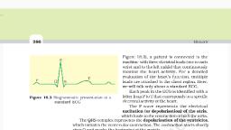
SURYA CLASSES, , LECTURE NOTES BIOLOGY [PAPER-II], , XII SCIENCE, , 3. The third electrode connected to upper or lower limbs. The third, electrode is reference connection., 4. Conducting gel applied between skin and electrodes to detect tiny, impulse of few millivilts., 5. The electrical activity of heart represented in the form of graph as, time[X axis] against voltage displacement[Y axis] ., • Normal ECG: The normal ECG is series of ridges and furrows., , The Normal ECG represents following waves, 1., , P wave: It is small upward wave. It represents impulse generation by, SA node. It causes arterial depolarization resulting in atrial contraction, , 2., , QRS wave /complex: It begins as downward wave(Q) ,continues, as large upward and triangular wave(R) and ends as downward wave, (S).It represents spreading of impulse from SA node to AV node and, then bundle of His and purkinje fibers., It causes ventricular depolarization resulting ventricular, contraction., , 3., , T wave: It is broad, upward wave .It causes ventricular repolarization, resulting ventricular relaxation., • Uses :, 1. Detection of coronary artery disease, 2. Detection of heart blocks, 3. Detection of angina pectoris, 4. Detection of ischemic heart disease, 5. Detection of tachycardia, 23, , RESPIRATION & CIRCULATION, , | SIGN OUT BY DR. VIRESH SHELKE
Page 24 :
SURYA CLASSES, , LECTURE NOTES BIOLOGY [PAPER-II], , XII SCIENCE, , 6. Detection of cardiac arrest, 7. Detection of myocardial infraction, , ❖, , Pacemaker:, , o It is electronic device surgically fitted to SA node by wires., o The artificial pacemaker is programmed device and powered by Ni-cd, batteries., o The life span of pacemaker is 3-7 years., , ❖ Lymphatic system: A network of vessels that conveys, electrolytes, water, protein and electrolytes in the form of lymph is called, as lymphatic system, , The lymphatic system consist of :-, , 1. Lymph:, o, o, o, o, o, , Lymph is colorless, liquid connective tissue., Lymph is blood minus RBC’s, platelets and some plasma proteins., The lymph contains carbon dioxide and metabolic wastes., The lymph present in tissue spaces or intracellular spaces of tissue., The lymph bathes the cells., , 2. Lymphatic capillaries:, o Lymphatic capillaries, spaces., o Lymphatic capillaries, o Lymphatic capillaries, connected to them., o Lymphatic capillaries, end., , are thin walled vessels present in all tissues, lined by endothelium., interwoven with blood capillaries but not, are wider than blood capillaries and blind at one, , 3. Lymphatic vessels :, o Lymphatic capillaries unite to form large tubes called as lymphatic, vessels., A] Thoracic/ Left lymphatic duct:, 24, , RESPIRATION & CIRCULATION, , | SIGN OUT BY DR. VIRESH SHELKE
Page 25 :
SURYA CLASSES, , LECTURE NOTES BIOLOGY [PAPER-II], , XII SCIENCE, , o It receives lymph from left side of the head, neck, chest, left upper, arm and entire body below the ribs., o It is principal lymphatic duct., o Lacteals: It is lymphatic vessels coming from intestine .It contains, absorbed fat so milky in appearance., B] Right lymphatic duct:, o It receives lymph from upper right side of body., , 4. Lymph nodes:, o Small ,oval or bean shaped bodies present in the course of lymphatic, vessels called as lymph nodes, o Lymph nodes scattered throughout the body., o Lymph nodes are maximum in neck, armpits and groin region., o Lymph nodes act as filters because it contains macrophages which, remove bacteria, foreign material and cell debris., o Lymph nodes also produce lymphocytes., , ❖ Functions of Lymphatic system:, 1., 2., 3., 4., 5., , Drainage of excessive tissue fluid, Carries carbon dioxide and metabolic wastes from tissue to blood, Transport of absorbed fat from intestine to the blood, Production of lymphocytes, Destruction of micro organisms and foreign particle., , 25, , RESPIRATION & CIRCULATION, , | SIGN OUT BY DR. VIRESH SHELKE