Page 1 :
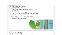
Nikhil Kashinath Vadhan, , RESPIRATION AND CIRCULATION, 1., , Respiration: Respiration : It is a biochemical process of oxidation of organic, compounds in an orderly manner for the liberation of chemical energy in the, form of ATP., All living organisms require energy to carry out various life processes. The energy, that is stored in the body in the form of complex organic compounds (potential, energy) is however not usable by the organisms unless it is converted into usable, form. This conversion is achieved through the process of respiration., , C H 06 + 602 6 CO2 + 6H2O + 38 ATP, For this, the process of gaseous exchange takes place between the organism and, the environment. The site of gaseous exchange is called the respiratory surface., , 2., , Organs of Respiratory Exchange :, Respiratory exchange is a simple physical process., For efficient gaseous exchange, the respiratory surface should have the, following features ., a. It should have a large surface area., b. It should be thin, highly vascular and permeable to allow exchange of gases., c. It should be moist., , Gaseous exchange in plants :, The shape and structure of plants facilitate gaseous exchange by diffusion., A terrestrial flowering plant has many air spaces between the cells of stem,, leaf and root., These air spaces are continuous., Oxygen diffuses into the air space through stomata (the pores on leaves and, young stems), carbon dioxide and water vapour diffuse out., , 1
Page 2 :
Nikhil Kashinath Vadhan, , , In the aerated soil,, the oxygen dissolves in the, form of moisture or water, around the root tissue and, enters it by diffusion., , Woody flowering, plants (trees and shrubs), have an external, impervious bark., , Here, gaseous, exchange occurs through small pores in the stem surface, called lenticels., Respiration in Animals :, As compared to plants, animals show wide variety of respiratory surfaces or, organs., The respiratory surfaces differ in various animals., In animals, depending upon the complexity of organization and the, surrounding medium, certain parts of the body have become specialized into, different types of respiratory organs., In the higher animals, these respiratory organs are also associated with a, transport system., , 3., , Human Respiratory system:, The respiratory system brings about, inspiration, expiration and exchange of, gases in the lungs., These are then transported by blood from the lungs to the different tissues, and parts of the body., The respiratory system can be divided into an upper respiratory system, having external nares, nasal cavities, internal nares, nasopharynx, nose, throat, and associated structures., , 2
Page 3 :
Nikhil Kashinath Vadhan, , , The lower respiratory, system refers, to the larynx,, trachea, bronchi, bronchioles, and lungs., , Nose:, , The nose has a pair of slit, like openings called external, nares or nostrils for entry of, air into the nasal cavity., , The nasal cavity is, divisible into right and left, nasal chambers by a, mesethmoid cartilage., , Each nasal chamber is, further divided into three, regions., i., Vestibule, ii., iii., , Respiratory part, Olfactory or sensory chamber, , i., , Vestibule :, It is the proximal part about the nostrils. Its skin has hair for filtering, the air and traping the dust and suspended particles in the inhaled air., , ii., , Respiratory part (conditioner) :, The middle thin walled highly vascular pan for warming and, moistening the inhaled air., , iii. Olfactory or sensory chamber :, The uppermost part is lined by olfactory epithelium for detection of, smell., , Pharynx :, , 3
Page 4 :
Nikhil Kashinath Vadhan, It is divisible into three parts., The nasopharynx is the uppermost part from the nasal chamber it leads into, oropharynx (common passage for food and air). This continues below as the, laryngopharynx. Between the nasopharynx and oropharynx is the palate, bone., The pharynx has a set of lymphoid organs called tonsils., Larynx :, It is called voice box., It is the part of the respiratory tract which contains vocal cords for producing, sound., , The larynx extends from, the laryngo-pharynx and the, hyoid bone to the trachea., , It is a hollow, tubular, structure., , Its wall is made up of, cartilage plates held by, membranes and muscles., , Internally, it is lined by a, pair of folds of elastic vocal, cords ., , The larynx opens into, the layngopharynx through a slit like opening called glottis., This opening of the trachea or wind pipe is guarded by a leaf like Hap called, epiglottis. It prevents the entry of food into trachea., , Trachea (wind pipe) :, It is a long tube 10 to 12 cm in length., It runs through the neck in front of the oesophagus and extends into and upto, the middle of thoracic cavity., It is supported by ‘C’ shaped 16 to 20 rings of cartilage which prevent the, collapse of trachea., It is lined internally with ciliated, pseudostratified epithelium and mucous, glands that trap the unwanted particles preventing their entry into the lungs., , 4
Page 5 :
Nikhil Kashinath Vadhan, Bronchi :, The trachea divides into right and left primary bronchi us it reaches the, middle of the thoracic cavity., The bronchi are supported internally by ‘C’ shaped incomplete rings of, cartilage., The primary bronchi divide to form secondary and tertiary bronchi which lead, into terminal bronchioles ending into alveoli., , Lungs :, These are the main respiratory organs of humans., One pair of spongy and elastic lungs are present in the thoracic cavity., Each lung is enclosed and protected by a double pleural membrane, outer, parietal and inner visceral membrane., Between the two pleura is a pleural cavity filled with a lubricating fluid called, pleural fluid., It is secreted by the membranes., The right lung is larger and divided into 3 lobes, while the left lung is smaller, and divided into 2 lobes. Each lobe of the lung has the terminal bronchioles, ending in a bunch of air sacs, each with 10 to 12 alveoli., , Alveoli :, These are thin walled lobulated structures, like a bunch of grapes., Each alveolus is surrounded by a network of capillaries of pulmonary artries, and veins., There are about 700 million alveoli in the lungs and they provide the surface, area for exchange of gases., , Diaphragm : It is a muscular septum that separates the thoracic and abdominal, cavity. It is dome shaped and on contraction it becomes flattened., , 4., , Mechanism of respiration :, Respiration is a biological process involving exchange of gases between the, atmosphere and the lungs and it results in the formation of ATP. It includes the, following processes:, , 5
Page 6 :
Nikhil Kashinath Vadhan, A., B., C., D., , Breathing, External respiration, Internal respiration, Cellular respiration, , A., , Breathing :, , It is a physical process by which gaseous exchange takes place between the, atmosphere and the lungs., It involves inspiration and expiration., Both these steps involved parts of the thoracic cage, the ribs, sternum and the, intercostal muscles and muscles of the diaphragm., Inspiration, During inspiration, the atmospheric air, is taken in to the lungs., It is an active process., The diaphragm becomes flat and goes, downward, the external intercostal, muscles contract so the ribs and, sternum move upward and outward., , Expiration, During expiration, the air from lungs, release throughout the body, It is passive process., The diaphragm become dome shaped, and goes upward, the external, intercostals muscles relax so the ribs, and sternum move downward and, inward., This leads to an increase in the thoracic This leads to an decrease in the, volume and a decrease in pressure of, thoracic volume and increase in, thorax and the lungs., pressure of thorax and lungs., To equalize the low pressure inside the To equalize the high pressure inside, lungs, air from the atmosphere rushes, the lungs, air from lungs release, into lungs. This is inspiration., outside the body. This is Expiration., , B., , External respiration / Exchange of gases at the alveolar level, , 6
Page 7 :
Nikhil Kashinath Vadhan, An alveolus consists of a layer of simple squamous epithelium resting on a, basement membrane., It is intimately associated with a dense network of capillaries., The capillary wall is also made up of simple squamous epithelium resting on a, thin basement membrane., Both the layers have similar structure and are thin walled., Together they make up the respiratory membrane through which gaseous, exchange occurs i.e. between, the alveolar air and the blood., , Diffusion of gases will, take place from an area of, higher partial pressure to an, area of lower partial pressure, until the partial pressure in the, two regions reaches, equilibrium., , The partial pressure of, carbon-dioxide of blood, entering the pulmonary, capillaries is 45 mmHg while, partial pressure of carbon dioxide in alveolar air is 40 mmHg. Due to this, difference, carbon dioxide diffuses from the capillaries into the alveolus., Similarly, partial pressure of oxygen of blood in pulmonary capillaries is 40, mmHg while in alveolar blood it is 104 mmHg. Due to this difference oxygen, diffuses from alveoli to the capillaries., , Pulmonary volumes and capacities (Normal values), Lung Volumes :, Tidal volume (T.V.) : It is the volume of air inspired or expired during, normal breathing. It is 500 ml., Inspiratory reserve volume (IRV) : The maximum volume of air, or the, extra volume of air, that is inspired during forced breathing in addition to, TV. Its value is 2000 to 3000ml., , 7
Page 8 :
Nikhil Kashinath Vadhan, Expiratory reserve volume (ERV) : The maximum volume of air that is, expired during forced breathing after normal expiration. Its value is 1000, to 1100ml., Dead space (DS) : The volume of air that is present in the respiratory, tract (from nose to the terminal bronchioles), but not involved in gaseous, exchange. It is 150 ml., Residual volume (RV) : The volume of air that remains in the lungs and, the dead space even after maximum expiration. It is 1100 to 1200ml., , Lung capacities :, Total Lung capacity : The maximum amount of air that the lungs can hold, after a maximum forceful inspiration (5200 to 5800ml)., Vital capacity (VC) : The maximum amount of air that can be breathed out, after a maximum inspiration. It is the some total of TV, IRV and ERV and is, 4100 to 4600ml., , C., , Internal respiration :, , The two main components of blood involved in transport of the respiratory gases, CO2 and O2 are the RBCs and the plasma., , i., , Transport of 0xygen :, , Of the total oxygen transported only 3% is transported in a dissolved state by, the plasma. The remaining 97% is bound to the haemoglobin (Hb) present in, the RBCs., Haemoglobin acts as the respiratory carrier., It has a high affinity for O2 and combines with it to form oxyhaemoglobin., Theoretically, one molecule of Hb has 4 Fe++, each of which can pick up a, molecule of oxygen (O2)., , Hb, , +, , 4O2, , Hb (4O2), , Oxyhoemoglobin is transported from lungs to the tissues where it readily, dissociates to release 02., , 8
Page 9 :
Nikhil Kashinath Vadhan, Hb (4O2), , Hb, , +, , 4O2, , Carbon monoxide poisoning :, Affinity of haemoglobin for carbon monoxide is about 250 times more, than, for oxygen., In the presence of carbon monoxide, haemoglobin readily combines to form a, stable compound carboxy-haemoglobin., The haemoglobin is blocked by carbon monoxide, preventing oxygen from, binding with haemoglobin., Thus, less haemoglobin is available for oxygen transport depriving the cells, of oxygen., This is carbon monoxide poisoning., , ii., , Transport of CO2 :, , Carbon dioxide is readily soluble in water and is transported by RBCs and, plasma in three different forms., a. By plasma in solution form (7%): Only 7% of CO2 is transported in a, dissolved form as carbonic acid (which can breakdown into CO2 and H2O)., , CO2, , +, , H2 O, , H2 CO3, , b. By bicarbonate ions (70%) : Nearly 70% of carbon dioxide released by, the tissue cells diffuses into the plasma and then into the RBCs., In the RBCs, CO2 combines with water in the presence of a Zn containing, enzyme, carbonic anhydrase to form carbonic acid., Carbonic anhydrase enzyme is found in the RBCs and not in the plasma., The rate of formation of carbonic acid inside the RBC is very high as, compared to its formation in the plasma., Carbonic acid being unstable almost immidiately dissociates into HCO-3, and H+ in the presence of the enzyme carbonic anhydrase (CA) leading, to large accumulation of HCO-3 inside the RBCs., , 9
Page 10 :
Nikhil Kashinath Vadhan, CO2 + H2O, , H2CO3, , H+, , +, , HCO-3, , It thus moves out of the RBCs. This would bring about inbulunce 0f the, charge inside the RBCs, , HCO3 that comes into the plasma joins to Na+/ K+ forming NaHCO3 /, KHCO3 (to maintain pH of blood)., , HCO-3, , +, , Na+, , NaHCO3, , Carbonic acid + Sodium, , Sodium Bicarbonate, , H+ is taken up by protein (haemoglobin)., , Hb, , +, , H+, , HHb, Reduced Hb, , These H+ ions might be expected to lower blood pH, but they are buffered, by haemoglobin by the formation of deoxyhaemoglobin (reduced, haemoglobin)., At the level of the lungs in response to the low partial pressure of carbon, dioxide (ppCO2) of the alveolar air, hydrogen ion and bicarbonate ions, recombine to form carbonic acid and under the influence of carbonic, anhydrase yields carbon dioxide and water., , H+, , + HCO-3, , H2CO3, , CO2, , +, , H2O, , c. By red blood cells (23%) :, Carbon dioxide binds with the amino group of the haemoglobin and form a, loosely bound compound carbaminohaemoglobin., This molecule readily decomposes in region where the partial pressure of, , 10
Page 11 :
Nikhil Kashinath Vadhan, carbon dioxide (ppCO2) is low (alveolar region), releasing the carbon, dioxide., , Hb, , +, , CO2, , HbCO2, , D. Cellular Respiration :, It is the last step taking place inside the cell where food is oxidized and ATP, is generated. It can be shown by two steps:, a. Oxidation :, Breaking down of complex organic molecules into simple inorganic, molecules with release of heat energy., , C6 H12 O6, , +, , 6O2, , 6CO2, , +, , 6H2O, , +, , 686 Kcal, , b. Phosphorylation :, It involves trapping the heat energy in the form of high energy bond, of ATP molecule., ATP is used to carry out vital life processes and so is called as energy, currency of the cell., , ADP, , 5., , +, , iP, , +, , 7.3 Kcal, , ATP, , Regulation of Breathing :, Respiration is under dual control : nervous and chemical., Human adults breathe about 12 times/minute while a new born about 44, times/ minute., Normal breathing is an involuntary process., Steady rate of respiration is controlled by neurons located in the pons and, medulla and are known as the respiratory centres., It regulates the rate and depth of breathing., It is divided into three groups :, a. Dorsal group of neurons in the medulla (inspiratory center), b. Ventro lateral group of neurons in medulla (inspiratory and expiratory, , 11
Page 12 :
Nikhil Kashinath Vadhan, , , , , , , , center), c. Pneumotaxic center located in pons (primarily limits inspiration, slow, wave sleep and rapid eye movement sleep)., Apneustic center in the medulla is antagonistic to the neumotaxic center. It, controls non rapid eye movement sleep and wakefullness., , During inspiration, when the lungs expand to a, critical point, the stretch, receptors are stimulated and, impulses are sent along the, vagus nerves to the, expiratory centre., , It then sends out, inhibitory impulses to the, inspiratory center., , The inspiratory muscles, relax and expiration follows., , As air leaves the lungs, during expiration, the lungs, are deflated and the stretch, receptors are no longer, stimulated., , Thus, the inspiratory, centre is no longer inhibited, and a new respiration begins., , These events are called, the Hering-Breuer reflex., , The Hering-Breuer, reflex controls the depth and rhythm of respiration. It also prevents the lungs, from inflating to the point of bursting., The respiratory centre has connections with the cerebral cortex which means, we can voluntarily change our pattern of breathing., Voluntary control is protective because it enables us to prevent water or, irritating gases from entering the lungs., But the ability to stop breathing is also limited by the buildup of carbon, , 12
Page 13 :
Nikhil Kashinath Vadhan, dioxide in the blood., , 6., , Modified Respiratory Movements., Some respiratory movements are different from the normal movements and, help express emotion or clear the air passage., Of these movements some may be reflexes, but others can be initiated, voluntarily e.g. coughing and yawning., , 13
Page 14 :
Nikhil Kashinath Vadhan, 7., , Common disorders of respiratory system:, The given table shows a list of some common respiratory disorders, their, symptoms, cause and treatment., , Artificial ventilation :, It is also called artificial respiration., It is the method of inducing breathing in a person when natural respiration, has ceased or is faltering., If used properly and quickly, it can prevent death due to drowning, choking,, suffocation, electric shock, etc., The process involves two main steps : establishing and maintaining an open, air passage from the upper respiratory tract to the lungs and force inspiration, , 14
Page 15 :
Nikhil Kashinath Vadhan, and expiration as in mouth to mouth respiration or by mechanical means like, ventilator., , Ventilator :, A ventilator is a machine that supports breathing and is used during surgery,, treatment for serious lung diseases or other conditions when normal, breathing fails., It is mainly used in hospitals as pan of life support system., Ventilators do the following,, 1. Get oxygen into the lungs., 2. Remove carbon dioxide from the lungs., 3. Help the patient breathe., , 9., , Transportation in living organisms., All living organisms, whether unicellular or multicellular show an important, property of exchange of material with their surrounding as well as between, various parts of their cell or body., Organisms take up oxygen and nutrients from the surrounding, these are, circulated within the body for various metabolic activities., The wastes generated within are given out into the surrounding., Transportation in organisms and animals occurs by diffusion and by active, transport between the cells., This mechanism is suitable where the surface area of body is large and the, distance between parts of the body in the organism is extremely small., Cyclosis is the streaming movement of the cytoplasm shown by almost all, living organisms e.g. Paramoecium, Amoeba, root hair cells of many plants, and WBCs in animals., It is for transportation within the cell or intracellular transport., In sponges and coelenterates the surrounding water is circulated through the, body cavities., In flat worms there is parenchymal circulation. In round worms there are no, , 15
Page 16 :
Nikhil Kashinath Vadhan, blood vessels and the body fluid is moved around the viscera by contraction, of body wall and muscles. This is extracellular transport., , 9., , Circulation in animals., In higher animals the circulation is carried out by special fluids blood and, lymph., , Blood vascular system :, Higher animals from Annelida to chordata have a special circulating fluid, the, blood which is pumped to the tissues by the heart through the blood vessels., Types of blood vascular system :, 1. Open circulation, In animals having an open circulation,, blood is circulated through the body, cavities (haemocoels)., The visceral organs lie in the blood, filled body cavity., , 2. Closed circulation, In all the vertebrates, higher molluscs, and annelids, blood is circulated all, over the body through a network of, blood vessels, In this type of circulation, blood flows, within the blood vessels and does not, come in direct contact with cells and, body tissues., , Exchange of material takes place, directly between blood and cells or, tissues of the body., , Exchange of material between blood, and body tissues is through an, intermediate fluid called lymph., , The blood flows with low pressure and, usually does not contain any, respiratory pigment like haemoglobin,, so it does not transport respiratory, gases., e.g. Arthropods (cockroach) and, Molluscs., , Blood flows with high pressure and, contains respiratory pigments like, haemoglobin for transportation of, respiratory gases., , 16, , e.g Human being
Page 17 :
Nikhil Kashinath Vadhan, The closed circulation can be divided into two main types :, o Single circulation., o Double circulation., , Single circulation :, In single circulation, the blood passes through heart only once during each, cycle as in fishes., Deoxygenated blood is, pumped from heart towards gills,, where it undergoes oxygenation., This oxygenated blood, moves towards various body, parts, gets deoxygenated and, returns back to heart for next, cycle., Since, the heart of fish, carries only deoxygenated blood,, it also called ‘venous heart’., , Double circulation :, In double circulation, blood passes through heart twice during each cycle; it, occurs in birds and mammals., In these animals, heart pumps deoxygenated blood to lungs for oxygenation, and it returns to heart as oxygenated blood., This is ‘pulmonary circulation’., The oxygenated blood is pumped from the heart towards various body parts, (except lungs) and returns back to the heart as deoxygenated blood., , 17
Page 18 :
Nikhil Kashinath Vadhan, , This is ‘systemic, circulation’., Human heart shows, double circulation., , 10., , Circulatory System in Human., The human circulatory system is composed of blood vascular and lymphatic, system., , Blood vascular system :, In human beings it consists of blood, heart and blood vessels. It is responsible for, various functions like transport, homeostasis and protection., , Blood Composition and Coagulation :, , , , , , , , Study of blood is called haematology., An average adult has about 4 to 6 liters of blood., It is a red coloured fluid connective tissue derived from embryonic mesoderm., It is slightly alkaline (pH 7.4), salty and viscous fluid., It is heaviere then water., It has two main components the fluid plasma (55%) and the formed elements, i.e. blood cells (44° 0)., These can be separated by centrifugation., , 18
Page 19 :
Nikhil Kashinath Vadhan, Plasma :, It constitutes 55% of the blood., It is a straw-coloured, slightly alkaline, viscous fluid and consists of, following:, , Formed elements :, The blood cells, that are produced in the, body are collectively, called formed, elements., Human blood, contains three types of, formed elements as:, i. Red blood, corpuscles (RBCs), ii., iii., , 11., , White blood corpuscles (WBCs), Platelets., , Red blood corpuscles / Erythrocytes :, Erythrocytes are the most abundant cells in the human body., They are circular, biconcave and enucleated., The red colour or RBCs is due to an oxygen carrying pigment, the, haemoglobin, in their cytoplasm., In males, their average number is about 5.1-5.8 million/mm3 (per µL) and in, females about 4.3-5.2 million/mm3. This is called total RBC count., Life span of RBCs - 120 days., The process of formation of RBCs is called erythropoiesis., Origin - Bone marrow., Vitamin B12, folic acid and heme protein are required for production of RBCs, , 19
Page 20 :
Nikhil Kashinath Vadhan, The old and worn out RBCs are, destroyed in the liver and spleen, (graveyard of RBCs)., Condition with increase in the, number of RBCs is called polycythemia, and with decrease in number of RBCs is, called as erythrocytopenia., The hormone erythropoietin, produced by the kideny cells stimulates the bone marrow for production of, RBCs., Mature erythrocyte is devoid of nucleus, mitochondria or other membrane, bound cell organelles., Its cytoplasm (stroma) is rich in haemoglobin and O2 carrying proteinaceous, pigment that gives red colour to the RBCs and blood., , Function:, Erythrocytes are responsible for the transport of respiratory gases O2 and, CO2, maintaining pH and viscosity of blood., They also contribute in the process of blood clotting., , 12., , White blood corpuscles / Leucocytes:, Leucocytes are colourless, nucleated and amoeboid cells larger than RBCs., Due to their amoeboid movement they can move out of the capillary walls by a, process called diapedesis., A normal adult has on an average, 5000-11000 WBCs per mm3 of blood., Decrease in number of WBCs (<4000) is called leucopenia (common in HIV,, AIDS and TB patients or those exposed to radiations, shock, etc)., Temporary increase in number of WBCs is called as leucocytosis. It is due to, infection. It also occurs during pregnancy and in newborn babies., Uncontrolled increase in number of WBCs is a type of blood cancer called, leukemia., Function:, protection., , WBCs are mainly concerned with defense mechanism i.e., , 20
Page 21 :
Nikhil Kashinath Vadhan, , Types of WBCs : They can be classified into two main types such as :, A .Granulocytes, , B. Agranulocytes., , A. Granulocytes :, These are WBCs with a granular cytoplasm, also called Polymorpho, nuclear leucocyte (PMN) cells., They have lobulated nuclei in different shapes., Granulocytes are formed from myeloid stem cells and once formed, do not, divide., Granulocytes constitute about 72% of total WBCs., Depending upon staining property of the granules, these granulocytes are, classified into three types as :, a. Neutrophils, b. Basophils, c. Eosinophills., , a. Neutrophils :, o Granules are very fine, large in number, evenly distributed and stained, with neutral stains (dyes)., o Neutrophils are about 70% of total WBCs. These cells are spherical and, nucleus is several lobed (27)., o These are able to perform amoeboid movements and phagocytosis., o Function : They are responsible for destroying pathogens by the process, of phagocytosis., o ‘Pus’ is mixture of dead neutrophils, damaged tissues and dead microbes., , b. Basophils / Cyanophils :, o These cells have very few granules of large size, and stain with basic, stains like methylene blue., o Basophils are non-phagocytic, small, spherical cells and are about 0.5-1%, of total WBCs., o Nucleus is twisted., , 21
Page 22 :
Nikhil Kashinath Vadhan, o They are present in infected and allergic conditions only., o Function : Basophils secrete heparin, histamine and serotonin., , c. Eosinophils / Acidophills :, o Acidophills contain lysosomal granules that are stained to red colour with, acidic stains like eosin., o Eosinophils are about 1- 3 % of total WBCs., o Nucleus is bilobed., o They destroy antigenantibody complex by phagocytosis. They are also, responsible for detoxification as they produce antitoxins., , B. Agranulocytes:, , , , , , Agranulocytes are about 2800 of total WBCs., Cytoplasm of these leucocytes is without granules., They are formed from lymphoid stem cells and can divide by mitosis., Nuclei of agranulocytes are large in size but are not lobulated like the, granulocytes., There are two types of agranulocytes :, a. Lymphocytes, b. Monocytes., , a. Lymphocytes :, o Lymphocytes are the smallest of all WBCs and have a large spherical, nucleus., o They constitute about 25-30% of total WBCs., o Depending upon function, two types of lymphocytes are present as Blymphocytes and T-lymphocytes., o B-lymphocytes mature in bone marrow and are responsible for antibody, production/humoral immunity., o T-lymphocytes mature in thymus and are responsible for cell-mediated, immunity., o Helper T-cells, killer T-cell, memory T-cells and suppressor T-cells are, four main subtypes of T-lymphocytes., , 22
Page 23 :
Nikhil Kashinath Vadhan, b. Monocytes :, o, o, o, o, o, , 13., , Monocytes are the largest of all the WBCs., Its nucleus is large and bean or kidney shaped., They form 3-5% of WBCs., Monocytes are actively motile and give rise to macrophages., Function : They are mainly phagocytic and destroy the bacteria and dead, or damaged tissue by phagocytosis., , Thrombocytes / Platelets:, Thrombocytes are cellular fragments formed from the large cells called, megakaryocytes., These are produced in bone marrow., They are very small, oval shaped cell fragments without nucleus., Normal count of thrombocytes in human blood is about 2.5 - 4.5 lakh / mm3, of blood., If number 0f thrombocytes decreases than normal, condition is called as, thrombocytopenia. This condition causes internal bleeding (haemorrhage)., Function:, o Platelets secrete platelet factors which are essential in blood clotting., o They also seal the ruptured blood vessels by formation of platelet, plug/ thrombus. T, o hey secrete serotonin a local vasoconstrictor., , 23
Page 24 :
Nikhil Kashinath Vadhan, Blood Clotting/ Coagulation of blood :, Clotting or coagulation is the process of converting the liquid blood into a solid, jelly., This process may be initiated by contact of blood with any foreign surface, (intrinsic process) or with damaged tissue (extrinsic process)., Intrinsic and extrinsic processes involve interaction of various substances, called clotting factors by a step wise or cascade mechanism., , There are in all twelve, clotting factors numbered as I, to XIII (factor VI is not in active, use)., , Interaction of these, factors in a cascade manner, leads to formation of the, enzyme thrombin., , Thromboplastin, helps in, the formation of enzyme, prothrombinase., , This enzyme inactivates, heparin and it also converts, inactive prothrombin into its, active thrombin., Thrombin converts soluble blood protein~ fibrinogen into insoluble fibrin., Fibrin forms a mesh in which platelets and other blood cells are trapped to, form the clot., Blood clotting occurs as shown in the following flowchart., , 14., , Heart :, Heart is the main pumping organ of the circulatory system., It is reddish brown in colour, hollow, muscular organ, roughly the size of one’s, fist., Its average weight is about 300gm in males and 250gm in females., It is conical in shape and lies in mediastenum-i.e. the space between two lungs., , 24
Page 25 :
Nikhil Kashinath Vadhan, It is broader at upper end (base) and conical at lower end (apex)., , Conical end is slightly, tilted to left side and rests, above the diaphragm., , Heart is enclosed in a, membranous sac called, pericardium., , Pericardium is, formed of two main layers, outer fibrous and inner, serous pericardium., , Serous pericardium, is soft, moist and elastic., , It is formed of, squamous epithelium and, is further divisible into two, layers as: parietal and visceral layer., Parietal and visceral layers of serous pericardium are separated by a, pericardial space., This space is filled with pericardial fluid (about 50ml) which acts as a shock, absorber and protects the heart from mechanical injuries., It also keeps the heart moist and acts as lubricant., , Heart wall :, The heart is mesodermal in origin., Its wall is formed of three layers:, - Outer epicardium, - Middle myocardium, - Inner endocardium, Epicardium is thin and formed of a single layer of flat squamous epithelium, resting on basement membrane., Myocardium is the middle thick layer formed of cardiac muscles., Endocardium is a single thin layer formed of squamous epithelium., Functions : The epicardium and endocardium are protective in function, , 25
Page 26 :
Nikhil Kashinath Vadhan, whereas myocardium is responsible for contraction and relaxation of heart., , 14.1 External structure of heart :, The human heart is four chambered., a. The two superior chambers are called atria (auricles), b. The inferior two are called ventricles., , Dorsal view, Externally, the atria are separated from ventricles by a transverse groove, called coronary sulcus or atrioventricular groove., The two ventricles are externally separated from each other by two grooves,, the anterior and posterior inter-ventricular sulci., Coronary arteries and coronary veins run through these sulci., Pulmonary trunk arising from right ventricle and aorta from left ventricle are, present on anterior surface of heart., The pulmonary trunk bifurcates into, - right pulmonary artery., - left pulmonary arteri., , 26
Page 27 :
Nikhil Kashinath Vadhan, Aorta (systemic aorta) is divisible into three regions as :, - ascending aorta, - systemic arch /aortic arch, - descending aorta, , Ventral view, The Ligamentum arteriosum joins pulmonary trunk and aortic arch., It is the remnant of an embryonic duct called ductus arteriosus., The aortic arch gives out three arteries viz. brachiocephalic (innominate), artery, left common carotid and left subclavian., The right atrium recieves superior and inferior vena cava along its dorsal, surface., Pulmonary veins open into left atrium along the dorsal surface of heart., , 27
Page 28 :
Nikhil Kashinath Vadhan, 14.2 Internal structure of heart:, Atria :, These are the thin-walled receiving chambers of heart., They are separated from each other by inter-auricular septum., Two atria are there : Right atrium, Left atrium, Superior vena cava (precaval), inferior vena cava (postcaval) and coronary, sinus open into the right atrium., Opening of the postcaval is guarded by a Eustachian valve while the Thebesian, valve guards the opening of coronary sinus into right atrium., Four pulmonary veins open into the left atrium. These openings are without, valves., Both the atria open into the ventricles of their respective sides by, atrioventricular apertures., These openings are guarded by cuspid valves., The tricuspid valve is present in the right AV aperture and bicuspid valve, (mitral valve) is present in the left AV aperture., All these heart valves help in maintaining a unidirectional flow of blood., They also avoid back flow of blood., , Ventricles :, -Right Ventrical, - Left ventrical, These are inferior, thick-walled pumping chambers of the heart., The right and left ventricles are separated by an interventricular septum., Wall of the left ventricle is more muscular and about 3-times thicker than the, right ventricle., The lumen of ventricles also shows inelastic fibers called chordae tendinae., These attach the bicuspid and tricuspid valves to the ventricular wall, (papillary muscles) and regulate their opening and closing., The right ventricle opens into the pulmonary aorta and left ventricle opens, into the aorta., , 28
Page 29 :
Nikhil Kashinath Vadhan, These openings are guarded by three semilunar valves each., These valves prevent the backward flow of blood into the ventricles., , 14.3 Pumping action of heart, The heart acts as the main pumping organ of the circulatory system., The pumping action is brought about by a rhythmic contraction and relaxation, of the cardiac muscles or heart muscles., Contraction of heart muscles is systole and relaxation of heart muscles is, diastole., A single systole followed by diastole makes one heart beat., The heart beats 70 to 72 times per minute. This is called heart rate., During each heart beat ventricles pump about 70 ml of blood this is called, stroke volume., It means heart pumps about 72 (heart rate) x 70 ml (stroke volume) = 5040 ml, ≈5 liters of blood per minute this is called cardiac output., , 14.4 Conducting tissue of heart:, The human heart is myogenic i.e. the heart is capable of generating a cardiac, contraction independent of nervous input., It also shows auto rhythmicity i.e. it can generate its own rhythm by, specialized muscles., A specialized cardiac musculature called the nodal tissue is distributed in the, heart., A part of this nodal tissue is present in the upper right corner of the right, atrium. It is called SA Node or Sinoatrial node. It lies at the base of opening of, superior vena cava., Another mass of nodal tissue, the modified muscular fibers also called, autorhythmic fibers (conducting tissue) control the beating rate of heart., Conducting (nodal) tissue consists of :, - SA node, - AV node, - Bundle of His, - Purkinje fibers, , 29
Page 30 :
Nikhil Kashinath Vadhan, Conducting system of the heart :, SA node (sino-atrial node):, SA node (sino-atrial node) is present in the right atrium., It acts as pacemaker of heart because it has the power of generating a new, wave of contraction and making the pace of contraction., , AV node(atrio- ventricular node):, SA node passes the contraction to the left ventricle and also to the AV node., AV node (atrio-ventricular node) is present in the right atrial wall near the, base of interatrial septum. It acts as pace setter of heart., Bundle of His, Bundle of His/ Tawara branches start from AV node and pass through, interventricular septum., Bundle of His forms two branches, the right and left bundles, one for each, ventricle., Purkinje fibers, These branches form network in ventricular walls and these are called, Purkinje fibers., Bundle of His and Purkinje fibers spread impulses in ventricles., As a result both the ventricles contract simultaneously., , 30
Page 31 :
Nikhil Kashinath Vadhan, 15., , Working mechanism of human heart :, , 15.1 Cardiac Cycle, , , , , , Human heart alternately contracts and relaxes., Contraction is called systole and relaxation is called diastole., Atria and ventricles contract alternately., Consecutive systole and diastole constitutes a single heartbeat or cardiac, cycle., It is completed in 0.8 sec. On an average, 72 beats are completed in one, minute in an adult, at rest., a. Atrial systole (AS):, Right atrium receives deoxygenated blood and left atrium receives oxygenated, blood., When both the atria are completely filled with blood, pressure is exerted on, the wall., In response to this pressure, SA node gets excited and generates cardiac, impulse., Due to this, cardiac muscles in the atrial wall contract causing atrial systole., During atrial systole, blood is pumped into ventricles., Blood is prevented from going back to the veins and coronary sinus by, Eustachian and Thebesian valve respectively., After completing systole the atria go into diastole., In normal conditions, atrial systole is for 0.1 sec. and atrial diastole (AD) is for, 0.7 sec., b. Ventricular systole, (VS):, , The impulse which, started from SA node now, reaches the AV node and it gets, excited., , AV node sends impulses, to bundle of His and from bundle of His to Purkinje fibers., , 31
Page 32 :
Nikhil Kashinath Vadhan, Purkinje fibers spread impulses all over the wall of ventricles., Due to this, ventricular wall contracts causing ventricular systole., During ventricular systole, right ventricle pumps deoxygenated blood into, pulmonary trunk and left ventricle pumps oxygenated blood into aorta., During ventricular systole the cuspid valves close both the atrioventricular, apertures preventing blood flow into atria (lubb sound is heard)., In normal conditions, ventricular systole lasts for 0.3 sec. and ventricular, diastole (VD) lasts for 0.5 sec., During ventricular diastole, semilunar valves are closed, preventing backflow, of blood from pulmonary trunk and systemic aorta into ventricles (dub sound, is heard)., c. Joint diastole, For about 0.4 second, both atria and ventricles are in diastole. When all the, chambers of heart are in diastole, this condition is called joint diastole or, complete diastole., Thus, duration of one cardiac cycle is 0.8 sec., Right side of heart contains deoxygenated and left contains oxygenated blood., Total volume of blood pumped during one ventricular systole is called stroke, volume (SV) and it is it approximately 70 ml., Cardiac output (CO):, It is the volume of blood pumped out per min. For a normal adult human being, it is calculated as follows :, (CO) = SV x HR, = 70 x 72, = 5040 ml/min, , 16., , Blood vessels ., There are three main types of blood vessels in the human circulatory system, viz,, - Arteries, - Veins, , 32
Page 33 :
Nikhil Kashinath Vadhan, - Capillaries., , Arteries :, , , , , , , , , , These blood vessels, carry blood from heart to, various parts/organs of the, body, there they branch into, arterioles and further into tine, capillaries., , They normally carry, oxygenated blood to all ports, of the body (except the, pulmonary artery which, carries deoxygenuted blood)., They are usually situated deep in the body except a few like the radial,, brachial, femoral, etc. which are superficially located., In a T. S. of artery, its wall shows three layers., 1. Tunica externa or tunica adventitia, 2. Tunica media, 3. Tunica interna or intima, The outermost tunica externa is a thick, tough layer of collagen fibers., The tunica media is made up of smooth muscles and elastic fibres., The innermost tunica interna is a single layer of flat compact endothelial cells, surrounding the lumen., , Veins :, Veins are thin walled, mostly superficial vessels which carry blood from the, organs towards the heart., The capillaries around the various organs join to form the veins., Except for the pulmonary veins or other veins of the body carry deoxygenated, blood towards the heart., Portal vein : A portal vein e.g. hepatic portal vein, differs from the other, normal veins in that its starts as capillaries from one organ and capillarises in, some intermediate organ e.g. liver, before taking the blood towards the heart., , 33
Page 34 :
Nikhil Kashinath Vadhan, Histologically, the veins also show the three layers like in the arteries., - The tunica externa, - Tunica media, - Tunica interna, However, the tunica media is comparatively thinner and their lumen is wide, and narrow., Internal valves at regular intervals can be seen., Blood flows with flow pressure and the valves prevent backflow of blood., , Capillary, These are a network of minute blood vessels., They are thin walled having a single layer of flat squamous epithelium resting, on a single basement membrane., They are mainly involved in exchange of materials., Wall of capillaries is formed of single layer of squamous epithelium and it is, stretchable., Blood flows through the capillaries under high pressure., Wall of capillaries bear small endothelial pores or fenestrae through which, blood cells (WBCs) can escape by the process called as diapedesis., , Pulse :, It is a series of pressure waves that travel through the arteries due to, ventricular systole., It is the strongest in arteries closer to the heart and gradually becomes weak, in arteries away from heart., It can be felt easily in the superficial arteries like radial artery in the wrist, and carotid artery in the neck., The pulse can be felt at particular points on the body., All locations where the pulse can be felt are shown in the figure., Pulse rate is equal to heart rate., Pulse rate higher than normal (above 100 beats/min) is called tachycardia, , 34
Page 35 :
Nikhil Kashinath Vadhan, and slower pulse rate (below 60, beats/min) than normal is called, bradycardia., , 17., , Blood pressure (B. P.), The pressure exerted by blood on the wall of the blood vessels is called blood, pressure., It is measured by the sphygmomanometer., It is usually measured from the arteries., , Arterial Blood Pressure:, Pressure exerted by blood on the wall of artery is arterial blood pressure., Pressure on arterial wall during ventricular contraction (systole) is systolic, pressure (SP)., For a normal healthy adult the average value is 120 mmHg., Pressure on arterial wall during relaxation of ventricles is diastolic pressure, (DP)., For a normal healthy adult it is 80 mmHg., B P = SP/ DP = 120/80 mmHg, Blood pressure is normally written as 120/80 mmHg., Difference between systolic and diastolic pressure is called pulse pressure., Normally, it is 40 mmHg., Deviations from normal blood pressure value indicate malfunctioning of heart., , 35
Page 36 :
Nikhil Kashinath Vadhan, It may be due to high or low blood volume, arterial inelasticity or hardening of, arteries (arteriosclerosis), deposition of fats like cholesterol in the arteries, (atherosclerosis), renal diseases and emotion induced hormonal changes,, obesity, etc., Blood pressure lower than normal i.e. below 90/60 mmHg is called, hypotension and blood pressure higher than normal i.e. above 140/90 mmHg, is hypertension., Various factors that affect the blood pressure are cardiac output, peripheral, resistance, blood volume, length and diameter of blood vessels, viscosity of, blood, age, gender, venous return, sleep, emotions, exercise, anxiety, etc., Blood pressure increases with age due to increase in inelasticity of blood, vessels., Amount of blood brought to the heart via the veins per unit time is called the, venous return and it is directly proportional to blood pressure., Blood pressure is also directly proportional to the total length of the blood, vessel., Blood pressure can also be affected by vaso constriction or vaso dilation., Females have slightly lower BP than males her age before menopause., However, the risk of high B. P. increases in the females after menopause sets, in., , Measurement of blood, pressure:, , Blood pressure is measured with the, help of an instrument called, sphygmomanometer., , This instrument consists of inflatable, rubber bag cuff covered by a cotton cloth., , It is connected with the help of tubes, to a mercury manometer on one side and a, rubber bulb on the other side., , During measurement, the person is, asked to lie in a sleeping position., , 36
Page 37 :
Nikhil Kashinath Vadhan, The instrument is placed at the level of heart and the cuff is tightly wrapped, around upper arm., The cuff is inflated till the brachial artery is blocked due to external pressure., Then pressure in the cuff is slowly lowered till the first pulsatile sound is, heard., At this moment, pressure indicated in manometer is systolic pressure. Sounds, heard during measurement of blood pressure are called as Korotkoff sounds., Pressure in the cuff is further lowered till any pulsatile sound cannot be heard, due to smooth blood flow., At this moment, pressure indicated in manometer is diastolic pressure. An, optimal blood presure (normal) level reads 120/80 mmHg., , Hypertension :, Persistently raised blood pressure higher than the normal is called, hypertension., 140/90 mmHg is called as threshold of hypertension and the 180/120 mmHg, and higher readings are dangerous to the health., It may damage the heart, brain and kidneys., Under the condition of hypertension, heart uses more energy for pumping, which causes angina pectoris - the chest pains due to lowered blood supply to, cardiac muscles and may lead to myocardial infarction., There are more chances of brain hemorrhage due to hypertension as arteries, in brain are less protected by surrounding tissues as compared to other, organs., In kidney, hypertension may cause kidney failure., , , , Coronary Artery Disease (CAD) :, , It is also known as atherosclerosis., In this, calcium, fat cholesterol and fibrous tissues gets deposited in blood, vessels suppling blood to the heart muscles making the lumen narrow., , Angina Pectoris :, , 37
Page 38 :
Nikhil Kashinath Vadhan, It is the pain in the chest resulting from a reduction in the blood supply to the, cardiac muscles because of atherosclerosis or arteriosclerosis., It is charactarized by severe pain and heaviness in the chest., The pain may spread to the neck, lower jaw, left arm and left shoulder., The pain usually results from exertion, when there is more demand of oxygen, by the heart, but the supply does not meet the requirement., , Angiography :, X-ray imaging of the cardiac blood vessels to locate the position of blockages is, called angiography., Depending upon the degree of blockage, remedial procedures like angioplasty, or by-pass surgery are performed., In angioplasty, a stent is inserted at the site of blockage to restore the blood, supply while in by-pass surgery, the atherosclerotic region is by-passed with, part of vein or artery taken from any other suitable part of the body, like hands, or legs., , Heart Transplant :, Replacement of severely damaged heart by normal heart from brain-dead or, recently dead donor is called heart transplant., Heart transplant is necessary in case of patients with end-stage heart failure, and severe coronary arterial disease., , Silent Heart Attack :, Silent heart attack, also known as silent myocardial infarction is a type of heart, attack that lacks the general symptoms of classic heart attack like extreme, chest pain, hypertension, shortness of breath, sweating and dizziness., Symptoms of silent heart attack are so mild that a person often confuses it, for regular discomfort and thereby ignores it., It has been studied that men are more affected by silent heart attack than, , 38
Page 39 :
Nikhil Kashinath Vadhan, women., , 18., , Electrocardiogram, Graphical recording of electrical variations detected at the surface of body, during their propagation through the wall of heart is electrocardiogram (ECG)., This recording may be in the form of printout or onscreen display., The instrument used for this recording is the ECG machine or, electrocardiograph., This instrument detects and amplifies the signals., Various electrodes are used for recording of signals., Four electrodes are positioned on limbs; two on arms and two on legs. These, are limb electrodes., Six electrodes are positioned on chest. These are chest electrodes., In a normal record, three different waves are recognized as :, o P-wave, o QRS complex, o T-wave, , , P-wave is a small, upward deflection from, baseline of graph. It, represents the atrial, depolarization., , The QRS complex, starts as a slight downward, deflection from baseline,, continues as sharp and, large upright wave and, ends as a downward wave. QRS complex represents the ventricular, depolarization., , 39
Page 40 :
Nikhil Kashinath Vadhan, T-wave is small, wide and upwardly elevated wave. It represents the, ventricular repolarization., , 19., , Lymphatic System :, Lymphatic system consists of lymph, lymphatic vessels, some organs and, tissues., The word ‘lymph’ means ‘clear water’ and it is a fluid connective tissue with, almost similar composition to the blood except RBCs, platelets and some, proteins., Fluid from intercellular spaces of the body tissue enters into the lymphatic, vessels, from here it is discharged into the blood vessels (veins) through the, thoracic duct and the right lymphatic duct., , 40