Notes of 12, Biology MOLECULAR BASIS OF INHERITANCE.pdf - Study Material
Page 1 :
MOLECULAR BASIS OF, INHERITANCE
Page 2 :
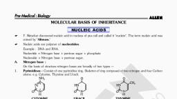
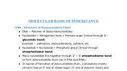
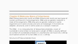
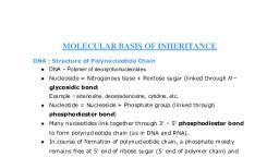
THE DISCOVERY OF DNA, • The discovery of DNA has, evolved from the discovery of, nucleic acid., • In 1869, Friedrich Miescher, separated a cellular substance, from the nuclei of pus cells and, called it Nuclein., • Chemically, nuclein has a high, phosphorus content and, showed acidic properties., Hence it was named as nucleic, acid., • There are two kinds of nucleic, acids found in cells i.e, DNA(deoxyribonucleic acid), and RNA (ribonucleic acid)
Page 3 :
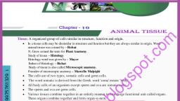
GRIFFITH’S EXPERIMENT, • In 1928, Frederick Griffith, performed an experiment, on bacterium Streptococcus, pneumoniae that causes, pneumonia in humans and, other mammals., • Griffith used two strains of, Streptococcus i.e, I. Virulent, Smooth,, pathogenic and, encapsulated S-type, II. Non-virulent, Rough, nonpathogenic and noncapsulated R-type., Griffith concluded that the R-type bacterium must have taken up, to what he called a, “transforming principle” from the heat- killed S- bacterium which allowed R-strain to, get transformed into Smooth-coated bacterium and become virulent.
Page 4 :
AVERY, McCARTY MACLEOD’S EXPERIMENT, • In 1944, after some 10, years of research and, experimentation,, Oswald Avery, Collin, MacLeod and Maclyn, McCarty first, evidenced to prove the, DNA is a genetic, material through their, experiments., These experiments prove that the tranforming principle is DNA, but all biologists were not convinced.
Page 5 :
HERSHEY-CHASE EXPERIMENT, • Hershey and Chase, worked with viruses that, infect bacteria i.e, bacteriophages, which, are composed of DNA, and protein. They used, radioactive phosphorus, 32, P in the medium for, some viruses and, radioactive sulphur 35S for, some others.
Page 6 :
PACKAGING OF DNA IN PROKARYOTES, 1. In prokaryotes like E.coli,, cell size is almost 2-3µ, long., 2. They do not have well, organized nucleus., 3. It is without nuclear, membrane and nucleolus., 4. The nucleoid is small,, circular, highly folded, naked ring of DNA which, is 1100µ long in, perimeter, containing, about 4.6 million base, pairs.
Page 7 :
5) The 1100µ long nucleoid is to be fitted or, packaged into a cell which is hardly 2-3µ long., Hence the negatively charged DNA becomes, circular, reducing the size to 350µm in diameter., 6) This is further reduced to 30µm in diameter, because of folding/looping., 7) 40-50 domains(loops) are formed., 8) Each domain if further coiled and supercoiled, thereby reducing the size down to 2µ in diameter., 9) This coiling is assisted by positively charged, HUprotein(Histone like DNA binding proteins)and, enzymes like DNA Gyrase and Topoisomerase I, for, maintaining super coiled state.
Page 8 :
PACKAGING OF DNA IN EUKARYOTES, , Beads- on- string magnified
Page 9 :
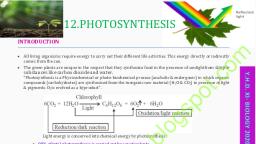
1. The organization of DNA is much more complex, in eukaryotes., 2. Histones are required for the packaging of DNA., 3. Histones are proteins that are rich in the basic, amino acid residues lysine and arginines which, carry positive charge in their side chain., 4. Eight molecule of histones (two each of H 2A,, H2B, H3 and H 4) get organized to form histone, octamer., 5. DNA is negatively charged and it is wrapped, around the positively charged histone octamer, forming a structure known as Nucleosome.
Page 10 :
6. H1 protein binds the DNA thread where it, enters and leaves the Nucleosome., 7. Under the electron microscope, nucleus shows, Chromatin network, the nucleosomes in Chromatin, are seen as ‘ Beads-on- string’., 8. Around the octamer, DNA molecule is wrapped as 1, and 3/4th turn. This DNA is called Core DNA and it, consists of about 146bp (base pairs)., 9. Adjacent Nucleosomes are linked with small, segment of DNA called Linker DNA; of about 54bp., 10. This ‘beads-on-string’ structure gets condensed into, nucleosome fiber which is coiled like a telephone, wire to make Solenoid fiber with diameter 30nm or, 300Å.
Page 12 :
• The packaging of chromatin at higher levels, need additional set of proteins that are, collectively called Non-Histone Chromosomal, (NHC) proteins., • A loosely packed region of chromatin that, stains light, is called Euchromatin and densely, packed region that stains dark is called, Heterochromatin., • Euchromatin is considered as transcriptionally, active chromatin, while Heterochromatin is, inactive.
Page 13 :
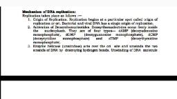
DNA REPLICATION, • The DNA molecule regulates and controls all the, activities of the cell. As a carrier of genetic, information, DNA has to perform two important, functions:A. Heterocatalytic function:- when DNA directs the, synthesis of chemical molecules other than itself,, then such functions of DNA are called heterocatalytic, functions., Eg. Synthesis of RNA(Transcription), synthesis of, protein (Translation), etc., B. Autocatalytic function:- when DNA directs the, synthesis of DNA itself, then such function of DNA is, called autocatalytic function.
Page 14 :
• The process by which DNA, duplicates itself is called, replication. Through replication,, it forms two copies that are, identical to each other., • In eukaryotic organisms,, replication of DNA takes place, only once in the cell cycle., • It occurs in the S-phase of, Interphase in the cell cycle., • DNA replicates through, semiconservative mode of, replication., • The model for Semiconservative, replication was proposed by, Watson and Crick, on the basis, of antiparallel and, complementary nature of DNA, strands.
Page 16 :
2.Point of origin or Initiation point, DNA replication begins, at certain specific sites, called ‘O’-Origin., In prokaryotes, there is, only one origin, however in Eukaryotes,, there are more than, one origin., At the point ‘O’,, enzyme Endonuclease, nicks one of the, strands of DNA,, temporarily., , Point of origin /, initiation point
Page 17 :
3. Unwinding of DNA molecule, The enzyme DNA, helicase breaks the, hydrogen bonds due to, which the strands of DNA, separate and unwind., Now the DNA molecule, appears as inverted ‘Y’shaped structure called, Replication fork., The separated strands, are prevented from, recoiling by SSBP(single, strand DNA binding, protein)
Page 18 :
4. Synthesis of new strands, Each separated strand acts as a, template or mould for the, synthesis of new complementary, strand., It takes place with the help of a, small RNA molecule called RNA, Primer., RNA Primer get associated with, the 3' end of template strand, and attracts complementary, nucleotides from surrounding, Nucleoplasm., The synthesis of new, complementary strand is, catalyzed by DNA Polymerase., The new complementary strand, is always formed in 5', 3', direction.
Page 19 :
5. Leading and lagging strand, The Template strand with free 3' end is, called Leading template and with free 5', end is called Lagging template., As both the strands of the parental DNA are, antiparallel, new strands are always formed, in 5', 3' direction., One of the newly synthesized strand, develops continuously towards replicating, fork is called leading strand., Another new strand develop, discontinuously away from the replicating, fork is called Lagging strand., DNA synthesis on lagging template takes, place in the form of small fragments, called, Okazaki fragments. Okazaki fragments are, joined by enzyme DNA Ligase., RNA primers are removed by DNA, polymerase and replaced by DNA sequence, with the help of DNA polymerase – I in, Prokaryotes and DNA polymerase – α in, Eukaryotes., Finally, DNA Gyrase enzyme forms double, helix to form daughter DNA molecules.
Page 20 :
6. Formation of daughter DNA molecule, At the end of the replication, two daughter, DNA molecules are formed., In each daughter DNA, one strand is parental, and the other one is totally newly synthesized., Thus, 50% is contributed by mother DNA., Hence, it is described as Semiconservative, replication.
Page 21 :
Experiment to confirm the Semiconservative, nature of DNA Replication, • In newly formed DNA, molecule, one strand is, old and other strand is, newly synthesized. Thus,, it is called, Semiconservative mode, of replication., • It was experimentally, proved by Matthew, Meselson and Franklin, Stahl(1958) by using, equilibrium – density –, gradient – centrifugation, technique.
Page 22 :
1. Meselson and Stahl cultured bacteria E.coli in the medium, containing 14 N(light nitrogen) and obtained equilibrium, density gradient band by using 6M CsCl 2. The position of, this band is recorded .
Page 23 :
2. E.coli cells were then, transferred to 15 N, medium (heavy, isotopic nitrogen) and, allowed to replicate, for, several generations. At, equilibrium point, density gradient band, was obtained, by using, 6M CsCl2. The position, of the band is, recorded.
Page 24 :
3. The heavy DNA ( 15N) molecule can be, distinguished from normal DNA by, centrifugation in a 6M Cesium chloride ( CsCl2), density gradient. The density gradient value of 6M, CsCl 2 and 15N DNA is almost same. Therefore,, , at the equilibrium point 15N DNA will form a, band. In this both the strands of DNA are, labelled with 15N.
Page 25 :
4. Such, , E.coli cells were then transformed to another medium, containing 14N i.e normal(light) nitrogen. After first generation,, the density gradient band for 14N 15N was obtained and its, position was recorded. After second generation, two density, gradient bands were obtained – one at 14N 15N position and, other at 14N position., 5. The position of bands after two generations clearly proved that, DNA replication is Semiconservative.
Page 26 :
PROTEIN SYNTHESIS, • Proteins are very important biomolecules., • They serve as structural components, enzymes and, hormones., • The cell needs to synthesize new protein molecules., • The process of protein synthesis includes, Trancription and Translation., • The process of copying genetic information from, one strand of DNA into a single stranded RNA, is, termed as Transcription and the process of, formation of protein from RNA is called Translation.
Page 27 :
CENTRAL DOGMA OF PROTEIN SYNTHESIS, • Double stranded DNA molecule gives rise to, mRNA which acts as a messenger to, programme the synthesis of a polypeptide, chain (protein)., • This type of unidirectional flow of information, from DNA to RNA to protein/proteins is, referred as central dogma of protein, synthesis., • It was postulated by F.H.C.Crick in 1958., Transcription, , DNA, , mRNA Translation Polypeptide
Page 28 :
The present concept of central, dogma in retroviruses or, riboviruses is given by Temin, (1970) and Baltimore(1970), DNA, , Transcription, , mRNA Translation Polypeptide, , Reverse Transcription, , Accordingly enzyme RNA dependent DNA, polymerase, synthesizes DNA from RNA.
Page 29 :
TRANSCRIPTION, • During Transcription, information of only one strand, of DNA is copied into RNA with the help of RNA, polymerase enzyme., • Transcription takes place in nucleus in Eukaryotes, whereas Translation occurs in cytoplasm., • DNA transfers information to mRNA which moves to, ribosomes., • Transcription occurs in the nucleus during G1 and G2, phases of cell cycle., • DNA has promotor and terminator sites. Transcription, starts at promotor site and stops at terminator site., • The process of Transcription in both Prokaryotes and, Eukaryotes, involves three stages viz. Initiation,, Elongation and Termination.
Page 30 :
Transcription Unit, • Each transcribed segment of DNA is called, transcription unit., • It consists of (i)Promotor (ii) The structural, gene (iii) Terminator., i. Promotor :- It is a DNA sequence that, provides binding site for enzyme RNA, polymerase. RNA polymerase binds to, specific Promotor. It is present towards 5', end i.e upstream. In prokaryotes, the, enzyme recognizes the promotor by its, sigma factor sub unit.
Page 31 :
ii. Structural genes:- Structural genes are the, segments of DNA, which carry codes for the synthesis, of proteins. The DNA strand which is used for, synthesis of RNA is called Template strand which is, oriented in 3', 5' direction. It is also called Antisense, stand while the other strand, not involved in RNA, synthesis is called Coding strand. It is oriented in, 5' 3'direction. It is also called Sense strand.
Page 32 :
iii. Terminator :- It is a small DNA, sequence which terminates the, transcription process. It is present, towards 3' end/downstream.
Page 33 :
During transcription the enzyme RNA, polymerase binds to the promotor site, and brings about the initiation of the, process.
Page 34 :
The two strands of DNA separate from each other and, according to the base sequence present on the, template strand, the complementary RNA nucleotides, are selected and joined one after the other to form the, mRNA strand (elongation). A small part of RNA, remains attached to the enzyme.
Page 35 :
• As the enzyme reaches to, the terminator region,, both the enzyme and, newly constructed RNA, falls off. This is the, termination of, transcription., • As the mRNA grows, the, transcribed region of, DNA molecule becomes, spirally coiled and, attains(regains) double, helical form.
Page 37 :
Transcription unit and the gene, • The DNA sequence coding for mRNA/tRNA or rRNA is, defined as a gene., • Cistron is a segment of DNA coding for a polypeptide., • A single structural gene in transcription unit is said to, be monocistronic where as a long segment of DNA, having set of various structural genes in one, transcription unit is referred as polycistronic., • Structural genes in eukaryotes have interrupted noncoding sequences called introns., • The coding sequences or express- sequences are, defined as exons. Only exons appear in processed, mRNA in Eukaryotes.
Page 38 :
Processing of hnRNA, • In eukaryotes, forms of, RNA transcribed from, DNA are called primary, transcripts., • Such transcripts undergo, changes called, processing or maturation, before becoming, functional., • Primary transcript if non, functional and contains, both exons and introns., • During processing only, introns are removed by, the process called, splicing.
Page 39 :
• Exons are joined in a definite sequence(order), by DNA ligase enzyme., • Heterogeneous nuclear RNA, undergoes the, process of capping and tailing., • In capping, methylated guanosine, triphosphate is added to 5' end of hnRNA., • In tailing, polyadenylation take place at 3', end., • It is the fully processed hnRNA, now called, mRNA.
Page 40 :
GENETIC CODE, • DNA is a master molecule of a cell that initiates,, guides, regulates and controls the process of, protein synthesis., • The information for the synthesis of proteins is, located in the DNA itself., • About, 20 different types of amino acids are, involved in the process of synthesis of proteins., • DNA molecule has 4 types of nitrogen bases to, identify these 20 different type of amino acids., • But how is it possible that 20 types of amino, acids are encoded by 4 types of nitrogen bases ?
Page 41 :
• According to Crick, this information is stored in the, form of coded language (Cryptogram) called genetic, code, that contains code words (Codons) each one, specifying (representing) specific amino acids., • If each codon has only one nitrogen base then there, will be 4 1 = 4 codons are possible (not enough to, code 20 different amino acids)., • If each codon has two nitrogen bases then there will, be 4 2 = 4*4=16 codons are possible (not enough to, code 20 different amino acids)., • If each codon has three nitrogen bases then there will, be 4 3 = 4*4*4=64 codons are possible (more than, enough to code 20 different amino acids)., • Hence G. Gamov (1954) suggested that in a codon,, there must be combination of three consecutive, nitrogen bases that will be sufficient to specify 20, different types of amino acids.
Page 43 :
Characteristics of Genetic code, i. Genetic code is a triplet code :, Sequence of three consecutive bases constitute, codon, which specifies one particular amino acid., In every living organism genetic code is a triplet, code., ii. Genetic code has distinct polarity :, Genetic code shows definite polarity i.e. direction., It, therefore, is always read in 5' 3' direction, and not in 3' 5' direction. Otherwise message, will change., e.g. 5' AUG 3'
Page 44 :
iii. Genetic code is non-overlapping :, code is non overlapping i.e. each single base is a part, of only one codon. Adjacent codons do not overlap. If, non-overlapping, then with 6 consecutive bases only, two amino acid molecules will be in the chain. Had it, been overlapping type, with 6 bases, there would be, 4, amino acid molecules in a chain.
Page 45 :
iv. Genetic code is commaless :- there is no gap or punctuation, mark between successive /consecutive codons., V. Genetic code has degeneracy :- usually single amino acid is, encoded by single codon. However, some amino acids are encoded, by more than one codons. E.g. Cysteine has two codons, while, Isoleucine has three codons. This is called degeneracy of the code.
Page 46 :
vi. Genetic code is universal :- By and large in all living organisms the, specific codon specifies same amino acid. E.g. codon AUG always specifies, amino acid methionine in all organisms from bacteria up to humans., vii. Genetic code is non-ambiguous :- specific amino acid is encoded by, a particular codon. Alternatively, two different amino acids will never be, encoded by the same codon.
Page 47 :
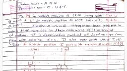
viii. Initiation codon and Termination codon :AUG is always an initiation codon in any and every, mRNA. AUG codes for amino acid methionine. Out, of 64 codons, three codons viz. UAA, UAG and UGA, are termination codons which terminate/stop the, process of elongation of polypeptide chain, as they, do not code for any amino acid., ix. Codon and Anticodon :- codon is a part of DNA, e.g. AUG is codon. It is always represented as 5' AUG 3'., Anticodon is a part of tRNA. It is always represented as, 3' UAC 5'.
Page 48 :
Mutations and Genetic Code, • Mutation is a phenomenon in, which sudden change in the DNA, sequence takes place., • It results in the change of, genotype (i.e. character)., • Along with recombination,, mutation is a raw material for, evolution as it results in, variations., • During mutation, possibility of, loss (deletion) or gain (insertion/, duplication) of a segment of DNA, results in alteration in the, chromosomes., • Mutation can also occur due to, change in a single base pair of, DNA. This is known as point, mutation. Eg. Sickle cell anaemia.
Page 49 :
• Deletion or insertion of, base pairs of DNA causes, frame – shift mutations or, deletion mutation., • Insertion or deletion of, one or two bases changes, the reading frame from, the point of insertion or, deletion., • Insertion or deletion of, three or multiples of three, bases (insert or delete), results in insertion or, deletion of amino acids, and reading frame remains, unaltered from that point, onwards.
Page 50 :
t-RNA- the adapter molecule, • Scientists considered that there has to be, a mechanism in which tRNA will read the, codon and also simultaneously binds the, amino acid as amino acid does not have, any special capacity to read the codon., So tRNA is considered as an adapter, molecule., • Cloverleaf structure of tRNA possess an, anticodon loop that has bases, complementary to the codon. It is called, anticodon., • It shows amino acid acceptor end (3', end) having unpaired CCA bases to which, amino acid binds., • For every amino acid, there is specific, tRNA. Initiator tRNA is specific to, methionine. There is no tRNA’s for stop, codons., • In the actual structure, the tRNA, molecule looks like inverted L (3, dimensional structure)
Page 51 :
Translation – protein synthesis, • Translation :- It is, the process in which, the sequence of, codons on the mRNA, strand is read/, decoded and, accordingly the, amino acids are, joined to each other, to form a, polypeptide chain, that makes protein.
Page 53 :
(B) Formation of the polypeptide chain :- it is the actual, translation process which involves following steps:, i. Initiation :It begins with the formation of, initiation complex which requires the, mRNA having codons for a, polypeptide, the smaller (30s) and, larger (50s) sub-units of ribosomes,, the initial AA1 -tRNA complex and ATP, and GTP as source of energy., The process starts with the binding of, mRNA on the smaller 30s sub-unit of, ribosome., AUG is present on mRNA which, initiates the process of protein, synthesis., Initiator tRNA binds with initiation, codon (AUG) by its anticodon (UAC), through hydrogen bonds. It carries, activated amino acid methionine (met), in Eukaryotes or formyl methionine, (f-met) in prokaryotes.
Page 54 :
Now the large subunit of, ribosome joins with the, smaller subunits, that, requires Mg ++ ions., The ribosome has three, sites namely : aminoacyl, (A) site, peptidyl site (P), and exit (E) site., The empty tRNA leaves, from E site. Only the AA 1tRNA complex binds at P, site directly while all the, other incoming tRNA, complexes get attached, first at A site and then, are shifted to P site.
Page 55 :
ii) Elongation :During this process,, activated amino acids are, added one by one to first, amino acid (methionine)., This amino acid binds, with amino acid binding, site of tRNA. This result, in formation of tRNA, amino acid complex.
Page 56 :
Addition of amino acid occurs in 3 steps cyclea) Codon recognition:-Amino acyl tRNA molecule enters, the ribosome at A-site. Anticodon binds with the codon, by hydrogen bonds., b) Amino acid on the first initiator tRNA at P-site and, amino acid on tRNA at A-site join by peptide bond. At, this time first tRNA at ‘P’ site is kicked off.
Page 57 :
c) Translocation :-The tRNA at Asite carrying a dipeptide at A-site, moves to the P-site. This process, is called translocation. In, translocation, both the subunits, of ribosome move along in, relation to tRNA and mRNA., Hence, tRNA carrying dipeptide, now gets positioned at ‘P’ site of, ribosome, making ‘A’ site vacant., At this site, then next charged, tRNA molecule carrying amino, acid will be received. During this, process, first uncharged tRNA is, discharged from E-site., This process is repeated as, amino acids are added to, polypeptide. It takes less than 0.1, second for formation of peptide, bond.
Page 58 :
iii) Termination and release of polypeptide :At the end of mRNA, there is a, stop codon (UAA/UAG/UGA)., It is exposed at the A-site. It is, not read and joined by, anticodon of any tRNA., The release factor binds to the, stop codon, thereby, terminating the translation, process. The polypeptide is, now released in the cytoplasm., Two subunits of ribosome, dissociate and last tRNA is set, free in the cytoplasm., Finally mRNA is also released, in the cytoplasm. It get, denatured by nucleases, immediately. Hence mRNA is, short lived.
Page 59 :
Regulation of gene expression, • It is a multistep process by which a, gene is regulated and its product is, synthesized. Thus, gene expression, results in the formation of a, Polypeptide., • Genes of a cell are expressed to, perform different functions. For eg., An enzyme β-galactosidase is, synthesized by E.coli. It is used for, hydrolysis of lactose into glucose and, galactose., • Certain bacteria like E.coli adapt to, their chemical environment by, synthesizing certain enzymes, depending upon the substrate, present. Such adaptive enzyme is, called inducible enzyme. A set of, genes will be switched on when, there is necessity to metabolise a, new substrate. This phenomenon is, called induction and small molecule, responsible for this, is known as, inducer. It is positive control.
Page 60 :
Structure of the operon, , • The clusters of gene with related, iii. The operators are present between, functions are called operons. They, the promoters and structural genes., usually transcribe single mRNA, The repressor protein binds to the, molecule., operator region of the operon., • In E.coli some 260 genes are grouped in, Regulatory genes are responsible, 75 different operons., for formation of repressors which, • Each operon is a unit of gene, interact with operators., expression and regulation which, includes the structural genes and their, control elements (promoters and, operators)., i. The structural genes code for, proteins, rRNA and tRNA required by, the cell., ii. Promoters are the signal sequences, in DNA that start RNA synthesis., These are the sites where the RNA, polymerases are bound during, transcription.
Page 61 :
Lac operon, • Francois Jacob and J. Monod,, proposed the classical model of Lac, operon which can explain properly, gene expression and regulation in E., coli., • Lactose or Lac operon of E.coli is, inducible operon. The operon is, switched on when a chemical inducer, – lactose is present in the medium., • The metabolism of lactose in cells, requires three enzymes- Permease, βgalactosidase and transacetylase. The, enzyme permease is needed for entry, of lactose in the cell, β-galactosidase, brings about hydrolysis of lactose into, glucose and galactose, while, transacetylase transfers an acetyl, group from acetyl Co-A to galactoside.
Page 62 :
•, , •, •, , •, , The Lac operon has promoter sites (p),, regulatory site (i) and operator site (o)., Besides this it has three structural genes, namely z, y and a. the ‘z’ gene codes for βgalactosidase, ‘y’ gene codes for, permease and ‘a’ gene codes for, transacetylase., In Lac operon a polycistronic structural, gene is regulated by common promoter, and regulatory genes., When the cell is using its normal energy, source glucose, the ‘i’ gene transcribes a, repressor mRNA, after translation of, which, a repressor protein is produced. It, binds the operator region of the operon, and prevents the RNA polymerase from, transcribing the operon. As a result βgalactosidase is not produced., In absence of glucose, if lactose is, available as energy source for the, bacteria then following events occur in, the cell. The lactose enters the cell as a, result of the activity of permease enzyme., It act as inducer and interacts with the, repressor to inactivate it., , •, , •, , As the repressor is inactivated, the RNA, polymerase can bind itself to the, operator site and transcribe the operon, to produce lac mRNA which enables, formation of all the required three, enzymes needed for lactose metabolism., this regulation of lac operon by the, repressor is an example of negative, control of transcription initiation., , (a)Inducer absent, , (b)Inducer present
Page 63 :
Genomics, • The term Genome (introduced by, H. Winker in 1920) is the total, genetic constitution of an, organism. Alternatively, it is a, complete copy of genetic, information (DNA) or one, complete set of chromosomes, (monoploid or haploid) of an, organism., • The term Genomics (term coined, by T.H. Roderick in 1986) is the, study of genomes through, Genomics study may be classified, analysis, sequencing and mapping into two types:, of genes along with the study of, a. Structural genomics: It involves, their functions., mapping,sequencing and analysis, • The sequencing of Yeast,, Drosophila and mouse genome, of genome., was done in order to facilitate, b. Functional genomics: It deals with, comparative studies between, the study of functions of all gene, humans and other organisms, sequences and their expression in, commonly used for genetic, studies, in laboratory., organisms.
Page 64 :
Application of genomics, Structural and functional genomics is used for, different purposes in the improvement of crop plant,, human health and livestock. The knowledge and, understanding acquired from genomics research can be, applied in a number of different sectors, including, medicine, biotechnology and social sciences. It helps in, the treatment of genetic disorders through gene, therapy., 1. Genomics is used in agriculture to develop, transgenic crops having more desirable characters., 2. Genetic markers developed in genomics, have, applications in forensic analysis., 3. Genomics can lead to introduce new gene in, microbes to produce enzymes, therapeutic proteins, and even biofuels.
Page 65 :
Human Genome Project, • The human genome project, was initiated in 1990. This, project was co-ordinated, by the US department of, Energy and National, institute of health., • The Human Genome, Project began in 1990 and, was completed in 2003., • The Human Genome, Project is a multinational, research project to, determine the genomic, structure of humans.
Page 66 :
The main aims of project are:I. Mapping the entire human genome at the, level of nucleotide sequences., II. To store the information collected from the, project in databases., III. To develop tools and techniques for analysis, of the data., IV. Transfer of the related technologies to the, private sectors, such as industries., V. Taking care of the legal, ethical and social, issues which may arise from project.
Page 67 :
•, •, •, , •, , •, , HGP (Human Genome Project) was closely associated with rapid development of a new, area in biology, called Bioinformatics., The work of human genome project has allowed researches to begin to understood the, blueprint in building and constructing the human genome., As researches learn more about the functions of genes and proteins, this knowledge will, have a major impact in fields like Medicine, Biotechnology and the life sciences., Therefore HGP is important., Human Genome Project was to provide a complete and accurate sequence of the 3, billion DNA base pairs that make up the humane genome and to find out the estimated, number of human genes. Now about 33000 genes have been estimated to be present in, humans., The secret of our complexity may not lie not in the number of our genes but how we use, them. It will lead to the understanding of gene structure and function in other species., Since we posses many of the genes same as flies, round worm and mice, such studies will, lead to a greater understanding of human evolution.
Page 68 :
DNA Fingerprinting, • Every individual has its, unique genetic make-up,, which may be called its, Fingerprint., • The technique developed, to identify a person with, the help of DNA restriction, analysis, is known as DNA, profiling or DNA, fingerprinting., • The technique of, fingerprinting was first, given by British geneticist,, Dr. Alec Jeffreys in 1984.
Page 69 :
• DNA fingerprinting technique, is based on identification of, nucleotide sequence present, in this wonder molecule., • About 99.9% of nucleotide, sequence in all persons, is, same. Only some short, sequences of nucleotides, differ from person to person ., , • In the population, every person, shows unusual sequences of, 20-100 base pairs, which are, repeated several times. They, are termed as Variable Number, of Tandem Repeats(VNTRs), • The length of the regions having, VNTRs is different in each, individual and hence is the key, factor in DNA Profiling.
Page 70 :
Steps involved in DNA finger printing, 1. Isolation of DNA : the DNA, must be recovered from, the cells or tissues of the, body (host). Only small, amount of tissue like, blood, hair roots, skin, etc., is required., 2. Restriction digestion : the, isolated DNA is treated, with restriction enzymes., The restriction enzymes, cut the DNA into small, fragments having variable, lengths. This phenomenon, is called Restriction, Fragment Length, polymorphism (RFLP).
Page 71 :
3. Gel electrophoresis : The, DNA samples are loaded for, agarose gel electrophoresis, under an electric influence., The DNA fragments, which, are negatively charged move, to the positive pole. The, movement of these, fragments depends on length, of the fragments. This results, in formation of bands., dsDNA splits into ssDNA by, alkali treatment., 4. Southern blotting : The, separated DNA fragments, are transferred to a nylon, membrane or a, nitrocellulose filter paper by, placing it over the gel and, soaking them with filter
Page 72 :
5. Selection of DNA Probe : A known, sequence of single- stranded DNA is, prepared. It is called DNA Probe. DNA, Probe is obtained from organisms or, prepared by cDNA preparation, method. The DNA Probe is labelled, with radioactive isotopes., 6. Hybridization :Probe DNA is added, to the nitrocellulose filter paper, containing host DNA. The singlestranded DNA probe pairs with the, complementary base sequence of the, host DNA strand. As a result DNA-DNA, hybrids are formed on the nitrocellulose, filter paper. Remaining single stranded DNA, probe fragments are washed off., 7. Photography : the nitrocellulose filter, paper is photographed on an X-ray film by, autoradiography. The film is analysed to, determine the presence of hybrid DNA.
Page 73 :
Application of DNA fingerprinting, 1. In forensic science, DNA fingerprinting is used to, solve problems of rape and some complicated, murder cases., 2. DNA finger printing is used to find out the, biological father or, mother or both, of, the child, in case of, disputed parentage., 3. DNA finger printing is, used in pedigree, analysis in cats, dogs,, horses and humans.
 Learn better on this topic