Page 1 :
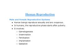
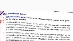
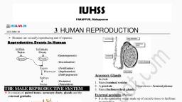
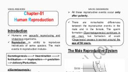
Mob No.:-99150-02300, , Manu Vatika, DAY BOARDING SR.SEC. PUNJABI MEDIUM SCHOOL,, , BUDHLADA-151502, Class:- 12th(Medical), , Sub:- Biology, , Teacher : Jaspreet kaur, , HUMAN REPRODUCTION, Basic Steps in Human Reproduction:, 1. Gametogenesis, 2. Insemination, 3. Fertilization, 4. Blastocyst development, 5. Implantation, 6. Embryo development, 7. Parturition, , 3.1: Male Reproductive System, The male reproductive system has four main parts:, 1. Testes, 2. Accessory ducts, 3. Glands, 4. External Genitalia, , Testes (singular Testis):, • Situated in the pelvic region outside the abdominal cavity within a pouch called as scrotum.
Page 2 :
• Scrotum is a small muscular sac that contains and protects the testes. It is a part of the external male genitalia and is, located behind the penis., • The lower temperature in testes is required for spermatogenesis as the normal human body temperature can lead to, mutation in the sperms., • Testis is oval in shape. It is 4-5cm long and 2-3cm wide., • Each testis has about 250 compartments called as Testicular lobules., • Each lobule contains 1-3 highly coiled seminiferous tubules., • Seminiferous tubules are the site for meiosis that leads to the formation of spermatozoa., , ➢ The inner lining of each seminiferous tubule consists of two types of cells: Spermatogonia and Sertoli cells., ➢ Spermatogonia are the immature male germ cells that undergo meiosis that leads to formation of sperms. Each, spermatogonium is diploid and contains 46 chromosomes., ➢ Sertoli cells provide nutrition to the spermatogonia., ➢ Interstitial spaces: These are the regions outside the seminiferous tubules. They contain small blood vessels, some, immunocompetent cells and interstitial cells or the Leydig cells., ➢ Leydig cells: These are the cells that synthesise and secrete testicular hormones called androgens., , Accessory Ducts:, The accessory ducts transport the sperms from the testes to the urethra for their release outside the body. There are, four accessory ducts in the male reproductive system:, • Rete Testis, • Vasa efferentia, • Epididymis, • Vas deferens, ➢ Rete testis:, These are the ducts that connect the seminiferous tubules of the testes to the vasa efferentia.
Page 3 :
➢ Vasa efferentia:, These ducts leave the testes and open into the epididymis that is located in the posterior surface of each testis., ➢ Epididymis:, It is located on the posterior surface of each testis and opens into the vas deferens., ➢ Vas deferens:, It is a duct that ascends into the abdominal cavity and loops over the urinary bladder. It receives a duct from the seminal, vesicle and opens into the urethra as ejaculatory duct., ➢ Ejaculatory duct, , : They store and transport the sperms from the testis to the outside through the urethra., ➢ Urethra, : It is a thin muscular tube. It originates from the urinary bladder. It then passes through the penis to its external opening, called as the urethral meatus., , Accessory Glands:, They are:, • Seminal vesicles, • Prostate gland, • Bulbourethral glands, These glands secrete products that mix with the sperms to nourish and protect them., • Seminal vesicles, : They contribute approximately 60-75% of the fluid in semen. The secretions are rich, in proteins, enzymes, fructose, vitamin C, phosphoryl choline and prostaglandins. The high fructose content provides nutrient, energy for the spermatozoa., • Prostate Gland:, It secretes a slightly alkaline milky fluid. This helps in the survival of sperms in the acidic vaginal environment. The, secretions also improve the motility of the sperm.s, • Bulbourethral glands:, The secretions of this gland lubricate the penis and neutralise any residual acidity in the urethra., 3.2: Female Reproductive System, The human female reproductive system is specialised to support the process of gametogenesis, ovulation, fertilisation,, pregnancy, birth and child care. The parts of the female reproductive system are:
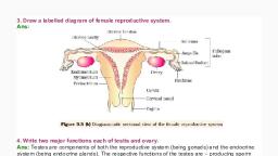
Page 4 :
1. A pair of ovaries, 2. A pair of oviducts, 3. Uterus, 4. Cervix, 5. Vagina, 6. External genitalia, 7. Mammary glands, , Ovaries:, • They are the primary female sex organs8, • They produce the female gamete- ovum, • They also produce steroid hormones (Ovarian hormones)., • 2-4cm in length and connected to the pelvic wall and uterus by ligaments., • Each ovary is covered by a thin epithelium enclosing an ovarian stroma., • Ovarian stroma- It is the matrix of the ovary and is divided into two zones, ➢ Peripheral cortex., , Accessory Ducts:, They are the oviducts, uterus and the vagina., , Oviduct (Fallopian Tube):, • Each fallopian tube is 10-12 cm long, • It extends from the periphery of each ovary to the uterus., • Infundibulum: It is the funnel shaped part closer to the ovary., • Fimbriae: They are the finger like projections at the edges of the infundibulum that aid in collecting the ovum, after ovulation., • Ampulla: It is the wider part of the oviduct after the infundibulum., • Isthmus: It is the last part of the oviduct. It has a narrow lumen and joins the uterus., , Uterus:, • It is also called as the womb, • It is shaped like an inverted pear, • It is attached to the pelvic wall by ligaments., • The uterus is the part where the embryo develops into the foetus., • The uterus opens into the vagina through a narrow cervix., • Uterine wall has three layers of tissues: perimetrium, myometrium and the endometrium.
Page 5 :
• Perimetrium: It is the thin external membranous layer., • Myometrium: It is the thick middle layer made up of smooth muscle. It exhibits strong contractions during the, delivery of the baby., • Endometrium: It is the inner glandular layer that lines the uterine cavity. It undergoes periodic changes during, the menstrual cycle., , Cervix:, • It is a narrow canal connecting the uterus to the vagina., • Cervical canal: The cavity of the cervix is called as the cervical canal., • Birth canal: The cervical canal along with the vagina forms the birth canal., , External Female Genitalia:, They consist of: mons pubis, labia majora, labia minora, hymen and clitoris, Mons pubis: It is a mass of fatty tissue covered by skin and hair Labia majora: They are fleshy folds of tissue that extend from, the mons pubis and cover the vaginal opening Labia minora:, , Hymen, : It is a membrane that partially covers the opening of the vagina. It is often torn during the first intercourse or, coitus. It can also be broken by active participation in some sports like horseback riding, cycling, etc., a sudden fall or, jolt, insertion of a vaginal tampon, etc. In some women the hymen can persist even after coitus. The presence or absence of, hymen is not a reliable indication of virginity or sexual experience., Clitoris: It is a tiny finger-like projection that lies at the junction of the labia minora above the urethral opening., , Mammary Glands:, The presence of functional mammary glands is characteristic of all female mammals., • They are paired structures of breasts that contain glandular tissue and variable amounts of fats., • Glandular tissue in each mammary gland consists of 15-20 mammary lobes., • The mammary lobes have clusters of cells called alveoli., • The cells of alveoli secrete milk., • The milk is stored in the lumen or cavities of the alveoli., • The alveoli open into mammary tubules., • The tubules of each lobe join to form a mammary duct., • Several mammary ducts join to from a wider mammary ampulla., • The mammary ampulla is connected to a lactiferous duct through which milk is sucked out., 3.3: Gametogenesis
Page 6 :
Gametogenesis is the process by the primary male and female sex organs produce the male and female gametes, respectively., , Spermatogenesis: The process by which the immature male germ cells or spermatogonia produce mature sperm cells, in the testis., , Oogenesis: The process by which the immature oogonia in the ovaries produce mature ovum., Spermatogenesis:, The process of spermatogenesis begins at puberty and proceeds as follows:, 1. The spermatogonia multiply by mitosis to increase in number. They are present in in the inner wall of seminiferous tubules., Each spermatogonium is diploid. Each spermatogonium contains 46 chromosomes., 2. Some spermatogonia called as the primary spermatocytes periodically undergo meiosis to form two equal,, haploid cells called as the secondary spermatocytes. They contain 23 chromosomes., 3. The secondary spermatocytes produce four equal haploid cells after they undergo second meiotic division., They are called as the spermatids. They contain 23 chromosomes., 4. The spermatids undergo spermiogenesis to form spermatozoa or sperms., 5. The sperm heads are embedded in the Sertoli cells., 6. They are finally released from the seminiferous tubules by the process of spermiation., , • Spermatogonia are the immature male germ cells that undergo meiotic divisions to form sperms. Each spermatogonium is, diploid and contains 46 chromosomes., • Primary spermatocytes are spermatogonia that undergo periodic meiosis to form two equal haploid cells, called as the secondary spermatocytes. The primary spermatocytes contain 46 chromosomes, • Secondary spermatocytes are haploid cells arising from the primary spermatocytes as a result of meiosis I., They contain 23 chromosomes., • Spermatids are the haploid cells that arise from the secondary spermatocytes as a result of meiosis II. They contain 23, chromosomes, • Spermiogenesis is the process by which spermatids mature to form spermatozoa., • Spermiation is the process by which mature spermatozoa are released from the seminiferous tubules., Hormones affecting spermatogenesis:, Gonadotropin releasing hormone (GnRH): It is a hormone secreted by the hypothalamus. Its levels increase, significantly at puberty. The increased levels of GnRH stimulates the release of two gonadotropins – Follicle Stimulating, Hormone (FSH) and Luteinizing Hormone (LH), from the anterior pituitary.
Page 7 :
Follicle Stimulating Hormone (FSH): FSH acts on the Sertoli cells and stimulates the release of some factors that help in the, process of spermatogenesis.Luteinizing Hormone (LH): LH acts on the Leydig cells and stimulates the synthesis and secretion, of androgens which in turn stimulate the process of spermatogenesis., , Structure of Sperm:, , • It consists of a head, neck, middle piece and a tai.l, • Plasma membrane covers the whole body of the sperm., • The head portion of the sperm contains a haploid nucleus., • The anterior portion of the head contains an acrosome., • Acrosome is a structure that is filled with digestive enzymes that help in the dissolving the membrane of, the egg cell and help in fertilization of the ovum., • Middle Piece:, It contains numerous mitochondria that produce energy for the movement of the tail. This is important for the motility of the, sperm which is essential for fertilization., • During ejaculation 200-300 million sperms are released. At least 60% of them should have normal shape and size and at, least 40% should show vigorous motility., • Semen: Semen is a milky white organic fluid released by the penis during ejaculation. It consists of the sperms and the, fluids secreted by the accessory ducts and the accessory glands such as the epididymis, vas deferens, seminal vesicles,, prostate and the bulbourethral glands., • The testicular hormones (androgens) maintain the functions of the male accessory ducts and glands, ., , Oogenesis:, The process of formation of a mature female gamete is oogenesis and it is initiated during the embryonic development
Page 8 :
stage. During this stage a couple of million gamete mother cells or oogonia are formed in the foetal ovary. No more oogonia, are formed and added after birth., • The oogonia form primary oocytes. The oogonia start the process of meiosis and get arrested at the stage of Prophase I., These cells originating from the oogonia that are arrested at the prophase I are called as primary oocytes., • Each primary oocyte gets surrounded by a layer of granulosa cells and is now called as the primary follicle., • A large number of primary follicles get degenerated between birth and puberty. At puberty, therefore, there are only about, 60,000-80,000 primary follicles in the ovary., • These remaining primary follicles get surrounded by more layers of granulosa cells as well as a new theca., They now form the secondary follicle., • The secondary follicle then gives rise to the tertiary follicle. The tertiary follicle is characterised by the presence of a fluid, filled cavity called as antrum. The theca is organised into two layers-inner theca interna and the outer theca externa. The, primary oocyte grows in size and completes the first meiotic division. It is an, unequal division forming a large secondary oocyte and a tiny first polar body., • Secondary oocyte retains much of the nutrient rich cytoplasm., • The tertiary follicle changes into the mature Graafian follicle., • The secondary oocyte forms a new layer called as the zona pellucida around it., • The Graafian follicle now ruptures to release the secondary oocyte (ovum) from the ovary by the process of ovulation.
Page 9 :
3.4: Menstrual Cycle:, • Menstrual Cycle: The reproductive cycle in the female primates like monkeys, apes and human beings is called as the, menstrual cycle. The cycle of events starting from one menstruation till the next one is called menstrual cycle. This cycle is, essential for the production of oocytes and for the preparation of uterus for pregnancy. The cycle repeats 28-35 days and, normally one egg is released per cycle, ., • Menstruation: Menstruation is the process by which blood and mucosal tissue are regularly discharged in a periodic, manner from the inner lining of the uterus through the vagina. It is also known as a period or monthly., • Menarche: Menarche is the first menstruation for a human female. It begins at puberty. The actual age for, menarche varies from person to person. Menarche signals the beginning of reproductive age in females., • Menopause: Menopause is the permanent ceasing of menstrual cycle in females due to the depletion of oocytes as a result of, aging. The age of menopause varies from person to person. The average age ofmenopause is between 45-50 years., , Menstrual cycle follows four phases:, 1. Menstrual phase, 2. Follicular phase, 3. Ovulation, 4. Luteal Phase, 1. Menstrual Phase: This is the period of menstruation when the menstrual flow occurs., • It typically lasts from 3-5 days., • There is breakdown of the endometrial lining of the uterus and its blood vessels which forms a liquid substance that comes, out of the vagina., • It occurs when the ovum released by the ovary is not fertilised., • Lack of menstruation may indicate pregnancy, • Menstruation may also be affected due to stress, poor diet, poor health, etc., 2. Follicular Phase: This is the phase of maturation of follicle, • The primary follicles in the ovary grow to form a fully mature Graafian follicle.
Page 10 :
• The endometrium of the uterus regenerates through proliferation., • These changes in ovary and uterus are induced by ovarian hormones- Luteinising Hormone (LH) and the Follicle, Stimulating Hormone (FSH)., • The levels of gonadotropins gradually increase through the follicular phase, • The increased levels of-LH and FSH gonadotropins stimulate follicular development. They also stimulate secretion of, oestrogens by the growing follicles., 3. Ovulation/Ovulatory Phase: This is the phase of release of ovum from ovary, • The levels of LH and FSH reach their peak at mid-cycle, around 14th day., • LH surge: The rapid increase in LH mid cycle leading to maximum LH level mid cycle is called as the LH surge. This induces, the rupture of the Graafian follicle and thereby the release of ovum., • Ovulation: The process by which an ovum is released by a mature Graafian follicle from the ovary. It, is a result of the LH surge which occurs mid-cycle around the 14th day., 4. Luteal Phase: This is the phase of formation of corpus luteum, • The remaining parts of the Graafian follicle after ovulation form the corpus luteum. Corpus luteum is therefore the structure, that is formed the ruptured Graafian follicle., • The corpus luteum secretes large amounts of progesterone which is essential for the maintenance of endometrium. The, endometrium is necessary for the implantation of the fertilised ovum and other, events of pregnancy., ❖ During pregnancy all events of menstrual cycle stop and there is no menstruation., ❖ In absence of pregnancy the corpus luteum degenerates. This causes breakdown of the endometrium leading, to menstruation and thus starting the new cycle, , 3.5: Fertilisation and Implantation:, • Insemination: During coitus or copulation semen is released into the vagina by the penis., • The motile sperms swim rapidly through the cervix, enter into the uterus and finally reach the ampullary region of the, fallopian tube., • The ovum released by the ovary is also transferred to the ampullary region of the fallopian tube., • Fertilisation occurs in the fallopian tube only if the ovum and sperms are simultaneously transferred into the ampullary, region of the fallopian tube., • Fertilisation: It is the fusion of the sperm and the egg. During fertilisation the sperm induces changes in the zona, pellucida layer of the ovum that block the entry of additional sperms ensuring that only one sperm can fertilise an ovum., • The secretions of the acrosome help the sperm enter the ovum through the zona pellucida and the plasma membrane., • This induces the secondary oocyte to complete meiosis. This is again an unequal division. It results in the formation of a, second polar body and a haploid ovum., • The haploid nuclei of the sperm and the ovum fuse to form a diploid zygote. The zygote contains 46 chromosomes., • The sex of the foetus is determined by the sex chromosome present in the sperm. As the female is XX the ovum will always, carry the X chromosome. Males are XY and therefore, the sperm can contain either X or Y. Therefore, half of all the sperms, carry the X chromosome and the other half carry the Y chromosome. Depending on whether the X-containing sperm or the, Y-chromosome fuses with the ovum, the zygote will be female or male., • The zygote undergoes mitotic cleavage as it moves along the isthmus of the oviduct towards the uterus. It forms 2, 4, 8 and, 16 daughter cells called as blastomeres., • Morula: The embryo with 8-16 blastomeres.
Page 11 :
• The morula continues division as it moves further along into the uterus. The blastomeres arrange in to an outer layer called, as the trophoblast. The inner cell mass is attached to the trophoblast., • The trophoblast attaches to the endometrium, • The inner cell mass differentiates to form the embryo, • The cells of the uterus divide rapidly and cover the blastocyst. The blastocyst thus embeds in the uterine wall. This is called, as implantation. This leads to pregnancy., , 3.6: Pregnancy and Embryonic Development:, • After implantation finger-like projections called as chorionic villi appear on the trophoblast., • The chorionic villi are surrounded by uterine tissue and maternal blood. It causes the chorionic villi and the uterine tissue, to become interdigitated with each other and jointly form a structural and functional unit between the developing foetus and, the maternal body called as the placenta., • Placenta supplies oxygen and nutrients to the developing embryo and removes the carbon dioxide and excretory waste from, the foetus., • The placenta is connected to the embryo through umbilical cord. The embryo transports nutrients and wastes, to and from the placenta through the umbilical cord., • Additionally the placenta functions as an endocrine gland. It produces several hormones such as human chorionic, gonadotropin (hCG), human placental lactogen (hPL), oestrogens, progesterone, etc., • Later in the pregnancy another hormone called as relaxin is produced by the ovary., • During pregnancy production of various hormones is increased. These hormones include oestrogens, progesterone, cortisol,, thyroxine, prolactin, etc. High levels of these hormones are essential for supporting foetal growth, metabolic changes in, pregnancy as well as maintenance of pregnancy., • Soon after implantation the embryo differentiates into the outer ectoderm and the inner endoderm. The mesoderm develops, soon after. These three tissues soon give rise to the tissues in the body., • The inner cell mass contains stem cells that have the ability to give rise to all the tissues and organs in the body., • Foetal, , development:, , .1 month- heart is formed., .2 month- limbs and digits are formed., .3 months- most of the major organ systems are formed., .5 months- appearance of hair on the head and the first movements of the foetus., . 6 months- body is covered with fine hair, eyelids separate, eyelashes are formed.
Page 12 :
.9 months – foetus is fully developed and ready for delivery., , 3.7: Parturition and Lactation:, .Gestation Period: The average duration of pregnancy is called as gestation period. In humans the gestation, period is 9 months., .Parturition: The process of childbirth by which the foetus is expelled or delivered due to vigorous contractions, of the uterus., .A fully developed foetus and placenta induces parturition. This causes mild uterine contractions called foetal, ejection reflex., .This triggers the release of oxytocin from the mother’s pituitary., . Oxytocin stimulates stronger uterine contractions which stimulates more production of oxytocin, .The stimulatory positive feedback reflex continues between the uterine contractions and oxytocin secretion., This leads to stronger and stronger contractions till the baby is expelled out of the uterus through the birth, canal., .Soon after the foetus is delivered the placenta is also delivered., .Lactation: The production of milk by the mammary glands of the females towards the end of the pregnancy is called, lactation, . Colostrum: The milk produced during the initial few days of lactation is called as the colostrum. It is rich in antibodies, that provide resistance and immunity to the new-born., ***************