Notes of Plus One Zoology, Zoology Notes. All Chapters - Study Material
Page 4 :
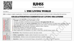
PROPERTIES OF LIVING ORGANISMS, 1., 2., 3., 4., 5., , Growth: Increase in number & mass of cells by cell division., Reproduction: Production of progeny having features similar to those of parents., Metabolism: All biochemical reactions taking place inside a living system., Cellular organization: Organisms are made up of one or more cells., Consciousness: Ability to sense their environment and respond to environmental stimuli., , DIVERSITY IN THE LIVING WORLD, Taxonomy: Study of identification, classification & nomenclature of organisms., Basic processes of taxonomy: Characterization, Identification, Classification & Nomenclature., Binomial nomenclature: Proposed by Carl Linnaeus., Botanical names are based on International Code for Botanical Nomenclature (ICBN)., Zoological names are based on International Code for Zoological Nomenclature (ICZN)., , Universal rules of Binomial nomenclature, •, , •, , •, , Scientific names are in Latin or Latinised and written in italics. When handwritten, they are, underlined., Genus name (Generic name) starts with capital letter and species name (specific epithet) starts, with small letter. E.g. Homo sapiens- Homo is the genus name and sapiens is the species name., Name of the author (in abbreviated form) appears at the end of the biological name., E.g., Mangifera indica Linn. (Linn. = Linnaeus)., , TAXONOMIC CATEGORIES, Taxonomic category (Rank), Kingdom, , Taxon (E.g.), Animalia, , ↑, Phylum/Division, , Chordata, , ↑, Class, , Mammalia, , ↑, Order, , Primata, , ↑, Family, , •, , Taxon: A unit of classification., • Kingdom: Highest category., • Species: Lowest category., , Hominidae, , ↑, Genus, , Homo, , ↑, Species, , sapiens, Vijayabheri, Malappuram Dist. Panchayat Educational Project, , 4
Page 10 :
ANIMAL TISSUES (Epithelial, Connective, Muscle & Neural Tissues), 1. Epithelial tissues (Epithelium), Types, a. Simple, (single, layered), , Location, Walls of blood vessels & alveoli., , Squamous, , Cubical (cuboidal) Ducts of glands and nephrons., Columnar, , b. Compound (Multi-layered), , Function, Diffusion., Secretion & absorption, , Lining of stomach and intestine., , Secretion & absorption, , Skin, buccal cavity, pharynx etc., , Protection., , Modification of columnar or cuboidal cells, Ciliated epithelium, - Bear cilia., - Seen in bronchioles & fallopian, tubes., - Function: move substances, over epithelium., , Glandular epithelium: For secretion., 2 types: Unicellular (E.g. Goblet cells) & Multicellular, (E.g. salivary glands)., Based on mode of secretion, glands are 2 types:, ▪ Exocrine glands: have ducts. E.g. Salivary gland., ▪ Endocrine glands: Ductless. Produce hormones., , Cell junctions: The junctions that provide link between adjacent cells. 3 types:, a. Tight junction: Stop substances from leaking across a tissue., b. Adhering junction: Perform cementing to keep neighbouring cells together., c. Gap junction: For communication b/w adjoining cells by connecting cytoplasm for rapid, transfer of ions, molecules etc., , 2. Connective tissues, Types, , Location/ Features/ Function, , Loose CT, Areolar, Loosely packed Fibres, Adipose, and fibroblasts., , Under skin. Support for epithelium., Under skin. Its cells (adipocytes) store fats., , Vijayabheri, Malappuram Dist. Panchayat Educational Project, , 10
Page 11 :
Dense CT, Compactly packed, Fibres and fibroblasts., , Dense regular, (Tendon &, Ligament), Dense irregular, Cartilage, , Specialized CT, , Bone, Blood, , Collagen fibres are regular., Tendon: Attach muscles to bones., Ligament: Attach bone to bone., Fibroblasts & fibres are irregular. Present in skin., Pliable due to chondroitin salts., Cartilage cells → chondrocytes., Non-pliable. Rich in calcium salts., Bone cells → osteocytes., Function: Protection, support, locomotion., Fluid CT. Circulation., , Areolar, tissue, , Adipose, tissue, , Dense, regular, , Dense, irregular, , Cartilage, , Bone, , Attached to bones. Striations present., Involuntary & fusiform. No striations., Visceral (Non-striated/ smooth), Found in blood vessels, stomach, intestine., Involuntary. Seen in heart., Communication junctions (intercalated, Cardiac, discs)., Skeletal (striated or voluntary), , 3. Muscle, tissues, , Muscle tissues:, a. Skeletal, b. Smooth, c. Cardiac, , 4. Neural tissue: Neural system. Made up of neurons & Neuroglia., Vijayabheri, Malappuram Dist. Panchayat Educational Project, , 11
Page 12 :
MORPHOLOGY OF COCKROACH (Periplaneta americana), Chitinous exoskeleton (cuticle)., Body has 3 regions – head, thorax and, abdomen., • Head: Antennae, compound eyes. Biting, & chewing mouth parts., Mouthparts: labrum (upper lip), 2, mandibles, 2 maxillae, hypopharynx, (tongue) & a labium (lower lip)., • Thorax: 3 parts: prothorax, mesothorax, & metathorax., 2 pairs of wings:, o Forewings (mesothoracic) or, tegmina: Opaque, dark., o Hind wings (metathoracic):, Transparent, used in flight., • Abdomen: 10 segments., Mouth parts, Differences between male & female cockroaches (Sexual dimorphism), Male, i. Wings beyond the tip of the abdomen., ii. Anal styles present, , Female, Wings up to the tip of abdomen., Absent, , ANATOMY OF COCKROACH, Digestive system: Alimentary canal, has 3 parts: foregut, mid gut &, hindgut., • Foregut: Mouth → pharynx →, oesophagus → crop (to store food), → gizzard (proventriculus- for, grinding food)., •, , Mid gut (Mesenteron): 6-8 tubules, (hepatic or gastric caecae) are seen, at the junction of foregut & mid gut., They secrete digestive juice., , •, , Hindgut: It includes ileum, colon & rectum., , Excretory system: Uricotelic. Excretory organ is Malpighian tubules., Respiratory system: Trachea with 10 pairs spiracles. Branches of tracheal tubes are, tracheoles. They carry oxygen from the air to all parts., Circulatory system: Open type., Vijayabheri, Malappuram Dist. Panchayat Educational Project, , 12
Page 13 :
Haemolymph (blood)= colourless plasma + haemocytes., Blood from sinuses (haemocoel) → ostia → heart → anterior aorta → sinuses., Nervous system: 3 ganglia in thorax and 6 in the abdomen., - The head holds only a bit of nervous system. So, if the head of cockroach is cut off, it will still, live for one week., - Supra-oesophageal ganglion (brain)., - Sense organs: Antennae, eyes, maxillary palps, labial palps, anal cerci etc., - Each compound eye has 2000 ommatidia. Cockroach can receive several images of an object, (mosaic vision)., , Reproductive system, Male reproductive system: 2 testes, seminal vesicles,, accessory glands & external genitalia (male, gonapophysis or phallomeres)., Testis → vas deferens → seminal vesicle →, ejaculatory duct → male gonopore., ▪ Seminal vesicles: To store sperms. Sperms →, spermatophores., ▪ Accessory glands: mushroom gland & phallic, gland. They nourish the sperms., Female reproductive system: 2 large ovaries,, oviducts, spermatheca, genital chamber,, Colleterial glands etc., ▪ Each ovary has 8 ovarian tubules (ovarioles), containing developing ova., ▪ Oviducts unite into a median oviduct (vagina), → genital chamber., ▪ A pair of spermatheca is present. Fertilised eggs, are encased in oothecae., Development is paurometabolous (nymphal stage- 13 times moulting)., , , , , , , , VIDEO, CLASS, , SLIDES OF, THIS CHAPTER, , QUESTION, BANK, , Vijayabheri, Malappuram Dist. Panchayat Educational Project, , 13
Page 15 :
4. NITROGEN BASES, a. Purines: Adenine (A) & Guanine (G)., b. Pyrimidines: Cytosine (C), Thymine (T) & Uracil (U)., Nitrogen base + Sugar → Nucleoside, E.g. Adenosine, Guanosine, Cytidine, Thymidine, Uridine., N. base + Sugar + Phosphate → Nucleotide, E.g. Adenylic acid,, Guanylic acid,, Cytidylic acid,, Thymidylic acid,, Uridylic acid., , BIOMACROMOLECULES (MACROMOLECULES), 1. PROTEINS, They are heteropolymer of amino acids to form polypeptides. i.e., amino acids linked by peptide, bonds., Structural levels of protein, Primary structure: Sequence of amino acids, i.e. the positional information in a protein., o Secondary structure: Polypeptide folded as helix., o Tertiary structure: Helical polypeptide, chain is further folded giving 3-D view., o Quaternary structure: Assembly of 2 or, more polypeptide or subunits. E.g., Haemoglobin., Functions of proteins:, o For growth and tissue repair., o Transport nutrients across cell membranes. E.g. GLUT-4., o Acts as intercellular ground substance. E.g. collagen., o Acts as antibodies, receptors, hormones, enzymes, pigments etc., o, , Most abundant protein in animal world: Collagen, Most abundant protein in biosphere: RuBisCO, , 2. POLYSACCHARIDES (COMPLEX CARBOHYDRATES), Polymers of sugars (monosaccharides). E.g., ▪, , Starch, Cellulose, Glycogen: Homopolymers of glucose, , ▪, , Inulin: Homopolymer of fructose., , ▪, , Chitin: Homopolymer of N-acetyl glucosamine., , Glycosidic bond: Formed b/w monosaccharides., Vijayabheri, Malappuram Dist. Panchayat Educational Project, , 15
Page 16 :
Diagrammatic representation of glycogen, , 3. NUCLEIC ACIDS (DNA & RNA), Heteropolymer of nucleotides. i.e. polynucleotide., Structure of DNA (Watson - Crick Double Helix Model), - 2 polynucleotide strands arranged antiparallelly., - Steps are formed of Nitrogen base pairs., - Nitrogen bases: A, G, C & T. Uracil absent., - A pairs with T (A=T) by 2 hydrogen bonds., G pairs with C (G≡C) by 3 hydrogen bonds., Bond b/w sugar (deoxyribose) and phosphate is, phosphodiester bond., , METABOLISM, Anabolic (Biosynthetic) pathway, Simple molecules → complex structures., It consumes energy., E.g. acetic acid → cholesterol,, Amino acids → protein., , Catabolic pathway, Complex molecules → simple structures., It releases energy (stored as ATP - energy, currency), E.g. glycolysis, respiration etc., , Metabolites (intermediate products of metabolism)., •, , Primary metabolites: Have identifiable functions in physiological processes. E.g. amino, acids, sugars, nucleic acids, lipids, vitamins etc., , •, , Secondary metabolites: They are not directly involved in growth, development or, reproduction. E.g. Pigments (Carotenoids, Anthocyanins etc), Alkaloids (Morphine,, Codeine), Terpenoids, Essential oils (Lemongrass oil etc.), Drugs (Vinblastine, curcumin, etc.), Polymers (Rubber, gums, cellulose etc.)., , ENZYMES (Biological catalysts), Almost all enzymes are proteins. Carbonic anhydrase is the fastest enzyme., Ribozymes: Nucleic acids (RNA) that behave like enzymes., Vijayabheri, Malappuram Dist. Panchayat Educational Project, , 16
Page 17 :
Nature of enzyme action (catalytic cycle): E + S → ES → EP → E + P, •, , The substrate binds to the active site of enzyme (E+S)., , •, , Formation of enzyme- substrate complex (ES)., , •, , Formation of enzyme- product complex (EP)., , •, , Release of the products from enzyme (E+P)., , Activation energy is the additional energy to start a, chemical reaction. Enzymes lower the activation energy., As a result, speed of the reaction increases., , Factors affecting enzyme activity, a) Temperature & pH: Enzymes show highest activity at optimum temperature & pH. Activity, declines below and above optimum value., , b) Concentration of substrate: With the increase in substrate concentration, the velocity of, enzyme action rises at first and reaches a maximum velocity (Vmax). This is not exceeded by, further rise in concentration because enzyme molecules are fewer than the substrate molecules., c) Presence of Inhibitor: Binding of inhibitor shuts off enzyme activity. The inhibitor closely, similar to the substrate is called competitive inhibitor. It competes with substrate for the binding, site of the enzyme., E.g. Malonate is similar to the substrate succinate. So, it inhibits succinic dehydrogenase., , Classification and nomenclature of enzymes, Oxido-reductases, / Dehydrogenases, , Catalyze oxido-reduction b/w two substrates., S reduced + S’ oxidized → S oxidized + S’ reduced, , Transferases, , Catalyze transfer of a group. S-G + S’ → S’-G + S, , Hydrolases, Lyases, , Catalyze hydrolysis of ester, ether, peptide, glycosidic, C-C, C-halide or, P-N bonds., Catalyze removal of groups leaving double bonds. X-C-C-Y → X-Y +, C=C, , Isomerases, , Catalyze inter-conversion of optical geometric or positional isomers., , Ligases, , Catalyze the linking of 2 compounds together (joining of bonds like C-O,, C-S, C-N, P-O etc.)., Vijayabheri, Malappuram Dist. Panchayat Educational Project, , 17
Page 18 :
Co-factors, •, , Non-protein component bound to enzyme to make the enzyme catalytically active., , •, , Apo-enzyme: Protein portion of enzyme., , •, , Co-factor + Apoenzyme = Holoenzyme., , •, , Co-factors are 3 types:, Prosthetic group, Co-enzymes, Metal ions, , Organic. Tightly bound to apoenzyme. E.g. Haem., Organic. Transient binding to apoenzyme. Many co-enzymes contain, vitamins. E.g. NAD and NADP contain niacin., E.g. Zn is a cofactor for Carboxypeptidase., , , , , , , , VIDEO, CLASS, , SLIDES OF, THIS CHAPTER, , QUESTION, BANK, , Vijayabheri, Malappuram Dist. Panchayat Educational Project, , 18
Page 19 :
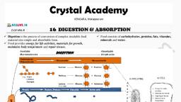
Alimentary canal:, Mouth → Buccal cavity → Pharynx → Oesophagus →, Stomach (cardiac → fundic → body → pyloric) →, Small intestine (Duodenum → Jejunum → Ileum) →, Large intestine (Caecum → Colon → Rectum) → Anus., Gastro-oesophageal sphincter: Between oesophagus, stomach., Pyloric sphincter: Between Stomach & small intestine., Anal sphincter: Guards anus., Rugae: longitudinal folds in stomach wall., Villi: Finger-like structures at the mucosa of small, intestine. It has capillary network and lacteal (lymph, vessel)., Vermiform appendix: finger-like structure arising from the caecum., , Stomach, , Transverse section of human gut, , Villi, , Human dentition is Thecodont, Heterodont & Diphyodont., • Thecodont: teeth are placed in the jaw sockets., • Heterodont: different kinds of teeth - incisors (I), canines, (C), premolars (PM) & molars (M)., • Diphyodont: teeth appear twice in lifetime - milk (deciduous), teeth (20) and permanent teeth (32)., Human dental formula (of permanent teeth):, , Digestive glands, , 2123, 2123, , 1. Salivary glands: Parotids, Submaxillary & Sublingual → Saliva, 2. Gastric glands: Secretes Gastric juice., • Mucus neck cells: Secrete mucus., • Chief (peptic) cells: Secrete pepsinogen & prorennin., • Oxyntic (parietal) cells: Secrete HCl & intrinsic factor., 3. Liver: Secretes Bile juice. Bile is transported, from liver to duodenum as follows:, Bile → hepatic duct → gallbladder → cystic duct, → common bile duct → common hepato-pancreatic, duct → duodenum., Hepato-pancreatic duct is guarded by sphincter, of Oddi., 4. Pancreas: Secretes Pancreatic juice., 5. Intestinal glands: Secretes intestinal juice (Succus entericus)., Vijayabheri, Malappuram Dist. Panchayat Educational Project, , 19
Page 20 :
Digestive glands &, Digestive enzymes/, Juice, components, 1. Salivary glands →, Salivary amylase, Saliva, (Ptyalin) & Lysozyme., Site of action:, Buccal cavity, 2. Gastric glands →, Pepsinogen, Gastric juice, Rennin, Site of action:, Gastric lipase, Stomach, No enzyme., 3. Liver → Bile, Bile pigments, Bile, Site of action: Small salts, Phospholipids, intestine, Cholesterol, , 4. Pancreas →, Pancreatic juice, Site of action: Small, intestine, , Role in digestion, , Rennin digests milk protein in infants., Chyme: Acidic pasty food formed in stomach., Emulsification of fats (fat → micelles). It, increases surface area for the action of lipase., Bile also activates lipase., , Trypsinogen, Chymotrypsinogen, Procarboxypeptidase, Pancreatic amylase, Pancreatic lipase, Nucleases, , Dipeptidase, Maltase, 5. Intestinal glands, → Intestinal juice Lactase, Sucrase, Site of action: Small Lipase, intestine, Nucleotidase, Nucleosidase, , ABSORPTION OF DIGESTED PRODUCTS, Absorption is 2 types- passive and active., a) Passive absorption (Passive transport):, Higher concentrated region to lower concentrated region. It includes osmosis & diffusion., Diffusion is 2 types:, i. Simple diffusion: E.g. glucose, amino acids, Cl-., ii. Facilitated diffusion: Diffusion with the help of carrier proteins. E.g. glucose, amino acids., b) Active absorption (Active transport):, Absorption against concentration gradient. E.g. absorption of amino acids, monosaccharides, like glucose, electrolytes like Na+ etc., Absorption of lipids:, Bile salts & phospholipids convert lipids to water-soluble droplets (micelles) → small protein, coated fat globules (chylomicrons) → transported into lacteals in the villi → lymph → blood., Vijayabheri, Malappuram Dist. Panchayat Educational Project, , 20
Page 21 :
Absorption in different parts of alimentary canal:, • Mouth: Certain drugs., • Stomach: Water, simple sugars, some drugs & alcohol., • Small intestine: All nutrients. It is the chief area of absorption due to villi, its length and, coiled nature., • Large intestine: Water, some minerals & drugs., Absorbed nutrients are incorporated into tissues (assimilation)., Undigested substances form faeces. It enters caecum through ileo-caecal valve., , DISORDERS OF DIGESTIVE SYSTEM, 1. Jaundice: Skin and eye turns yellow due to the deposition of bile pigments. It indicates liver, damage., 2. Vomiting: Ejection of stomach content through mouth., 3. Diarrhoea: Frequent elimination of watery faeces. It reduces the absorption of food., 4. Constipation: Infrequent elimination of dry stool. It is due to decreased peristalsis in colon., 5. Indigestion: Condition leading to feeling of fullness due to improper digestion., , , , , , , , VIDEO, CLASS, , SLIDES OF, THIS CHAPTER, , QUESTION, BANK, , Vijayabheri, Malappuram Dist. Panchayat Educational Project, , 21
Page 22 :
HUMAN RESPIRATORY SYSTEM, 1. Air passages, External nostrils → nasal passage → nasal chamber →, pharynx → glottis → larynx → trachea → primary, bronchi → secondary bronchi → tertiary bronchi →, bronchioles → terminal bronchioles → alveoli., Epiglottis closes glottis to prevent entry of food into, larynx., 2. Lungs, - Lungs are covered by double-layered pleura., - Alveoli (air sacs) are the structural and functional units of lungs., , MECHANISM OF BREATHING, a) Inspiration: Diaphragm & External intercostal muscles contract → thoracic volume, increases → pulmonary volume increases → intra-pulmonary pressure decreases → air into, lungs., b) Expiration: Intercostal muscles & diaphragm relax → thoracic volume decreases →, pulmonary volume decreases → intra-pulmonary pressure increases → air moves out., , Spirometer: To measure respiratory rate., Normal respiratory (breathing) rate: 12-16 times/min, Respiratory volumes/capacities, Tidal volume (TV): Volume of air inspired or expired during a normal, respiration., Inspiratory reserve volume (IRV): Additional volume of air that can inspire, by forceful inspiration., Expiratory reserve volume (ERV): Additional volume of air that can expire, by a forceful expiration., Residual volume (RV): Volume of air remaining in lungs after a forcible, expiration., Inspiratory capacity (IC): Total volume of air inspired after a normal, expiration (TV+IRV)., Expiratory capacity (EC): Total volume of air expired after a normal, inspiration (TV+ERV)., , Amount (ml), 500, 2500-3000, 1000-1100, 1100-1200, 3000-3500, 1500-1600, , Vijayabheri, Malappuram Dist. Panchayat Educational Project, , 22
Page 23 :
Respiratory volumes/capacities, Functional residual capacity (FRC): Volume of air in lungs after normal, expiration (ERV+RV)., Vital capacity (VC): Volume of air that can breathe in after a forced, expiration or Volume of air that can breathe out after a forced inspiration, (ERV + TV + IRV)., Total lung capacity (TLC): Volume of air in lungs after a maximum inspiration, (RV + ERV + TV + IRV or VC + RV)., , Amount (ml), 2100-2300, 3500-4500, 5000-6000, , GAS EXCHANGE, Gas exchange occurs by simple diffusion between 1. Alveoli & blood 2. Blood & tissues, Alveoli are the primary sites of gas exchange. Factors influencing gas exchange are:, •, , Pressure/ concentration gradient, , Atmospheric air Alveoli, pO2 (mm Hg), 159, 104, pCO2 (mm Hg), 0.3, 40, •, •, , •, , Deoxygenated blood Oxygenated blood Tissues, 40, 95, 40, 45, 40, 45, , Solubility of gases: Solubility of CO2 is 20-25 times higher than, that of O2., Thickness of diffusion membranes: 3 layers- Squamous, epithelium of alveoli + Endothelium of capillaries + Basement, substance. Its total thickness is very less → easy gas exchange., Surface area: Presence of alveoli increases surface area → gas, exchange increases., , GAS TRANSPORT (O2 TRANSPORT & CO2 TRANSPORT), 1. O2 TRANSPORT (from lungs to various tissues), a. By blood plasma (3%): O2 + plasma → tissues., b. As oxyhaemoglobin (97%): O2 + haemoglobin (Hb) → oxyhaemoglobin., , - In alveoli: pO2 high, pCO2, H+ ion and temperature are low → formation of oxyhaemoglobin., - In tissues: pO2 low, pCO2, H+ ions and temperature are high → Hb4O8 dissociates to release O2., Oxygen-haemoglobin dissociation curve, It is a sigmoid curve obtained when percentage saturation of Hb, with O2 is plotted against the pO2., It is used to study the effect of factors like pCO2, H+ concentration, etc., on binding of O2 with Hb., , 2. CO2 TRANSPORT (from tissues to lungs), a. As carbonic acid (7%): Through plasma., b. As carbamino-haemoglobin (20-25%): CO2 + Hb → carbamino-haemoglobin → lungs →, CO2 dissociates., Vijayabheri, Malappuram Dist. Panchayat Educational Project, , 23
Page 24 :
c. As bicarbonates (70%):, , In alveoli: Reaction in opposite direction., , REGULATION OF RESPIRATION, Respiratory centres in Brain:, • Respiratory rhythm centre: In medulla oblongata. It regulates respiratory rhythms., • Pneumotaxic centre: In Pons. It moderates functions of respiratory rhythm centre., • Chemosensitive area: Seen adjacent to the rhythm centre. Increase in the concentration of CO2, and H+ activates this centre., , DISORDERS OF RESPIRATORY SYSTEM, •, •, •, , Asthma: Difficulty in breathing due to inflammation of bronchi and bronchioles., Emphysema: Damage of alveolar walls → decreases respiratory surface. Major cause is, cigarette smoking., Occupational respiratory disorders: Exposure of industrial dusts → fibrosis of lungs → lung, damage., , , , , , , , VIDEO, CLASS, , SLIDES OF, THIS CHAPTER, , QUESTION, BANK, , Vijayabheri, Malappuram Dist. Panchayat Educational Project, , 24
Page 25 :
Types of circulation:, ➢ Single circulation: In fishes. Heart receives impure blood only (venous heart)., Deoxygenated blood → to heart → to gills → oxygenated blood → to body parts → deoxygenated, blood → to heart., ➢ Incomplete double circulation: In amphibians & reptiles. Left atrium gets oxygenated blood, from gills/ lungs/skin. Right atrium gets deoxygenated blood from other body parts. They get, mixed up in single ventricle. It pumps out mixed blood., ➢ Double circulation: In birds & mammals. Right atrium gets deoxygenated blood and passes to, right ventricle. Left atrium gets oxygenated blood and passes to left ventricle. The ventricles pump, it out separately., , BLOOD VASCULAR SYSTEM (HEART, BLOOD & BLOOD VESSELS), BLOOD (Plasma + Formed elements), Plasma, , •, , Constituents: Water, Plasma proteins, organic & inorganic components., , •, , Plasma proteins: Fibrinogen (blood coagulation), Globulins (act as antibodies), & Albumins (osmotic balance)., , (55%), •, , Serum= Plasma without clotting factors., , Formed elements (45%), RBC, - Biconcave non-nucleated cells., (Erythrocytes) - Count: 5 - 5.5 millions/ mm3., - Colourless nucleated cells., - Count: 6000-8000 /mm3., WBC, - Function: Part of immune system., (Leucocytes), Granulocytes:, • Neutrophils: 60-65%. Function:, Types of, Phagocytosis., WBC:, • Eosinophils: Resist infections., Granulocytes, Allergic reactions., &, • Basophils: Cause inflammation., Agranulocytes, Secrete histamine, serotonin,, heparin., PLATELETS - Count: 1.5 - 3.5 lakhs /mm3., , - Average lifespan: 120 days., - Function: CO2 and O2 transports., , Agranulocytes:, • Lymphocytes: Smallest WBC with, largest nucleus. Cause immune, responses., • Monocytes: Largest WBC. For, phagocytosis., , - Function: Blood clotting., , BLOOD COAGULATION, Clumped platelets & tissues release thrombokinase (Prothrombinase) → Thrombokinase, hydrolyses prothrombin to thrombin → Thrombin converts fibrinogen to fibrin → Fibrin trap dead, & damaged blood cells to form clot (coagulum)., Vijayabheri, Malappuram Dist. Panchayat Educational Project, , 25
Page 26 :
BLOOD GROUPS: ABO grouping, Antigens on Antibodies in, RBC, plasma, A, A, Anti-B, B, B, Anti-A, AB (Universal recipient), A, B, Nil, O (Universal donor), Nil, Anti-A & Anti-B, Blood group, , Can donate, blood to, A & AB, B & AB, AB only, A, B, AB & O, , Donor’s group, A, O, B, O, A, B, AB & O, O only, , Rh grouping based on Rhesus (Rh) factor (Antigen), - Rh+ve = presence of Rh factor and Rh-ve = absence of Rh factor. Anti-Rh antibodies are not, , naturally found., , -, , Erythroblastosis foetalis, Rh incompatibility between the Rh-ve blood of a pregnant mother and Rh+ve blood of the, foetus., During first delivery, maternal blood may be exposed to some foetal blood (Rh+ve) → Rh, antibodies in maternal blood., In her next pregnancies, Rh antibodies leak into the foetal blood (Rh+ve) and destroy the foetal, RBCs., It can be avoided by administering anti-Rh antibodies to the mother immediately after the first, delivery., , HEART, -, , It is protected by pericardium., 4 chambers- two atria and two ventricles., Walls of ventricles are thicker than that of atria., A tricuspid valve guards the opening b/w right, atrium & right ventricle., - A bicuspid (mitral) valve guards opening b/w, left atrium & left ventricle., - Opening of right ventricle to pulmonary artery, and opening of left ventricle to aorta have semilunar valves. They prevent backward flow of, blood., , CONDUCTING SYSTEM OF HEART, - It includes nodal tissues [Sino-atrial node (SAN) & Atrio-ventricular node (AVN)], bundles, & Purkinje fibres., - Fibres + bundles = Bundle of His., - SAN initiates contraction of heart by generating action potentials. So, it is called pacemaker., - Normal activities of heart are auto-regulated by nodal tissues. So, it is called myogenic heart., , CARDIAC CYCLE, Cyclic process of heart to pump blood. A cardiac cycle is completed in 0.8 second. It has 3 stages:, 1. Joint diastole: Relaxed state of all chambers. Blood from pulmonary vein and vena cava, flows into left & right ventricles through left and right atria. Semilunar valves are closed at, this stage., Vijayabheri, Malappuram Dist. Panchayat Educational Project, , 26
Page 27 :
Atrial systole: Contraction of atria due to action potential from SAN. This increases the flow, of blood into the ventricles., 3. Ventricular systole: Action potential from SAN → AVN→ AV bundle → bundle of His →, ventricular musculature. As a result, ventricles contract (ventricular systole). So semilunar, valves open and deoxygenated blood enters the pulmonary artery from right ventricle and, oxygenated blood enters the aorta from left ventricle., • One heartbeat = a cardiac cycle. So, normal heartbeat: 70-75 times/min., • Stroke volume: Volume of blood pumped out by each ventricle during a cardiac cycle. It is, about 70 ml., • Cardiac output: Stroke volume x heart rate (70 x 72). It is about 5000 ml. Cardiac output of, an athlete is very high., • Heart sounds: First sound (lub) is due to closure of tricuspid and bicuspid valves. Second sound, (dub) is due to closure of the semilunar valves. One heartbeat = a lub + a dub., 2., , ELECTROCARDIOGRAPH (ECG), Instrument used to get electrocardiogram (graphical representation of electrical activity of the, heart). ECG consists of the following waves:, o P-wave: Represents excitation (depolarization) of, atria during atrial systole., o QRS-complex:, Represents depolarization of, ventricles (Ventricular systole)., o T-wave: Represents the repolarisation of ventricles., Deviation in ECG indicates abnormality of heart. So, ECG has great clinical significance., , DOUBLE CIRCULATION, Blood flows through the heart twice for completing its circuit. It includes:, 1. Pulmonary circulation: b/w lungs and heart., Deoxygenated blood from right ventricle → to pulmonary, artery → to lungs → oxygenated blood → to pulmonary, veins → left atrium., Systemic circulation: b/w heart and various body parts., Oxygenated blood from left ventricle → to aorta → arteries, → arterioles → capillaries → tissues → deoxygenated, blood from tissues → venules → veins → vena cava → to, right atrium., Systemic circulation provides nutrients, O2 and other, substances to the tissues and takes CO2 and other harmful substances away for elimination., • Hepatic portal system: It is a system which includes hepatic portal vein that carries blood from, intestine to the liver., • Coronary circulatory system: It is a system of coronary vessels that circulate blood to and from, cardiac musculature., 2., , LYMPHATIC SYSTEM (Lymph, Lymph vessels & Lymph nodes), - The fluid filtered into tissues from blood through capillaries is called tissue fluid., - When tissue fluid enters lymphatic system, it is called lymph. It drains back to major veins., , Vijayabheri, Malappuram Dist. Panchayat Educational Project, , 27
Page 28 :
Functions of lymph, ▪, , Exchange nutrients, gases, etc. b/w blood and cells., ▪ Transports digested fats, hormones etc., ▪ Lymphocytes in lymph gives immunity., , DISORDERS OF CIRCULATORY SYSTEM, •, •, •, •, •, •, , Hypertension (High Blood Pressure): Normal BP is 120/80 mm Hg. BP >140/90 is called, hypertension. It causes heart diseases and affects vital organs., Coronary Artery Disease (CAD) or Atherosclerosis: Ca, fat, cholesterol etc. are deposited in, coronary arteries. So lumen of arteries becomes narrow affecting blood flow., Angina pectoris: An acute chest pain due to O2 deficiency to heart muscles. It occurs due to, improper blood flow., Heart Failure: Inability of heart to pump blood enough to meet the needs of the body., Congestion of the lungs., Cardiac arrest: Heart stops beating., Heart attack: Sudden damage of heart muscle due to inadequate blood supply., , , , , , , , VIDEO, CLASS, , SLIDES OF, THIS CHAPTER, , QUESTION, BANK, , Vijayabheri, Malappuram Dist. Panchayat Educational Project, , 28
Page 29 :
Types of Excretion, •, •, •, , Ammonotelism: Excretion of Ammonia (NH3). E.g. Aquatic invertebrates, bony fishes,, aquatic amphibians., Ureotelism: Excretion of urea. E.g. Cartilaginous fishes, amphibians, mammals., Uricotelism: Excretion of uric acid. E.g. Insects, terrestrial reptiles & birds., , HUMAN EXCRETORY SYSTEM, Includes kidneys, ureters, urinary bladder & urethra., Kidney: Covered by renal capsule. Blood vessels, nerves, ureter, etc. enter the kidney through hilum. Hilum leads to renal pelvis, with renal calyces., A kidney has outer cortex & inner medulla., Medulla consists of medullary pyramids., Nephron: Structural & functional units of kidney., A nephron has 2 parts:, o Glomerulus: Capillary network., o Renal tubule: Bowman’s, capsule + Proximal convoluted, tubule (PCT) + Henle’s loop +, Distal convoluted tubule (DCT)., Glomerulus + Bowman’s, capsule = Malpighian body., Types of nephrons: Cortical, (85%) & Juxtamedullary (15%)., , URINE FORMATION, 1. Glomerular filtration (ultrafiltration), - In glomerulus, blood is filtered through 3 layers- endothelium of glomerulus, basement, membrane & epithelium of Bowman’s capsule (contains podocytes that form filtration slits)., - Glomerular filtration rate (GFR): Amount of glomerular filtrate formed per minute., - Normal GFR = 125 ml/minute, i.e., 180 litres/day., 2. Reabsorption, - 99% of filtrate is reabsorbed by the renal tubules. So volume of urine released is 1.5 litre., - PCT reabsorbs most of the nutrients and 70-80% electrolytes & water., - In DCT: Conditional reabsorption of Na+ & water., - Collecting duct reabsorbs water to concentrate urine., 3. Tubular Secretion, - PCT, DCT & Collecting duct maintain ionic and acid-base balance (pH) of body fluids by, selective secretion of H+, K+ & NH3 into filtrate and absorption of HCO3- from it., Vijayabheri, Malappuram Dist. Panchayat Educational Project, , 29
Page 30 :
Mechanism of concentration of the filtrate, - Henle’s loop & vasa recta help to concentrate the urine., - Flow of filtrate in the 2 limbs of Henle’s loop and the flow of blood through the 2 limbs of, vasa recta are in opposite directions. This is called Counter current mechanism., - Thus osmolarity increases from cortex (300 mOsmolL-1) to the inner medullary interstitium, (1200 mOsmolL-1)., - This gradient is caused by NaCl & urea., - DCT & collecting duct produce urine four times concentrated than the initial filtrate formed., , MICTURITION, - It is the release of urine., - Filled urinary bladder → stretch receptors send impulses to CNS → motor messages → urinary, bladder contracts → micturition (1 - 1.5 litre urine (25-30 g urea) per day)., - Micturition reflex: Neural mechanism of micturition., - Urine analysis helps in clinical diagnosis of metabolic disorders and malfunctioning of the, kidney., , REGULATION OF THE KIDNEY FUNCTION, Regulation by ADH (vasopressin): Hypothalamus → release ADH → water reabsorption, from DCT & collecting duct. → prevents diuresis → increases body fluid volume., ADH → constricts blood vessels → BP increases → increases glomerular blood flow & GFR., 2. Regulation by JGA (Renin-Angiotensin mechanism): JGA (Juxta glomerular apparatus) is, a region in nephron. Fall in glomerular blood flow/GFR → activates JG cells → renin., Renin converts angiotensinogen → angiotensin I → angiotensin II (vasoconstrictor) →, increases glomerular BP & GFR., Angiotensin II → adrenal cortex → Aldosterone → reabsorption of Na+ & water from distal, parts of tubule., 3. Regulation by ANF: When blood flow increases, the atria of heart releases Atrial, Natriuretic Factor (ANF). It causes vasodilation → BP decreases., 1., , DISORDERS OF EXCRETORY SYSTEM, •, •, •, , Uremia: Accumulation of urea in blood due to kidney failure., Renal calculi: Stone of crystallized salts (oxalates, etc.) formed within the kidney., Glomerulonephritis: Inflammation of glomeruli., , Hemodialysis: Process of removal of urea in patients with uremia., Blood from artery (+ anticoagulant like heparin) → dialyzing unit → cellophane tube → passage, of molecules → Purified blood (+ anti-heparin) → pumped back through a vein., , Kidney transplantation: For acute renal failures., Receiving kidney from a close relative minimizes rejection by immune system of host., , , , , , , , VIDEO, CLASS, , SLIDES OF, THIS CHAPTER, , QUESTION, BANK, , Vijayabheri, Malappuram Dist. Panchayat Educational Project 30
Page 31 :
Types of, movement in, human being, , Types of, muscles, , - Amoeboid movement: By pseudopodia. E.g. Macrophages & leucocytes., - Ciliary movement: By cilia. E.g. trachea & oviducts., - Muscular movement: By muscles. E.g. limbs., Skeletal (striated), Attached to skeleton, Striations present, Voluntary, Rich blood supply, , Visceral (Non-striated), In visceral organs, Absent, Involuntary, Poor blood supply, , Cardiac, In heart wall, Present, Involuntary, Rich blood supply, , STRUCTURE OF STRIATED MUSCLE, , Skeletal muscle is made of muscle bundles (fascicles) containing muscle fibres., Muscle fibres are lined by plasma membrane (sarcolemma) enclosing the sarcoplasm., Each muscle fibre contains myofilaments (myofibrils)., Each myofibril has dark (Anisotropic or A-band) and light striations (Isotropic or I-band)., I-bands: Contain actin filaments. It is bisected by a dark band (Z-line). Region b/w 2 Z-lines is, called sarcomere (functional units of muscle contraction)., - A-bands: Contain actin & myosin. Its light middle region (H zone) is formed of myosin. Hzone has a dark line (M-line) at the centre., -, , Structure of contractile proteins, - An actin filament is made of 2 filamentous (F) actins., - F-actin is a polymer of Globular (G) actins., , Actin contains 2 other proteins (tropomyosin & troponin)., Troponin has 3 subunits., Each myosin filament is a polymer of Meromyosins., A meromyosin has 2 parts: Heavy meromyosin or HMM or cross arm & Light meromyosin, or LMM (tail)., - Head of cross arm is an ATPase enzyme., , -, , MECHANISM OF MUSCLE CONTRACTION, Sliding filament theory: Contraction of a muscle fibre occurs by the sliding of thin filaments over, thick filaments., Vijayabheri, Malappuram Dist. Panchayat Educational Project, , 31
Page 32 :
Steps:, Impulse from CNS → neuromuscular junction → Synaptic vesicles release Acetylcholine →, action potential in sarcolemma → release of Ca2+ from sarcoplasmic cisternae → Ca binds, troponin → unmask the active sites for myosin → energy from ATP hydrolysis → myosin head, binds to active sites on actin to form cross bridge → actin filaments pull towards centre of Aband → H-zone disappears → Z- line is pulled inwards → contraction of sarcomere., , ▪ Repeated, , activation of muscles → anaerobic breakdown of glycogen → accumulation of lactic, acid → muscle fatigue., Red (Aerobic) muscles, White muscle, Red colour due to more myoglobin, White colour due to less myoglobin, More mitochondria, Less mitochondria, Aerobic metabolism, Anaerobic metabolism, Slow & sustained contraction, Fast contraction for short period, , HUMAN SKELETAL SYSTEM (206 bones & few cartilages), AXIAL SKELETON (80 bones), Bones of head (29), • Skull (22): Cranial bones (8), + Facial bones (14)., • Ear ossicles (2x3=6), • Hyoid bone (1), Skull articulates with First, vertebra (atlas) by 2 occipital, condyles (dicondylic skull)., , Vertebral column, 26 vertebrae:, • Cervical (7), • Thoracic (12), • Lumbar (5), • Sacrum (1), • Coccyx (1), , Ribs (12 pairs), Sternum (1), th, • True ribs (1-7 pairs):, Breast, Connected to sternum by, bone, Hyaline cartilage., • False or vertebro-chondral, ribs (8-10th pairs): Join to the, 7th rib., • Floating ribs (11-12th pairs):, Not connected with sternum or, other ribs., , Vijayabheri, Malappuram Dist. Panchayat Educational Project 32
Page 33 :
APPENDICULAR SKELETON (126 bones), • Bones of forelimbs (2x30 =60): Humerus (1),, , Radius (1), Ulna (1), Carpals (wrist bones-8),, Metacarpals (Palm bones-5) & Phalanges (14)., • Bones of hind limbs (2x30 = 60): Femur (thigh, , bone -1), Patella (knee cap-1), Tibia (1), Fibula, (1), Tarsals (ankle bones-7), Metatarsals (5) &, Phalanges (14)., • Pectoral girdles (2x2=4): Clavicle (collar bones-, , 2) & Scapula (shoulder blades-2)., Clavicle articulates with acromion (elevated, ridge) of Scapula., Acromion has glenoid cavity into which humerus articulates to form shoulder joint., • Pelvic girdles (2x1=2 coxal bones): Formed by the fusion of Ilium, Ischium & Pubis., , At the point of fusion of Ilium, Ischium and Pubis is a cavity (Acetabulum) to which the, thigh bone articulates., The 2 halves of the pelvic girdle meet ventrally to form pubic symphisis., , JOINTS, 3 types:, • Fibrous (immovable) joints: E.g. sutures of skull., • Cartilaginous (Slightly movable) joints: E.g. Joints between the adjacent vertebrae., • Synovial (movable) joints: Have a fluid filled synovial cavity between 2 bones., Synovial Joints, Ball & socket, Hinge joint, Pivot joint, Gliding joint, Saddle joint, , Examples, b/w humerus & pectoral girdle., Knee joint, b/w atlas & axis., b/w carpals, b/w carpal & metacarpal of thumb, , DISORDERS OF MUSCULAR & SKELETAL SYSTEMS, •, •, •, •, •, •, , Myasthenia gravis: An auto immune disorder that affects neuromuscular junction. Fatigue and, paralysis of muscles., Muscular dystrophy: Progressive degeneration of skeletal muscles due to genetic disorder., Tetany: Rapid muscle spasm due to low Ca2+ in body fluid., Arthritis: Inflammation of joints., Osteoporosis: Age-related disorder. Decreased bone mass causing fractures. Low level of, estrogen is a common cause., Gout: Inflammation of joints due to accumulation of uric acid crystals., , , , , , , , VIDEO, CLASS, , SLIDES OF, THIS CHAPTER, , QUESTION, BANK, , Vijayabheri, Malappuram Dist. Panchayat Educational Project 33
Page 34 :
Neurons are structural & functional, unit of neural system., Neuron is made of Cell body,, Dendron (branches: dendrites) &, Axon (branches: axonites with, synaptic knob)., Types of Neurons, •, •, •, , Unipolar: No Dendron. Found in embryo., Bipolar: One dendron. Found in retina., Multipolar: 2 or more dendrons. Most, common., , •, , •, , Types of axons, Myelinated: Schwann cells with a myelin, sheath around axon. Gaps b/w adjacent, myelin sheaths are called nodes of Ranvier., Non-myelinated: Schwann cells without, myelin sheath., , GENERATION & CONDUCTION OF NERVE IMPULSES, - In a resting neuron, axoplasm has more K+ & –vely charged proteins and less Na+. The fluid, outside the axon contains low K+ & high Na+. This forms an ionic gradient., - Na+ - K+ pump maintains the ionic gradients. It transports 3 Na+ outwards for 2 K+ into cell., So membrane is polarized (outer +ve, inner -ve)., - Resting potential: Potential difference of resting, membrane., - If a stimulus is given, membrane at site A, becomes permeable to Na+ causing rapid influx, of Na+ and reversal of polarity (depolarization)., - Electrical potential difference during, depolarization is called action potential (a nerve, impulse)., - Action potential is conducted as current flow from site A to B and the process is repeated along, the axon., , Synapse:, Functional junction between two neurons. It is 2 types:, 1. Electrical synapse: In this, membranes of pre- & postsynaptic neurons are nearest. So impulse transmission is, same as along an axon. Impulse transmission is faster than in, chemical synapse., 2. Chemical synapse: It has synaptic cleft between pre- &, post-synaptic neuron. Presynaptic regions have Synaptic, knob. They contain synaptic vesicles filled with neurotransmitters., Impulse transmission in chemical synapse:, Impulse reaches at axon terminal → synaptic vesicles bind on plasma membrane → release of, neurotransmitter → diffuses across synaptic cleft → combine with receptors on post synaptic, membrane → opening of ion channels allowing entry of ions → action potential., Vijayabheri, Malappuram Dist. Panchayat Educational Project 34
Page 35 :
HUMAN NERVOUS (NEURAL) SYSTEM (CNS & PNS), CENTRAL NEURAL SYSTEM (CNS), A. BRAIN, - Covered by cranial meninges (outer dura mater,, , middle arachnoid mater and inner pia mater., - The subarachnoid space is filled with, , cerebrospinal fluid (CSF)., , Parts of Brain and their Functions:, , Forebrain, , Midbrain, , Motor area, Controls voluntary movements of muscles., Cerebrum, (2 Cerebral, Sensory area Controls functioning of sense organs, hemispheres with, Association, For intersensory association, memory &, cerebral cortex), area, communication, Coordinating centre (relay station) for sensory and motor, Thalamus, impulses., Regulates temperature, thirst, hunger & emotions. Secretes, Hypothalamus, hormones., Corpora, 4-lobed structure- Lobes of visual reflex (2) & Lobes of, quadrigemina, auditory reflex (2), Cerebellum, , Co-ordinates muscular activities and body equilibrium., , Co-ordinates the activities of eye and ear and regulates, respiration., Medulla, Controls respiration, cardiovascular reflexes, gastric, oblongata, secretions., Limbic system: Amygdala + hippocampus + hypothalamus etc. It regulates sexual behavior,, motivations, emotions., Hindbrain, , Pons varoli, , B. SPINAL CORD, - Conduction of impulses to and from the brain., - Centre of spinal reflexes., , Peripheral neural system (PNS)- Cranial & spinal nerves:, Somatic neural, system, Autonomic neural, system (ANS):, Transmits impulses, from CNS to, involuntary organs, & smooth muscles., , Relays impulses from the CNS to skeletal muscles., Sympathetic, nerves, , Prepares body to cope with emergencies, stresses &, dangers. It increases heartbeat, breathing rate,, constricts arteries and elevates BP., , Parasympathetic Returns the body to a resting state and slows down, nerves, heartbeat, dilates arteries, lowers BP etc., , Vijayabheri, Malappuram Dist. Panchayat Educational Project, , 35
Page 36 :
REFLEX ACTION, It is the rapid, involuntary and unconscious actions of body in response to a stimulus. E.g., , , , Withdrawal of hand when it touches a hot object., Knee jerk phenomenon., , Reflex arc: Pathway of impulses in a, reflex action., Receptor organ → Sensory (afferent), neuron → Interneuron in CNS →, Motor (efferent) neuron → effector, organ (muscle/gland)., , SENSE ORGANS, EYE, - Eyeball has 3 layers: outer Sclera, middle, -, , -, , Choroid (blood vessels) & inner Retina., Cornea: Anterior transparent portion of sclera., Iris: Pigmented portion of the eye., Pupil: Central opening of iris. It regulates the, amount of light entering the eye., Retina has 3 layers- inner ganglion cells middle, bipolar cells & outer photoreceptor cells., Photoreceptor cells are 2 types: rods and, cones. They contain photosensitive proteins (photopigments)., Cone cells: For Daylight (photopic) vision & colour vision. They contain photopsin., Rod cells: For Twilight (scotopic) vision. They contain rhodopsin. It contains a derivative of, Vitamin A., Blind spot: Region where there are no photoreceptor cells., Macula lutea: Yellowish pigmented spot with a central pit (fovea). In fovea, only cones are, seen. Greatest visual acuity., Space between cornea & lens (aqueous chamber) contains aqueous humor., Space between lens and retina (vitreous chamber) contains vitreous humor., , Mechanism of vision, - Light from object → cornea & lens → focus on retina → dissociation of retinal & opsin →, membrane permeability changes → potential difference in photoreceptor cells → action, potential in ganglion cells → impulses to optic nerves → brain → vision., , EAR, 3 divisions: External ear, middle ear & inner ear., •, , External ear: Consists of pinna & auditory meatus (ear canal) & tympanic membrane (ear, drum)., , Vijayabheri, Malappuram Dist. Panchayat Educational Project, , 36
Page 39 :
•, , Thyroid, Largest endocrine, gland, , Parathyroid, Thymus, , Adrenal, cortex, Adrenals, , Adrenal, medulla, , Pancreas (Islets of, Langerhans), , Testis (male gonad), , Ovary (female gonad), , Regulation of basal metabolic rate (BMR)., • Physical, mental and sexual, development., Thyroxin, • Support RBC formation., (tetraiodothyronine,, • Control metabolism of carbohydrates,, T4 ) &, proteins & fats, Triiodothyronine, Hypothyroidism (Goiter): deficiency of, (T3), iodine., Hyperthyroidism: Exophthalmic goiter, (Grave’s disease)., Thyrocalcitonin, Lowers blood calcium (hypocalcaemic, (TCT), hormone)., Parathyroid hormone Increases Ca2+ level in blood, (PTH), (hypercalcaemic hormone)., Gives immunity. Thymus is degenerated in, Thymosins, old people. So, thymosin production, decreases and immunity become weak., - For carbohydrate metabolism., Glucocorticoids, - Stimulate gluconeogenesis, lipolysis and, (mainly cortisol), proteolysis., Deficiency: Addison’s disease., Mineralocorticoids, Regulate water & ionic balance, osmotic, (mainly aldosterone), pressure and BP., Androgenic, For growth of axial hair, pubic hair and, corticoids, facial hair., Secreted during stress emergency, Adrenaline &, situations so called emergency hormones, Noradrenaline, (hormones of Fight or Flight)., • For glycogenolysis to increase blood sugar, Glucagon (from α, (hyperglycemia)., cells), • Stimulates gluconeogenesis., Hyperglycemic factor, • Reduces the cellular glucose uptake., • Decreases blood glucose (hypoglycemia)., Insulin (from β cells), • Glycogenesis., Hypoglycemic factor, Deficiency: Diabetes mellitus., • Maturation of accessory sex organs &, Androgens (mainly, sex characters., testosterone), • For spermatogenesis., • Development of secondary sex organs, & sex characters., Estrogen, • Development of ovarian follicles &, mammary glands., • Supports pregnancy., Progesterone, • Development of mammary alveoli &, milk secretion., , Vijayabheri, Malappuram Dist. Panchayat Educational Project, , 39
Page 40 :
HORMONES OF HEART, KIDNEY & GASTROINTESTINAL TRACT, Atrial wall, JGA of, kidney, , Atrial natriuretic factor (ANF), , Dilation of blood vessels to reduce the BP., , Erythropoietin, , Stimulates erythropoiesis., , Stimulates secretion of HCl & pepsinogen, from gastric glands., For secretion of water & bicarbonate ions, Secretin, from exocrine pancreas., For secretion of bile from gall bladder and, Cholecystokinin (CCK), pancreatic enzymes., Gastric inhibitory peptide (GIP) Inhibits gastric secretion., Gastrin, , Gastrointestinal, tract, , MECHANISM OF HORMONE ACTION, •, •, , A hormone binds to its specific receptor in target tissue to form hormone-receptor complex., It leads to biochemical changes in target tissue and thereby regulates metabolism and, physiological functions., , Interaction of hormones (e.g. protein hormone,, FSH) with Membrane-bound receptors., , Interaction of hormones (e.g. steroid hormones,, iodothyronines) with Intracellular receptors, , , , , , , , VIDEO, CLASS, , SLIDES OF, THIS CHAPTER, , QUESTION, BANK, , Vijayabheri, Malappuram Dist. Panchayat Educational Project, , 40
 Learn better on this topic