Page 1 :
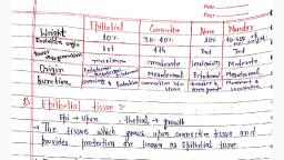
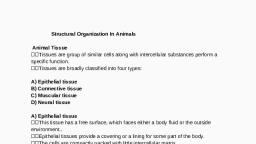
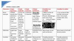
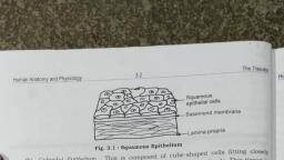
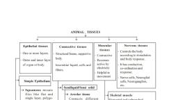
Chapter 7: Structural organization in animals, There are many cells in the body of an organism. Many organisms like plants and animals are made up of cells, while all of them form complex bodies. In order to do so these cells form a group- ‘The cells having same origin which may or may not have same structure and function form a tissue.’, Group of cells- Tissue, Group of tissues- Organs, Group of organs- Organ system, The body of an organism divides the functions performed by different parts of the body, therefore such arrangement is called ‘Division of labour’., A tissue was first seen in Coelentrates., Study of tissues- Histology., Term histology was first given by-Meyer, Term tissue proposed by- Bichat, Types of Animal Tissues, Based on Location, structure and function animal tissues are of following types:-, Epithelium- Forms the covering of both inside and outside parts of the body., Connective- Most amount of these tissues are present in an animal’s body. They connect different parts of the body., Muscular- It forms the muscles of the body, which help in locomotion and movement of the body., Nervous- It forms the brain, spinal cord etc. of the body along with the nerves of the body. It helps in transmission of messages., All of these are tissues in vertebrate animals, invertebrates do not have such arrangement., Epithelial Tissues, Epithelium is made up of two words- ‘Epi’-Upon, ‘Thelia’-Growth. Means a tissue which grows upon another tissue., When an embryo develops, epithelium is the first developed tissue where the outer lining of the body from the outside as well as inside is formed by Epithelium. Maximum amount of regeneration capacity is found in this tissue while least amount of it is found in nervous tissues., Term Epithelium introduced by Ruysch., The characteristic features of epithelial tissue are as follows, (i) The cells are compactly arranged., (ii) Intercellular spaces are narrow, 20-30 nm wide., (iii) Adjacent cells are held together by intercellular junctions., (iv) The epithelial tissue lies on a thin, non-cellular basement membrane., (v) Blood vessels are not present in the epithelial tissue., (vi) Materials are exchanged by diffusion between epithelial cells and the blood vessels of the connective tissues across the basement membrane., (vii) Nerve endings may penetrate the epithelial tissues., Epithelium tissue is present upon Connective tissue, in between which an acellular basement membrane is present which is highly permeable and secreted by both Epithelium and connective tissue. Since no blood capillaries or lymph vessels reach the epithelial layer therefore, these cells depend on connective tissue for their nutrients., Junctions Between Epithelial Cells, The common intercellular junctions may include tight junctions, gap junctions, desmosomes, intercellular bridges and interdigitations., Tight Junctions, The plasma membrane in the apical region of the adjacent epithelial cells become tightly packed together. These junctions check the flow of materials between the cells and are called occluding junctions., Adhering Junctions, Facilitate the cementing process so as to keep the * neighboring cells together. They include desmosomes and hemidesmosomes., Desmosomes, These are thick and strong junctions. They serve, anchoring functions., Gap Junctions, They are fine hydrophilic channels between adjacent cells formed with the help of protein cylinders called connexin. They help in chemical exchange between adjacent cells and hence are called communicating junctions., Surfaces of Epithelial Cells, Epithelial tissue is the only tissue which originates from all the three germinal layers i.e. Ectoderm, Mesoderm, and Endoderm., An epithelial cell typically has four surfaces:, The apical surface, which faces the lumen; also known as the ‘luminal border’, The two lateral surfaces, through which one epithelial cell communicates with the epithelial cells on each side, The basolateral surface, which is opposite to the apical surface and faces the basement membrane, Modification of Epithelial Cells, Cilia, villi, and microvilli, As mentioned above, the cilia are present on the columnar epithelium and produce a to-and-from motion to entrap the particles and move them in a specific direction parallel to the surface of the epithelium. Therefore, they are, in addition to other mechanical barriers, included in the primary defense of the body. The villi and microvilli are present on the absorptive surfaces as they increase the surface area for absorption without increasing the size of the epithelium., Types of Epithelial Tissue, On the basis of different layers present in the tissues, they are of two types-, Simple Epithelium, Compound Epithelium, Simple Epithelium, Simple Squamous Epithelium, Looks like Pavement on the floor., Single layer of flattened cells with irregular boundaries., Nuclei are at the centre of cells., Examples- alveoli of the lungs, Bowman’s capsule, Henle’s loop of uriniferous tubules, pericardial cavity, abdominal cavity, lining of various components of blood vascular system like endothelium of blood and lymph vessels, inner lining of heart wall,, Functions Simple squamous epithelium performs the function of protection, excretion, gas exchange and secretion of coelomic fluid., Simple Cuboidal Epithelium, Single layer of cube like cells., Single nucleus in a cell., This epithelium is also called Germinal epithelium because it is present in the lining of primary sexual organs like testis and ovary., Free end of the cells may be smooth or bearing Microvilli., This microvilli or brush borders increases the surface area of the tissue to multiple times., Example-vesicles of thyroid gland, acini of pancreas, sweat glands, iris, choroid, ciliary body of eye, tubular parts of nephrons in kidneys, ovaries seminiferous tubules of testes, PCT of nephron, neck of nephron and collecting duct etc., Functions The main function of this epithelium is protection, secretion, absorption, excretion and gamete formation., Simple Columnar Epithelium, Composed of a single layer of tall and slender cells., A single oval or elongated nucleus is situated near the base of the cell., Some of its cells produce mucus, called goblet cells., The brush border columnar epithelium occurs in the gall bladder. The mucus secreting goblet cells are found in the lining layer of stomach, intestine, respiratory tract, etc., Examples- Bile duct, liver, gall bladder, Stomach, colon, rectum, Duodenum, ileum, fallopian tube and brain ventricles., Functions The simple columnar epithelium helps in secretion, absorption and protection to the components of most glandular epithelia., Simple Ciliated Epithelium, If the columnar or cuboidal cells bear cilia on their free surface they are called ciliated epithelium., They move particles or mucus in a specific direction over the epithelium., The epithelium lies over a basement membrane. The number of cilia varies in different cellular forms., Examples- In sensory cells of internal ear, a cilium accompanies number of stereocilia., This epithelium is of two types, i.e., ciliated columnar and ciliated cuboidal., (a) Simple Ciliated Columnar Epithelium- It possess columnar cells that possess cilia over their free surface. It occurs in respiratory tract, fallopian tubes, parts of uterus and cervix, the different tubules of testes, etc., (b) Simple Ciliated Cuboidal Epithelium- It has cuboidal or cubical cells that bear cilia on their free surface. It occurs in many parts of ependyma of nervous system and parts of uriniferous tubules., Functions- The epithelium maintains a flow of mucus, liquid or suspended particles constantly in one direction., In the oviducts, cilia helps in the movement of egg towards the uterus. In respiratory tract, cilia helps to push the mucus towards the pharynx. In nephrons of kidney, cilia keep the urine moving., In nervous system, cilia of the ventricles of the brain and central canal of the spinal cord helps in the circulation of cerebrospinal fluid., Pseudostratified Epithelium, The epithelium is one-cell thick, but appears 2-layered because all the cells do not reach the free surface., The cells are attached to the basement membrance, hence they are called pseudostratified. The mucus secreting goblet cells also occur in this epithelium., This epithelium is of two types, (a) Pseudostratified Columnar Epithelium- It has columnar cells without cilia. It lines the respiratory epithelium of nasal cavity, like parotid salivary glands and the urethra of human male., (b) Pseudostratified Ciliated Columnar Epithelium- It has columnar cells. The tall cells bear cilia at the free surfaces and the short cells are without cilia., The epithelium lines the trachea and large bronchi. The movements of its cilia push the mucus laden with dust particles and bacteria towards the larynx., Functions- The pseudostratified epithelium helps in protection, movement of secretions from glands, urine and semen in urethra and mucus loaded with dust particles and bacteria in trachea., Compound Epithelium, Stratified Compound Epithelia, The stratified epithelia consist of many layer of cells., On the basis of the shape of the cells present in the superficial layers, the stratified epithelia are of four types-, Stratified Squamous Epithelium- The cells in the basal (deepest) layer are columnar or cuboidal with oval nuclei., It is called germinative layer. The cells in this region divide by mitosis to form new cells., The stratified squamous epithelium is further sub-divided as two main types, i.e., keratinised and non-keratinised., Keratinised Stratified Squamous Epithelium-. These layers of flat, dead cells are called stratum corneum or homy layer., The heavy deposits of keratin in the dead superficial cells makes the epithelium impervious to water and highly resistant to mechanical abrasions. This epithelium forms the epidermis of the skin in land vertebrates., Non-keratinised Stratified Squamous Epithelium- It provides moderate protection against abrasion., It lines the buccal cavity, pharynx, oesophagus, lower part of urethra, vocal cord, vagina, cervix (lower part of uterus), conjuctiva, cornea of eye and inner surface of eyelids., Stratified Cuboidal Epithelium- It has outer layer of cuboidal cells and basal layer of columnar cells., It forms the epidermis of fishes and many urodeles (tailed amphibians like salamanders). It also lines the sweat gland ducts and larger salivary and pancreatic ducts., Stratified Columnar Ciliated Epithelium- Its outer layer consists of ciliated columnar cells and basal layer of columnar cells., It lines the larynx and upper part of the soft palate., Stratified Columnar Epithelium- It consists of columnar cells in both superficial and basal layers., It covers the epiglottis and lines mammary gland ducts and parts of urethra., Transitional Compound Epithelium, The epithelium consists of more than one layer of cells, but is much thinner and more stretchable than the stratified epithelium., It contains cuboidal cells at the base, two or three layers of large polygonal or pear-shaped cells in the middle and a superficial layer of large, broad, rectangular or oval cells., The transitional epithelium lines the inner surface of urinary bladder, ureter and renal pelvis., They have thick membrane with thin regions that fold when the bladder contracts., Glandular Epithelia:, Glands are an organised collection of secretory epithelial cells., Most glands are formed during development by proliferation of epithelial cells so that they project into the underlying connective tissue., Some glands retain their continuity with the surface via a duct and are known as EXOCRINE GLANDS. They secrete Enzymes., Exocrine glands secrete substances through ducts or tubes. Saliva, mucus, earwax, oil, milk, digestive enzymes, and other cell products are secreted by these glands., Other glands lose this direct continuity with the surface when their ducts degenerate during development. These glands are known as ENDOCRINE glands. These secrete Hormones., Endocrine: They are devoid of ducts (ductless glands). Their byproducts are known as hormones, and they are directly secreted into the fluid that surrounds the gland., Heterocrine /Mixed glands- They secrete both Enzymes and hormones. Example- Pancreas and liver.