Page 1 :
JOHNSTONE HIGHER SECONDARY SCHOOL, Class – 11 (Biology) 2020-2021, , UNIT - V, CHAPTER - 19, EXCRETORY PRODUCTS AND THEIR ELIMINATION, Day-1 Class (Respective diagrams/figures should be mentioned from the textbook), , •, •, •, , •, •, , Excretion is the removal of metabolic waste products from the animal body to regulate, the composition of body fluids and tissues., Osmoregulation is a process that regulates the body’s salt and water concentration., Ammonia, urea and uric acid are the major forms of nitrogenous wastes excreted by, animals. These substances get accumulated in the animal body either by metabolic, activities or by other means like excess ingestion., Ammonia is the most toxic form, followed by urea and uric acid. It requires large, amount of water for its elimination., Uric acid, being least toxic, can be removed with a minimum loss of water., , Types of Excretion, •, •, , •, , •, , Depending upon the nature of excretory product, animals exhibit different processes of, nitrogenous excretion. They are as follow, (i) Ammonotelism: the process to eliminate ammonia is known as ammonotelism. The, organism that excrete ammonia are called ammonotelic. Many bony fishes, aquatic, amphibians and aquatic insects are ammonotelic in nature. Ammonia, as it is readily, soluble, is generally excreted by diffusion across body surfaces or through gill surfaces, (in fish) as ammonium ions. Kidneys do not play any significant role in its removal., (ii) Ureotelism: The process of excreting urea is called ureotelism. Mammals, many, terrestrial amphibians and marine fishes mainly excrete urea. Ammonia produced by, metabolism is converted into urea (in the liver) and release into the blood, which is, filtered and excreted out by the kidneys., (iii) Uricotelism: The process of excreting uric acid is called uricotelism. Uric acid, being, the least toxic nitrogenous waste can be removed with a minimum loss of water from, the animal body. Thus, it is excreted in the form of pellet or paste (i.e., semi-solid form)., , 1, , Johnstone Higher Secondary School | Class 11 Biology Notes by Dr. Dishma M
Page 2 :
•, , Normally, the animals which live in dry conditions exhibit uricotelism. Examples of, uricotelic animals are Reptiles, birds, land snails and insects., Some animals perform dual excretion, i.e., two modes of excretion. For example,, Earthworms excrete ammonia when sufficient water is available, while it excretes urea, in drier surroundings. Other examples are Xenopus (African toad), crocodiles, etc., , Different types of Excretory Organs, •, , •, , •, , •, •, , Different animal groups have a variety of excretory structures (organs) to, perform the process of excretion. In most of the invertebrates, these structures, are simple tubular form, whereas, vertebrates have complex tubular organs, called kidneys., Protonephridia or flame cells are excretory structures in Platyhelminthes,, rotifers, some annelids and the Cephalochordate – Amphioxus. Protonephridia, are primarly concerned with ionic and fluid volume regulation, i.e.,, osmoregulation., Nephridia are the tubular excretory structures of earthworms and other, annelids. It helps to remove nitrogenous wastes and maintain a fluid and ionic, balance., Malphigian tubules are the excretory structures of most of the insects including, cockroaches., Antennal glands or green glands are the excretory structures of crustaceans like, prawn., , Human Excretory System, •, , •, , 2, , Human excretory system consists, of a pair of kidneys, a pair of, ureters, a urinary bladder and a, urethra., (i) Kidney: These are reddish, brown, bean-shaped structures, located below the diaphragm on, the left and right sides. The right, kidney is lower and smaller than, the left kidney because the liver, , Johnstone Higher Secondary School | Class 11 Biology Notes by Dr. Dishma M
Page 3 :
takes up much space of the right side. Each kidney of an adult human measures, 10-12 cm in length, 5-7 cm in width, 2-3 cm in thickness with an average weight, of 120-170 gm., Structure of Kidney: The outer surface of each kidney is convex and inner, surface is concave, where it has a notch called hilum, through which ureter,, blood vessels and nerves, enter. the supply of, blood occurs, i.e., renal, artery and renal vein,, pass in and out of the, kidneys along with the, ureter and the nerve, supply of kidney. Kidney, is covered by three, layers:, renal, fascia, (outermost), the adipose, layer and then renal, capsule (innermost layer). These coverings protect the kidneys from external, shocks and injuries. Inner to the renal capsule, a kidney is divided into two, regions: an outer cortex and inner medulla. The medulla is divided into a number, of conical areas (medullary pyramids) projecting into calyces (sing.: calyx). The, cortex spreads in between medullary pyramids as renal columns called columns, of Bertini. The medullary pyramids are connected with minor calyces which in, turn lead into major calyces. The major calyces open into a funnel shaped, structure, the renal pelvis which in turn leads into the ureter., •, , •, , •, , 3, , (ii) Ureters: The ureter of each kidney leaves from the renal pelvis in the hilus, region and opens into the urinary bladder. They carry urine from the kidneys to, the urinary bladder., (iii) Urinary bladder: It is somewhat pear shaped, muscular, sac-like structure., The urinary bladdr has a triangular area, the trigone, between the three, openings through which the ureters enter the bladder and one opening through, which the urethra leaves the bladder. The urinary bladder stores urine, temporarily., (iv)Urethra: It is a canal like structure which extends from the neck of the, bladder and leads to exterior. In female, urethra is short and carries only urine., In male, urethra is much longer and carries both urine and semen., , Johnstone Higher Secondary School | Class 11 Biology Notes by Dr. Dishma M
Page 4 :
•, , Structure of Nephron: Nephrons are structural and functional units of kidneys., Each kidney contains about one million nephrons. Each nephron consists of two, parts, i.e., the Malpighian, corpuscle or renal corpuscle and, the renal tubule., (i), , Malpighian Corpuscle or, Renal, Corpuscle:, It, comprises glomerulus and, Bowman’s capsule., (a) Glomerulus It is a tuft, of capillaries. Blood enters, the glomerulus through an, afferent arteriole and, leaves it through an efferent arteriole. Glomerulus filtration takes place, in the glomerulus., (b) Bowman’s Capsule (Glomerular capsule) It is a double walled cup-like, structure that surrounds the glomerulus. The two layers of the Bowman’s, capsule are outer parietal layer and inner visceral layer. The parietal layer, consists of squamous cells. The visceral layer surrounds the glomerulus, and is composed of special type of cells, podocytes., , (ii), , Renal Tubules: It consists of proximal convulated tubule (PCT) and, Henle’s loop (Loop of Henle)., (a)Proximal Convoluted Tubule (PCT): The Bowman’s capsule leads into, the PCT, a highly coiled network which is situated in the cortical region of, the kidney., (b) Henle’s Loop: It is a hairpin shaped and starts at the end of PCT and, its major part lies in the medulla. It consists of a descending limb that, ends into the medulla and an ascending limb that extends back from the, medulla into the cortex. The ascending limb continues as another highly, coiled tubular region called distal convulated tubule (DCT). The DCTs of, many nephrons open into a straight tube called collecting duct, many of, which converge and open into the renal pelvis through medullary, pyramids in the calyces., , 4, , Johnstone Higher Secondary School | Class 11 Biology Notes by Dr. Dishma M
Page 5 :
UNIT - V, CHAPTER - 19, EXCRETORY PRODUCTS AND THEIR ELIMINATION, Day-2 Class (Respective diagrams/figures should be mentioned from the textbook), , Types of Nephrons, Based on the location in the kidney, nephrons are of following two types, (i) Cortical Nephrons: They form about 85% of total nephrons. In this nephrons,, the loop of Henle is too short and extends only very little into the medulla i.e., lie in, the renal cortex. Such nephrons are called cortical nephrons. Their glomeruli lie in, the outer cortex. Vasa recta is absent or highly reduced., (ii) Juxtamedullary Nephrons: In some of the nephrons (about 15%), the loop of, Henle is very long and runs deep into the medulla. These nephrons are called, juxtamedullary nephrons. Their glomeruli lie close to the inner margin of the cortex., They are associated with vasa recta., , Urine Formation, It involves three main processes namely. Glomerular filtration, reabsorption and, secretion., Glomerular Filtration: The first step of urine formation is the filtration of blood,, which is carried out by the glomerulus and is called glomerular filtration., Kidneys filter about 1100-1200 ml of blood per minute, which constitute roughly, 1/5th of the blood pumped out by each ventricle of the heart in a minute., The glomerular capillary blood pressure causes filtration of blood through three, layers, i.e., (i) the endothelium of glomerular blood vessels, (ii) the epithelium of, Bowman’s capsule, (iii) a basement membrane between the above mentioned two, layers. The podocytes (epithelial cells of Bowman’s capsule) are arranged in such a, manner so, as to leave some minute spaces called filtration slits or slit pores., Blood is filtered so finely through these membrane, that almost all the constituents, of the plasma except the proteins pass onto the lumen of the Bowman’s capsule., , 5, , Johnstone Higher Secondary School | Class 11 Biology Notes by Dr. Dishma M
Page 6 :
Selective Reabsorption: This is the second step in the formation of urine from, filtrate. The urine released is 1.5 L as compared to the volume of the filtrate formed, per day (180 L) which suggests that nearly 99% of the filtrate is reabsorbed by the, renal tubules. Thus, the process is called reabsorption. The tubular epithelial cells, in different segments of nephron perform this either by active or passive, transports. Substance like glucose and amino acids etc., are reabsorbed by active, transport whereas nitrogenous wastes by passive transport. Reabsorption of water, also occurs passively., Tubular Secretion: Certain chemicals in the blood that are not removed by filtration, from the glomerular capillaries are removed by this process of tubular secretion. It, helps in the maintenance of ionic and acid-base balance of body fluids by removing, chemicals like K+, H+, ammonia., , Glomerular filtration rate: The amount of the filtrate formed by the kidneys per minute, is called glomerular filtration rate (GFR). It is about 125ml/minute, i.e., 180 litres per, day., , Juxtaglomerular apparatus (JGA): JGA is a special sensitive region formed by cellular, modifications in the distal convoluted tubule and the afferent arteriole at the location of, their contact. GFR is regulated by one of the efficient mechanism carried out by, Juxtaglomerular Apparatus (JGA). A fall in the GFR can activate the JG cells to release, rennin which can stimulate the glomerular blood flow and thereby the GFR back in, normal., , 6, , Johnstone Higher Secondary School | Class 11 Biology Notes by Dr. Dishma M
Page 7 :
Functions of the Tubules, 1. Proximal Convoluted Tubule (PCT):, i. PCT is lined by simple cuboidal brush-border epithelium which increase the, surface area available for reabsorption., ii. Nearly all of the essential nutrients, 70-80% of electrolytes and water are, reabsorbed., iii. PCT helps to maintain the pH and ionic balance of the body fluids by selective, secretion of the hydrogen ions, ammonia and potassium ions into the filtrate and, by absorption of HCO3- ions from it., 2. Henle’s Loop, i) It plays an important role in maintaining the high osmolarity of medullary, interstitial fluid., ii) The descending Limb of Loop of Henle is permeable to water but almost, impermeable to electrolytes. This concentrates the filtrate as it moves down., iii) The ascending Limb of Loop of Henle is impermeable to water but permeable to, electrolytes. Therefore, as the concentrated filtrate pass upward, it gets diluted due, to the passage of electrolytes to the medullary fluid., 3. Distal Convoluted Tubule (DCT), i) Conditional reabsorption of sodium ions and water takes place in this segment., ii) DCT is also capable of reabsorption of HCO3– and selective secretion of hydrogen, and potassium ions and NH3 to maintain the pH and sodium potassium balance in, blood., 4. Collecting Duct, i) Large amount of water could be reabsorbed from this region to produce a, concentrated urine., ii) It allows passage of small amounts of urea into the medullary interstitium to, maintain the osmolarity., iii) It also plays an important role in the maintenance of pH and ionic balance of, blood by the selective secretion of H+ and K+ ions., , 7, , Johnstone Higher Secondary School | Class 11 Biology Notes by Dr. Dishma M
Page 8 :
Counter Current Mechanism, (Mechanism of concentrated, urine) Mammals have the, ability to produce concentrated, urine (hypertonic urine). This, ability is due to the presence of, counter-current, mechanism., Two, counter, current, mechanism occurs inside the, kidney. (i) The flow of filtrate in, the limbs of Henle’s loop is in, opposite directions and thus,, forms a counter current. (ii) The, flow of blood in the two limbs, of vasa recta also occur in the, counter current pattern. The, proximity between the loop of Henle’s and vasa recta, as well as the counter, current mechanism in them help in maintaining an increasing osmolarity towards, the inner medullary interstitial fluid, i.e., from 300 mOsmolL-1 in the cortex to about, 1200 mOsmolL-1 in the inner medulla. This gradient is mainly caused by NaCl and, urea. NaCl is transported by the ascending limb of loop of Henle which is exchanged, with the descending limb of vasa recta. NaCl is returned to the medullary, interstitium by the ascending part of vasa recta. Similarly, small amounts of urea, enter the thin segment of the ascending limb of loop of Henl which is transported, back to the medullary interstitium by the collecting tubule., The counter current mechanism helps in the maintenance of a concentration, gradient in the medullary interstitium.Presence of such gradient helps in an easy, passage of water from the collecting tubule resulting in the formation of, concentrated urine (filtrate), i.e., nearly four times concentrated than the initial, filtrate formed., The overall function of counter current mechanism is to concentrate sodium, chloride in the interstitial fluid and thereby cause water to diffuse out of the, collecting tubule and concentrate the urine., , 8, , Johnstone Higher Secondary School | Class 11 Biology Notes by Dr. Dishma M
Page 9 :
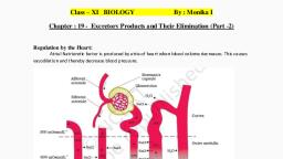
UNIT - V, CHAPTER - 19, EXCRETORY PRODUCTS AND THEIR ELIMINATION, Day-3 Class (Respective diagrams/figures should be mentioned from the textbook), Regulation of Kidney Functions, Three main parts are involved in the kidney regulation - hypothalamus, JGA, (Juxtaglomerular Apparatus) and the heart., (i) Regulation by the Hypothalamus Excessive loss of fluid from the body activates, osmoreceptors, which stimulate the hypothalamus of the brain to release, Antidiuretic hormone (ADH) or vasopressin from the neurohypophysis. ADH, facilitates water reabsorption from posterior parts of tubule. An increase in body, fluid volume can switch off the osmoreceptors and suppresses the ADH release to, complete the feedback. ADH also causes constrictory effects on blood vessels., This causes an increase in blood pressure, which in turn increase the glomerular, blood flow and thereby the GFR (Glomerular Filtration Rate)., (ii) Regulation by the Juxtaglomerular Apparatus (JGA) or Renin-Angiotensin, mechanism As the glomerular blood pressure/glomerular blood flow /GFR, decreases, the cells of the JGA release the enzyme renin. Renin converts, angiotensinogen in blood to Angiotensin I and Angiotensin II. Angiostensin II is a, vasoconstrictor which increases the glomerular blood pressure and thereby, GFR. It, also activates the adrenal cortex to release aldosterone. Aldosterone causes, reabsorption of Na+ and water from the distal parts of the tubule. This also leads to, an increase in blood pressure and GFR., (iii) Regulation by the Heart Atrial Natriuretic Factor (ANF) produced by the atria of, heart can cause vasodilation (dilation of blood vessels) and thereby, decrease the, blood pressure. ANF mechanism acts as a check on the renin-angiotensin, mechanism., Micturition, The process of release of urine from the urinary bladder is called micturition and, the neural mechanism causing it is called the micturition reflex., Mechanism Urine is produced and drained continuously by the nephron into the, urinary bladder where it is stored till a voluntary signal is given by the Central, Nervous System (CNS). As urine collects, this signal is initiated by the stretching of, the urinary bladder. The stretch receptors on the walls of the bladder send signals, to the (CNS). The CNS passes on motor messages to initiate the contraction of, , 9, , Johnstone Higher Secondary School | Class 11 Biology Notes by Dr. Dishma M
Page 10 :
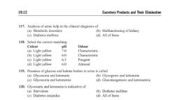
smooth muscles of the bladder and simultaneously relaxation of the urethral, sphincter causing the release of urine., Analysis of urine helps in clinical diagnosis of many metabolic disorders as well as, malfunctioning of the kidney. For example, presence of glucose (glycosuria) and, ketone bodies (ketonuria) in urine are indicative of diabetes mellitus and presence, of protein, blood and pus in the urine is called proteinuria, haematuria and pyuria, respectively., Role of Other Organs in Excretion, Other than the kidneys, there are some accessory excretory organs that help in the, elimination, of, excretory, wastes., They, are, as, follows:, 1. Lungs - About 18 L of CO2 per hour and 400 mL of water per day are eliminated, by human lungs., 2. Liver - It secretes bile containing substances like bilirubin, biliverdin, cholesterol,, degraded steroid hormones, certain vitamins and drugs., 3. Skin - The sweat and sebaceous glands in the skin can eliminate certain, substances through their secretions., (i) Sweat Glands – it secretes sweat., (ii) Sebaceous Glands (oil glands) – It eliminates sterols, waxes and hydrocarbons, through the sebum., Disorders of the Excretory System, Malfunctioning of kidneys can lead to several disorders of the excretory system., Some of these are as follows, (i) Uremia - It is the presence of an excessive amount of urea in the blood. Urea is, highly harmful as it poisons the cells at high concentration and may lead to kidney, failure., (ii) Kidney Failure (renal failure) – It is characterized by deterioration of functioning, of the kidneys which results in accumulation of toxic nitrogenous wastes like urea, leading to uraemia., (iii) Renal Calculi It is the formation of stone or insoluble mass of crystallised salts, (calcium, magnesium, phosphates and oxalates etc.), formed within the kidney., (iv) Glomerulonephritis - It is the inflammation of glomeruli of kidney., , 10, , Johnstone Higher Secondary School | Class 11 Biology Notes by Dr. Dishma M
Page 11 :
Haemodialysis, Artificial kidney (haemodialyser) is a machine that is used to filter the blood (to, remove urea and other nitrogenous wastes) of a person, whose kidneys are, damaged. The process is called haemodialysis., , 11, , Johnstone Higher Secondary School | Class 11 Biology Notes by Dr. Dishma M