Page 1 :
EXCRETORY PRODUCTS AND THEIR. TION 293 ©, , (PCT). A hairpin shaped Henle’s loop is the Afferent arteriole, next part of the tubule which has a, descending and an ascending limb. The, ascending limb continues as another highly, coiled tubular region called distal, convoluted tubule (DCT). The DCTs of many, nephrons open into a straight tube called, collecting duct, many of which converge and, open into the renal pelvis through medullary, pyramids in the calyces., , The Malpighian corpuscle, PCT and DCT, of the nephron are situated in the cortical, region of the kidney whereas the loop of Henle, dips into the medulla. In majority of, nephrons, the loop of Henle is too short and, extends only very little into the medulla. Such, nephrons are called cortical nephrons. In, some of the nephrons, the loop of Henle is, very long and runs deep into the medulla., These nephrons are called juxta medullary, nephrons., , The efferent arteriole emerging from the glomerulus forms a fine, capillary network around the renal tubule called the peritubular, capillaries. A minute vessel of this network runs parallel to the Henle’s, loop forming a ‘U’ shaped vasa recta. Vasa recta is absent or highly, reduced in cortical gsphrons., , Efferent, arteriole, , Bowman’s, capsule, , , , , Proximal, convoluted tubule, , Figure 19.4 Malpighian body (renal corpuscle), , , , Urine formation involves three main processes namely, glomerular, tration, reabsorption and secretion, that takes place in different parts of, , The first step in urine formation is the filtration of blood, which is carried, out by the glomerulus and is called glomerular filtration. On an average,, 1100-1200 ml of blood is filtered by the kidneys per minute which constitute, roughly 1/5" of the blood pumped out by each ventricle of the heart ina, minute. The glomerular capillary blood pressure causes filtration of blood, through 3 layers, i.e., the endothelium of glomerular blood vessels, the, epithelium of Bowman's capsule and a basement membrane between these, two layers. The epithelial cells of Bowman's capsule called podocytes are, arranged in an intricate manner so as to leave some minute spaces called, filtration slits or slit pores. Blood is filtered so finely through these, membranes, that almost all the constituents of the plasma except the, proteins pass onto the lumen of the Bowman's capsule. Therefore, it is, considered as a process of ultra filtration.
Page 2 :
294, , Bio.ocy, , The amount of the filtrate formed by the kidneys per minute is called, glomerular filtration rate (GFR). GFR in a healthy individual is, approximately 125 ml/minute, i.e., 180 litres per day !, , The kidneys have built-in mechanisms for the regulation of glomerular, filtration rate. One such efficient mechanism is carried out by juxta, glomerular apparatus (JGA). JGA is a special sensitive region formed by, cellular modifications in the distal convoluted tubule and the afferent, arteriole at the location of their contact. A fall in GFR can activate the JG, cells to release renin which can stimulate the glomerular blood flow and, thereby the GFR back to normal., , A comparison of the volume of the filtrate formed per day (180 litres, per day) with that of the urine released (1.5 litres), suggest that nearly 99, per cent of the filtrate has to be reabsorbed by the renal tubules. This, process is called reabsorption. The tubular epithelial cells in different, segments of nephron perform this either by active or passive mechanisms., For example, substances like glucose, amino acids, Na‘, etc., in the filtrate, are reabsorbed actively whereas the nitrogenous wastes are absorbed by, passive transport. Reabsorption of water also occurs passively in the initial, segments of the nephron (Figure 19.5)., , During urine formation, the tubular cells secrete substances like H’*,, K* and ammonia into the filtrate. Tubular secretion is also an important, step in urine formation as it helps in the maintenance of ionic and acid, base balance of body fluids., , 19.3 FuNcTION OF THE TUBULES, , Proximal Convoluted Tubule (PCT): PCT is lined by simple cuboidal, brush border epithelium which increases the surface area for reabsorption., Nearly all of the essential nutrients, and 70-80 per cent of electrolytes, and water are reabsorbed by this segment. PCT also helps to maintain, the pH and ionic balance of the body fluids by selective secretion of, hydrogen ions, ammonia and potassium ions into the filtrate and by, absorption of HCO, from it., , Henle’s Loop: Reabsorption is minimum in its ascending limb., However, this region plays a significant role in the maintenance of high, osmolarity of medullary interstitial fluid. The descending limb of loop of, Henle is permeable to water but almost impermeable to electrolytes. This, concentrates the filtrate as it moves down. The ascending limb is, impermeable to water but allows transport of electrolytes actively or, passively. Therefore, as the concentrated filtrate pass upward, it gets, diluted due to the passage of electrolytes to the medullary fluid., , Distal Convoluted Tubule (DCT): Conditional reabsorption of Na*, and water takes place in this segment. DCT is also capable of reabsorption, of HCO, and selective secretion of hydrogen and potassium ions and, NH, to maintain the pH and sodium-potassium balance in blood.
Page 3 :
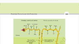
EXcCRETORY PRODUCTS AND THEIR ELIMINATION, , Proximal convoluted tubule, , NaCl Nutrients, , , , Cortex, Descending, limb of loop of, Henle, H,O, Medulla, , 295, , Distal convoluted tubule, , , , , , , , , , , nac1 #20 HCO,, , Thick segment of, ascending limb, , , , tT Nacl, Collecting, , duct, }—Thin segment of, ascending limb, t—> Nacl, i+—— Urea TO, , , , Figure 19.5 Reabsorption and secretion of major substances at different parts of, the nephron (Arrows indicate direction of movement of materials.), , Collecting Duct: This long duct extends from the cortex of the kidney, to the inner parts of the medulla. Large amounts of water could be, reabsorbed from this region to produce a concentrated urine. This segment, allows passage of small amounts of urea into the medullary interstitium, to keep up the osmolarity. It also plays a role in the maintenance of pH, and ionic balance of blood by the selective secretion of H* and K* ions, , (Figure 19.5)., , [Od nn, , Mammals have the ability to produce a concentrated urine. The Henle’s, loop and vasa recta play a significant role in this. The flow of filtrate in, the two limbs of Henle’s loop is in opposite directions and thus forms a, counter current. The flow of blood through the two limbs of vasa recta is