Page 1 :
Hosocns, , 19.1 Human, Excretory, System, , 19.2 Urine Formation, , 19.3 Function of the, Tubules, , 19.4 Mechanismof, , Concentration of, , the Filtrate, , 19.5 Regulation of, Kidney Function, , 19.6 Micturition, , 19.7 Role of other, Organs in, Excretion, , 19.8 Disorders of the, Excretory, System, , , , , XCRETORY Propucts AND, THEIR Evimanation, , Animals accumulate ammonia, urea, uric acid, carbon dioxide, water, and ions like Na*, Kt, Cr, phosphate, sulphate, etc., either by metabolic, activities or by other means like excess ingestion. These substances have, to be removed totally or partially. In this chapter, you will learn the, mechanisms of elimination of these substances with special emphasis on, common nitrogenous wastes. Ammonia, urea and uric acid are the major, forms of nitrogenous wastes excreted by the animals. Ammonia is the, most toxic form and requires large amount of water for its elimination,, whereas uric acid, being the least toxic, can be removed with a minimum, loss of water., , The process of excreting ammonia is Ammonotelism. Many bony fishes,, aquatic amphibians and aquatic insects are ammonotelic in nature., Ammonia, as it is readily soluble, is generally excreted by diffusion across, body surfaces or through gill surfaces (in fish) as ammonium ions. Kidneys, do not play any significant role in its removal. Terrestrial adaptation, necessitated the production of lesser toxic nitrogenous wastes like urea, and uric acid for conservation of water. Mammals, many terrestrial, amphibians and marine fishes mainly excrete urea and are called ureotelic, animals. Ammonia produced by metabolism is converted into urea in the, liver of these animals and released into the blood which is filtered and, excreted out by the kidneys. Some amount of urea may be retained in the, kidney matrix of some of these animals to maintain a desired osmolarity., Reptiles, birds, land snails and insects excrete nitrogenous wastes as uric, acid in the form of pellet or paste with a minimum loss of water and are, called uricotelic animals., , 2020-21
Page 2 :
Asurvey of animal kingdom presents a variety of excretory structures., In most of the invertebrates, these structures are simple tubular forms, whereas vertebrates have complex tubular organs called kidneys. Some, of these structures are mentioned here. Protonephridia or flame cells are, the excretory structures in Platyhelminthes (Flatworms, e.g., Planaria),, rotifers, some annelids and the cephalochordate — Amphioxus., Protonephridia are primarily concerned with ionic and fluid volume, regulation, i.e., osmoregulation. Nephridia are the tubular excretory, structures of earthworms and other annelids. Nephridia help to remove, nitrogenous wastes and maintain a fluid and ionic balance. Malpighian, tubules are the excretory structures of most of the insects including, cockroaches. Malpighian tubules help in the removal of nitrogenous, wastes and osmoregulation. Antennal glands or green glands perform, the excretory function in crustaceans like prawns., , 19.1 Human Excretory System, , In humans, the excretory system consists, , of a pair of kidneys, one pair of ureters, a, , urinary bladder and a urethra (Figure, , 19.1). Kidneys are reddish brown, bean, , shaped structures situated between the, , levels of last thoracic and third lumbar CoP, vertebra close to the dorsal inner wall of, , the abdominal cavity. Each kidney of an, adult human measures 10-12 cm in, length, 5-7 cm in width, 2-3 cm in, thickness with an average weight of 120170 g. Towards the centre of the inner, concave surface of the kidney is a notch, called hilum through which ureter, blood, vessels and nerves enter. Inner to the hilum, is a broad funnel shaped space called the {, renal pelvis with projections called calyces. Urinary, The outer layer of kidney is a tough bladder, capsule. Inside the kidney, there are two Urethra, zones, an outer cortex and an inner, , medulla. The medulla is divided into a few Mighte 19.1. Human Urinary system, , Adrenal gland, , , , , , , , Renal artery, , Renal vein, , Dorsal aorta, , Ureter, , conical masses (medullary pyramids), projecting into the calyces (sing.: calyx)., The cortex extends in between the
Page 3 :
292, , Medullary, pyramid, , Figure 19.2 Longitudinal section (Diagrammatic), of Kidney, , Afferent, arteriole, , Glomerulus, , Bowman's, capsule, , Descending limb, of loop of Henle, , Henle's loop, , Ascending limb, of loop of Henle, , Vasa recta, , , , , , , , , , medullary pyramids as renal columns called, Columns of Bertini (Figure 19.2)., , Each kidney has nearly one million, complex tubular structures called nephrons, (Figure 19.3), which are the functional units., Each nephron has two parts - the, glomerulus and the renal tubule., Glomerulus is a tuft of capillaries formed by, the afferent arteriole — a fine branch of renal, artery. Blood from the glomerulus is carried, away by an efferent arteriole., , The renal tubule begins with a double, walled cup-like structure called Bowman's, capsule, which encloses the glomerulus., Glomerulus alongwith Bowman's capsule, is, called the malpighian body or renal, corpuscle (Figure 19.4). The tubule, continues further to form a highly coiled, network — proximal convoluted tubule, , Efferent arteriole, , Proximal, convoluted, tubule, , Distal, convoluted, tubule, , Collecting duct, , Figure 19.3 A diagrammatic representation of a nephron showing blood vessels,, duct and tubule
Page 4 :
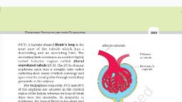
EXCRETORY PRODUCTS AND THEIR. TION 293 ©, , (PCT). A hairpin shaped Henle’s loop is the Afferent arteriole, next part of the tubule which has a, descending and an ascending limb. The, ascending limb continues as another highly, coiled tubular region called distal, convoluted tubule (DCT). The DCTs of many, nephrons open into a straight tube called, collecting duct, many of which converge and, open into the renal pelvis through medullary, pyramids in the calyces., , The Malpighian corpuscle, PCT and DCT, of the nephron are situated in the cortical, region of the kidney whereas the loop of Henle, dips into the medulla. In majority of, nephrons, the loop of Henle is too short and, extends only very little into the medulla. Such, nephrons are called cortical nephrons. In, some of the nephrons, the loop of Henle is, very long and runs deep into the medulla., These nephrons are called juxta medullary, nephrons., , The efferent arteriole emerging from the glomerulus forms a fine, capillary network around the renal tubule called the peritubular, capillaries. A minute vessel of this network runs parallel to the Henle’s, loop forming a ‘U’ shaped vasa recta. Vasa recta is absent or highly, reduced in cortical nephrons., , ae, , 19.2 Urine Formation, , Efferent, arteriole, , Bowman’s, capsule, , , , , Proximal, convoluted tubule, , Figure 19.4 Malpighian body (renal corpuscle), , Urine formation involves three main processes namely, glomerular, filtration, reabsorption and secretion, that takes place in different parts of, the nephron., , The first step in urine formation is the filtration of blood, which is carried, out by the glomerulus and is called glomerular filtration. On an average,, 1100-1200 ml of blood is filtered by the kidneys per minute which constitute, roughly 1/5" of the blood pumped out by each ventricle of the heart ina, minute. The glomerular capillary blood pressure causes filtration of blood, through 3 layers, i.e., the endothelium of glomerular blood vessels, the, epithelium of Bowman's capsule and a basement membrane between these, two layers. The epithelial cells of Bowman's capsule called podocytes are, arranged in an intricate manner so as to leave some minute spaces called, filtration slits or slit pores. Blood is filtered so finely through these, membranes, that almost all the constituents of the plasma except the, proteins pass onto the lumen of the Bowman's capsule. Therefore, it is, considered as a process of ultra filtration.