Page 1 :
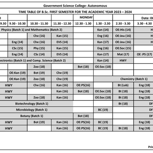
I B.SC. I SEMESTER, MICROBIOLOGY LABORATORY MANUAL, , I SEMESTER , PRACTICAL – 1, INTRODUCTION TO MICROBIOLOGY AND MICROBIAL, DIVERSITY, Experiments based on theory syllabus, 12 Practicals (one practical of 3hrs/week), Expt. No., 1. Microbiology good laboratory practices and biosafety., 2. Study of simple and compound microscopes, their handling including oil immersion, objective., 3-5 Preparation of micororganisms for light microscopic observation-simple (direct and, indirect) staining, differential staining (Gram staining), Structural staining (cell wall,, endospore of bacteria)., 6. Microscopic measurements of microorganisms/spores using stage and ocular, micrometer., 7. Study of cyanobacteria- Microcystis, Spirulina, Anabaena (any 2 for mounting), 8. Study of fungi - Rhizopus, Saccharomyces, Aspergillus, Penicillium, Agaricus using, temporary mounts (any 2 for mounting), 9. Study of Chlorella, Spirogyra, Diatoms and Gracilaria using temporary mounts, (any 2 for mounting), 10. Study of the following protozoans using permanent mounts/photographs: Euglena,, Paramaecium and Entamoeba., 11. Display of photographs of microscopes and scientists mentioned in the theory., 12. Study the principle and applications of important instruments (autoclave, hot air, oven, incubator, Inoculation loop, Inoculation needle, membrane filter, laminar air, flow system, colony counter. biological safety cabinets, BOD incubator, pH meter), used in the microbiology laboratory., INTERNAL ASSESSMENT, Break-up, , 20 marks, , Record, Continuous assessment, Test/Submissions, , 05 marks, 05 marks, 10 Marks, , *****, Department of Microbiology, Government Science College (Autonomous), Hassan, , 1
Page 2 :
I B.SC. I SEMESTER, MICROBIOLOGY LABORATORY MANUAL, , SCHEME OF PRACTICAL EXAMINATION, I B.Sc. /I SEMESTER/PRACTICAL I : INTRODUCTION TO, MICROBIOLOGY AND MICROBIAL DIVERSITY, Time : 3 hrs, , Max.marks : 30, , I. Measure the size of given material “A” using stage and ocular micrometer. Write, the principle, procedure and result, , 06 Marks, , (Principle: 1 marks, Procedure: 2 marks, Calibration: 2 marks, Result: 1 mark), , II. Stain the given material “ B ” by …………………….. method. Write the, principle, procedure and result. Leave the preparation for evaluation (Simple, direct/ indirect, Gram’s staining/ Structural staining), , 06 Marks, , (Preparation: 3 marks, Principle: 1 marks, Procedure:1 mark, Result: 1 mark), , III. Prepare a temporary mount of the given material “ C ” by, …………………….. method. Write the principle, procedure and result. Leave, the preparation for evaluation (Wet mounting of cyanobacteria/algae/fungi/ hanging, drop method), 06 Marks, (Preparation: 3 marks, Principle: 1 marks, Procedure:1 mark, Result: 1 mark), , IV. Write critical notes on “D”,”E”,”F”,”G”, , 3x4=12 Marks, , (Microscopes- charts/photographs/oil immersion/stains/laboratory equipment, /disinfectants /detergents/chromic acid) as per theory syllabus, , Department of Microbiology, Government Science College (Autonomous), Hassan, , 2
Page 3 :
I B.SC. I SEMESTER, MICROBIOLOGY LABORATORY MANUAL, , *****, , EXPERIMENT No. 01, MICROBIOLOGY GOOD LABORATORY PRACTICES, AND BIOSAFETY., Microbiology is a branch of biology which deals with microorganisms, which, are omnipresent but are invisible to naked eyes of human beings. They will be seen, through microscopes. Some of the important safety measures must be strictly, followed in the microbiology laboratory to avoid the occurrence of incidents which, may be harmful. The rules, regulations and safety measures which should be taken, in a Microbiology laboratory is as follows:, It should be assumed that the microorganisms with which we are working are, capable of producing diseases, allergies, itching, etc. Therefore, it is necessary to, take care of while handling the cultures containing test tubes, conical flasks, containing the media, and other equipment like microscopes etc., The glass slides, coverslips, pipettes, petri plates etc. should be cleaned before using, and discard into the jar of disinfectant solutions after use or completion of the, experiments., The laboratory coat or Apron with name plate should wear compulsorily while, entering into the lab. Practical record, pencil, pen, drawing aids and other, requirements should be brought to the laboratory., Working tables, laminar air flow system and hands are cleaned with water and then, swabbed with alcohol, Inoculation needles, loops should be properly sterilized before and after use by, incineration., Microbial cultures containing test tubes, conical flasks containing the media,, physiological saline etc. are plugged with sterile cotton and wrapped with craft or, brown paper., The test tubes should be kept upright in the test tube racks, never lay them on the, table. Never moist the labels with tongue., Students should not laugh, talk, eat, drink or smoke inside the laboratory. Accidents, such as spilled cultures, breakage of glass wares, any cuts or damages, should be, reported to the staff members immediately., Department of Microbiology, Government Science College (Autonomous), Hassan, , 3
Page 4 :
I B.SC. I SEMESTER, MICROBIOLOGY LABORATORY MANUAL, , Use instruments like hot air oven, incubators, balance, microwave oven and, microscopes etc. carefully. Hands should be thoroughly washed before leaving the, laboratory., Practical observation books should be completed in the laboratory itself and get, signed by batch teacher without fail., Practical record books should be completed in the laboratory itself or should be, submitted in next lab and get signed by batch teacher without fail., , Department of Microbiology, Government Science College (Autonomous), Hassan, , 4
Page 5 :
I B.SC. I SEMESTER, MICROBIOLOGY LABORATORY MANUAL, , EXPERIMENT No. 02, STUDY OF SIMPLE AND COMPOUND MICROSCOPES, THEIR, HANDLING INCLUDING OIL IMMERSION OBJECTIVE, Microscope is an optical instrument used in all biological laboratories to observe, the objects which cannot be seen through naked eyes of human beings. Microscopes, are of many types, namely bright field /compound microscope, dark field, phasecontrast, stereo, Transmission Electron Microscope and Scanning Electron, Microscopes., SIMPLE MICROSCOPE :, A simple microscope is a convex lens of short focal length. It is used to form, a virtual image of an object placed just inside its principal focus. It is made up of, a single convex lens or a combination of lenses which functions as a convex lens., The convex lens magnifies the object and also helps to produce a magnified image, of a near object which appears to be at the distance of distinct vision. Improved, simple microscopes, used by the biologists during field work, may magnify an, object even up to 100 times., A dissecting microscope is an example of a simple microscope used either for, dissecting the material or for viewing the magnified image of the material. It, consists of only one lens unit. This lens unit may even be an ordinary magnifying, glass., It is used either for dissecting the material or for less magnifications, that is,, only 6X, 12X or rarely 20X. For the light adjustment purposes, a mirror is attached, to the limb under the stage. Mirror can be moved vertically with the help of an, adjustment screw. At the tip of the limb is present a folded arm, on which a lens, of definite magnification (6X, 12X, etc.) is fitted. Folded arm is moved to keep, the lens in the desired position on the stage., The material to be viewed or dissected under a dissecting microscope is placed, on the stage. The eye is placed close to the lens. Folded arm is tilted to bring the, lens over the material. Light is adjusted by the movement of the mirror. Focusing, is done with the help of the adjustment screw of this microscope, Department of Microbiology, Government Science College (Autonomous), Hassan, , 5
Page 6 :
I B.SC. I SEMESTER, MICROBIOLOGY LABORATORY MANUAL, , COMPOUND MICROSCOPE:, The compound microscope consists of two important parts namely, optical parts, and mechanical parts., The optical parts include ocular (eye piece), objective lens, condenser and mirror., The mechanical parts include draw tubes, body tube, nose piece, rough/coarse, adjustment, fine adjustment, arm, stage, base/foot, inclination joint etc., THE OPTICAL PARTS:, Ocular lens (eyepiece): It consist of two magnifying lenses through which object, will be observed. It is available in magnification with 5X, 10X, 15X and 20X., Objective lens: They are used to magnify the objects and are available in three, magnifications, 10X (low power) 45X (high power) and 100X (oil immersion)., Condenser and iris diaphragm: It is present below the stage, moves up and down, and helps to open and close the shutter, so that the amount of light required can be, regulated. Abbey condenser is generally used., Mirror: It is fixed below the stage and is Plano-concave. The concave surface is, used for focusing., MECHANICAL PARTS:, Draw tube: It holds the ocular in proper position., Body tube: It is together with draw tube holds the ocular and objectives in proper, position at a distance from each other., Nose piece: It is a circular moving disk to which the objectives (10X, 45X & 100X, ) are fixed. It is present at the lower end of the body tube., Rough/coarse adjustment: It moves the body tube up and down over a greater, distance and brings the object in focus., Fine adjustment: It moves the body tube gently for focusing and clear image of the, object is seen., Stage: It supports the slide on which the object is mounted. It has central opening, through which light is passed on to the object. There are spring clips to hold the, slide., Base/foot: It is horse-shoe shaped and supports the entire body of the microscope., Inclination joint: It permits tilting of the upper part of the microscope towards the, eye to facilitate proper viewing., , Department of Microbiology, Government Science College (Autonomous), Hassan, , 6
Page 7 :
I B.SC. I SEMESTER, MICROBIOLOGY LABORATORY MANUAL, , USE OF OIL IMMERSION OBJECTIVE:, Ensure that the eyepiece, objective, condenser and the mirror of the microscope are, cleaned with muslin cloth or tissue paper., Focus the microscope and observe the object under low power(10X) and high power., Remove the high power objective and then place a drop of immersion oil or cedar, wood oil above the coverslip where the object is present and focus with oil, immersion objective(100X), gently using rough adjustment to view the object. Then, by using fine adjustment brilliant enlarged image of the specimen is seen., After viewing the image of the object, gently lift the oil immersion objective by, using rough adjustment, clean the objective lens with tissue paper., The specific magnification of the oil immersion objective is marked on it has 100X., The total magnification is calculated by multiplying the magnification of the, eyepiece and oil immersion objective is used., Magnification of the eyepiece x magnification of oil immersion objective, 10x X 100x = 1000x is the total magnification., , Department of Microbiology, Government Science College (Autonomous), Hassan, , 7
Page 8 :
I B.SC. I SEMESTER, MICROBIOLOGY LABORATORY MANUAL, , EXPERIMENT No. 03, PREPARATION OF MICROORGANISMS FOR LIGHT, MICROSCOPIC OBSERVATIONS, SIMPLE DIRECT OR POSITIVE STAINING, Aim: To stain the given bacteria by simple direct or positive staining., Requirements: Bacterial specimen, a basic stain (Crystal violet, Safranin,, Methylene blue etc.), Microscopic slides, Inoculation loop, Bunsen burner,, Microscope etc, Introduction: Simple staining implies the use of only a single stain, which is usually, sufficient to reveal the morphological features of most microbial cells, including, relative size, shape, and characteristic arrangements for groups of cells., Principle: A stain is a dye consisting of a colored ion (a chromophore) and a counter, ion to balance the charge. Attachment of the chromophore part of the dye complex, to a cellular component represents the staining reaction. Depending upon the dye,, the chromophore can be either positively charged (cationic) and have an affinity for, negative ions or negatively charged (anionic) with an affinity for positive ions., Bacteria carry a net negative charge at pH 7. Therefore, cationic dyes such as, methylene blue or crystal violet are useful for the direct staining of cells., Procedure :, Preparation of a smear and heat fixing, 1. Using a sterilized inoculating loop, transfer loopful of liquid suspension, containing bacteria to a slide or transfer an isolated colony from a culture plate to, a slide with a water drop., 2. Disperse the bacteria on the loop in the drop of water on the slide and spread the, drop over an area the size of a dime. It should be a thin, even smear., 3. Allow the smear to dry thoroughly., 4. Heat-fix the smear cautiously by passing the underside of the slide through the, burner flame two or three times. It fixes the cell in the slide. Do not overheat the, slide as it will distort the bacterial cells., Department of Microbiology, Government Science College (Autonomous), Hassan, , 8
Page 9 :
I B.SC. I SEMESTER, MICROBIOLOGY LABORATORY MANUAL, , Staining :, 1. Cover the smear with crystal violet or methylene blue and allow the dye to remain, in the smear for approximately one minute. Using distilled water wash bottle,, gently wash off the excess stain from the slide by directing a gentle stream of, water over the surface of the slide., 2. Wash off any stain that got on the bottom of the slide as well., 3. Wipe the back of the slide and blot the stained surface with a blotting paper., 4. Place the stained smear on the microscope stage smear side up and focus the smear, using the 10X objective., 5. Choose an area of the smear in which the cells are well spread in a monolayer., Center the area to be studied, apply immersion oil directly to the smear, and focus, the smear under oil with the 100X objective., Observation: The bacterial cells usually stain uniformly and the color of the cell, depends on the type of dye used. If Crystal violet is used bacterial cells will be, violet in colour., Result: If the observed bacterial cells are round in shape then its cocci and if its, rod shaped its bacilli. If the cocci cells are arranged in a series then its, Streptococci, if they are in cluster its Staphylococci., , Department of Microbiology, Government Science College (Autonomous), Hassan, , 9
Page 10 :
I B.SC. I SEMESTER, MICROBIOLOGY LABORATORY MANUAL, , EXPERIMENT No. 04, PREPARATION OF MICROORGANISMS FOR LIGHT, MICROSCOPIC OBSERVATIONS, SIMPLE INDIRECT OR NEGATIVE STAINING, Aim: To stain the given bacteria by simple indirect or negative staining., Requirements: Bacterial specimen, an acidic stain (Nigrosine), Microscopic slides,, Inoculation loop, Microscope etc, Introduction: The main purpose of Negative staining is to study the morphological, shape, size and arrangement of the bacteria cells that is difficult to stain., Eg: Spirilla. It can also be used to stain cells that are too delicate to be heat-fixed., Principle: India Ink or Nigrosin is an acidic stain. This means that the stain readily, gives up a hydrogen ion (proton) and the chromophore of the dye becomes, negatively charged. Since the surface of most bacterial cells is negatively charged,, the cell surface repels the stain. The glass of the slide will stain, but the bacterial, cells will not. The bacteria will show up as clear spots against a dark background., Procedure:, Preparation of a smear:, Using a sterilized inoculating loop, transfer loopful of liquid suspension, containing bacteria to a slide or transfer an isolated colony from a culture plate, to a slide with a water drop., Disperse the bacteria on the loop in the drop of water on the slide and spread, the drop over an area the size of a dime. It should be a thin, even smear., Staining:, Place few drops of Nigrosine beside the inoculum. Using another slide smear, the inoculum along with stain along the slide length forming an acute angle., Leave the slide for drying., Wipe the back of the slide with a blotting paper., , Department of Microbiology, Government Science College (Autonomous), Hassan, , 10
Page 11 :
I B.SC. I SEMESTER, MICROBIOLOGY LABORATORY MANUAL, , , Place the stained smear on the microscope stage smear side up and focus the, smear using the 10X objective and then on a high power objective., Observation: The bacterial cells appear transparent and background will be dark., Result: Bacterial cells appear transparent. If its round in shape, then its cocci and, if its rod shaped its bacilli. If the cocci cells are arranged in a series then its, Streptococci, if they are in cluster its Staphylococci., , Department of Microbiology, Government Science College (Autonomous), Hassan, , 11
Page 12 :
I B.SC. I SEMESTER, MICROBIOLOGY LABORATORY MANUAL, , EXPERIMENT No. 05, PREPARATION OF MICROORGANISMS FOR LIGHT, MICROSCOPIC OBSERVATIONS, DIFFERENTIAL STAINING: GRAM’S STAINING TECHNIQUE, Aim: To stain the given bacteria by Gram’s staining to differentiate as Gram positive, or Gram negative., Requirements: Bacterial specimen, Crystal violet, Gram’s iodine, 95% alcohol,, Safranin, Microscopic slides, Inoculation loop, Microscope, Bunsen burner, wash, bottle etc., Introduction: Gram Staining is the common, important, and most used differential, staining techniques in microbiology, which was introduced by Danish Bacteriologist, Hans Christian Gram in 1884. This test differentiates the bacteria into Gram Positive, and Gram Negative Bacteria, which helps in the classification and differentiations, of microorganisms., Principle: When the bacteria is stained with primary stain Crystal Violet and, fixed by the mordant, some of the bacteria are able to retain the primary stain and, some are decolorized by alcohol. The cell walls of gram positive bacteria have a, thick layer of protein-sugar complexes called peptidoglycan and lipid content is, low. Decolorizing the cell causes this thick cell wall to dehydrate and shrink,, which closes the pores in the cell wall and prevents the stain from exiting the, cell. So the ethanol cannot remove the Crystal Violet-Iodine complex that is, bound to the thick layer of peptidoglycan of gram positive bacteria and appears, blue or purple in colour., In case of gram negative bacteria, cell wall also takes up the CV-Iodine, complex but due to the thin layer of peptidoglycan and thick outer layer which is, formed of lipids, Crystal Violet-Iodine complex gets washed off. When they are, exposed to alcohol, decolorizer dissolves the lipids in the cell walls, which allows, the crystal violet-iodine complex to leach out of the cells. Then when again stained, with safranin, they take the stain and appears red in color., , Department of Microbiology, Government Science College (Autonomous), Hassan, , 12
Page 13 :
I B.SC. I SEMESTER, MICROBIOLOGY LABORATORY MANUAL, , Procedure:, 1. Take a clean, grease free slide., 2. Prepare the smear of suspension on the clean slide with a loopful of sample., 3. Air dry and heat fix, 4. Crystal Violet was poured and kept for about 30 seconds to 1 minutes and, rinse with water., 5. Flood the gram’s iodine for 1 minute and wash with water., 6. Then, wash with 95% alcohol for about 10-20 seconds and rinse with water., 7. Add safranin for about 1 minute and wash with water., 8. Air dry, Blot dry and Observe under Microscope., Observation: The bacterial cells appear with violet or red colour depending upon, the cell wall composition., Result: If the Bacterial cells appear violet or blue in colour, then it is Gram positive, bacteria. If it is red or pink in colour then its Gram negative bacteria., , Department of Microbiology, Government Science College (Autonomous), Hassan, , 13
Page 14 :
I B.SC. I SEMESTER, MICROBIOLOGY LABORATORY MANUAL, , EXPERIMENT NO. 06, MICROMETRY, AIM: Microscopic measurements of microorganisms/spores using stage and ocular, micrometer., REQUIREMENTS: Bright field microscope, Ocular micrometer, Stage, micrometer, cover glass, permanent slides of specimen, microscopic slide etc., , PRINCIPLE: Micrometry is the measurement of microorganism. Since the, microorganisms are very small, the dimension of these are usually expressed in units, that is, micrometer. One micrometer(µm) is 1000/mm that is 10 -3. The determination, of size of any kind of microorganism is one of the properties which is useful for, identification in laboratory. Since the microorganisms can be seen only under, microscope, their size can be measured by equipping it with an ocular micrometer, which is calibrated against stage micrometer., The ocular micrometer is the simple glass disc with etched lines. It has 100, equally spaced divisions which are marked from 0-10. The distance between the, gradation of an ocular micrometer does not have any standard value and the values, depending on the objectives are used. The distance between the gradation is found, out by calibrating it with a known scale that is stage micrometer. On this, 1mm, distance is etched into 100 equally spaced divisions. So, there are 1000 µm in 1mm., the distance of 1 division is thus 10µm or 0.01mm the distance of each stage µm, divisions become correspondingly enlarged under high power and oil immersion, objectives of microscope., The ocular micrometer after putting inside eye piece is calibrated by, superimposing the gradation of ocular micrometer over the stage micro micrometer, which is accomplished by rotating ocular lens. By determining the number of ocular, micrometer divisions coinciding with the number of divisions on stage micrometer,, the calibration factor for 1 ocular division is calculated by applying the formula as, follows., , Department of Microbiology, Government Science College (Autonomous), Hassan, , 14
Page 15 :
I B.SC. I SEMESTER, MICROBIOLOGY LABORATORY MANUAL, , One ocular division = Number of divisions on stage micrometer x 10, Number of divisions on ocular micrometer, After calibrating one ocular micrometer, it can be used to determine the size of, an organism. The structure of an organism in terms of length, breadth and diameter, can be calculated by the following formula., Size of microorganism = Number of ocular divisions x Calibration factor of the, objective used., PROCEDURE: Ocular micrometer is inserted in the place of eye piece. When, observed, ocular micrometer divisions are seen in sharp focus and there will be no, changes in lines and distances under different objective., Place the stage micrometer on microscope stage and bring its scale in the, microscopic field center under a sharp focus first using the 10x low power objective, Turn the ocular lens until the parallel lines of ocular micrometer coincide with, those of stage micrometer. Then count the number of divisions in both ocular and, stage micrometer between the two coinciding lines., , RESULT: Observations are recorded. Then the value of calibration factor for, 10x objective is………..µm, , Department of Microbiology, Government Science College (Autonomous), Hassan, , 15
Page 16 :
I B.SC. I SEMESTER, MICROBIOLOGY LABORATORY MANUAL, , EXPERIMENT No. 07, STUDY OF CYANOBACTERIA - MICROCYSTIS, Aim: Mounting of Microcystis., Requirements : Microcystis sample, glass slide, needle, forceps, safranin stain,, muslin cloth and microscope., , , , , , , , Procedure :, A cleaned glass slide is taken., A small amount of Microcystis sample is taken on the slide with the help of needle, and forceps., The material is spread on the slide using needle and forceps. A drop of safranin stain, is added on the material., A drop of glycerin is added and it is covered with a cover glass., The prepared slide is mounted on the stage of the microscope and observed under, low power, high power and oil immersion objectives (A drop of Cedar wood oil or, clove oil is used.), Systemic position:, CLASS, ORDER, FAMILY, GENUS, , , , , , , , : CYANOPHYCEAE, : CHROOCOCCALES, : MICROCYSTACEAE, : MICROCYSTIS, , Characteristic features :, Occurance :, It is a freshwater, planktonic cyanobacteria growing in ponds, pools, lakes, tanks and, in stagnant water. Examples of some species are Microcystis aeuroginosa, M.toxica, etc., It is unicellular planktonic water bloom., Structure of thallus :, It is a unicellular member which aggregate themselves into colonies., The colonies may be spherical, elongated, with common mucilaginous matrix packs, around the cells together., Cell structure :, , Department of Microbiology, Government Science College (Autonomous), Hassan, , 16
Page 17 :
I B.SC. I SEMESTER, MICROBIOLOGY LABORATORY MANUAL, , , , , , , , , In its general structure the cell is a typical cyanophycean type., The cell consists of outer cell wall and inner cytoplasm divided into peripheral, coloured chromoplasm where an incipient nucleus is present at the center., A number of gas vacuoles are present between the cell which helps the colonies to, float (buoyancy) on water., The colonies emit foul smell., It can produce neurotoxins and hepatotoxins, such as microcystin and cyanopeptolin, Reproduction :, The reproduction is by cell division which is seen to take place in all directions. The, whole colony also multiplies by fragmentation. Reproduction by nannocytes is also, reported in Microcystis flosaquae., , Department of Microbiology, Government Science College (Autonomous), Hassan, , 17
Page 18 :
I B.SC. I SEMESTER, MICROBIOLOGY LABORATORY MANUAL, , STUDY OF CYANOBACTERIA - SPIRULINA, Aim: Mounting of Spirulina, Requirements: Spirulina sample, glass slide, coverslip, needle, forceps, safranin, stain, muslin cloth and microscope, Procedure:, , , , , , , A clean glass slide is taken., A small amount of Spirulina sample is taken on the slide with the help of needle and, forceps., The material is spread on the slide using needle and forceps. A drop of safranin stain, is added on the material., A drop of glycerin is added and then it is covered with a cover slip., The prepared slide is mounted on the stage of microscope and observed under low, power, high and oil immersion objective (a drop of cedar wood oil or clove oil is, used)., Systemic position:, Class : Cyanophyceae, Order : Spirulinales, Family : Spirulinaceae, Genus : Spirulina, Characteristic features:, Occurrence:, , , , , It is a freshwater, microscopic, aquatic, blue green, unicellular alga growing in ponds,, pools, where the organic matter is more., It is also cultured in laboratories for its nutritive and medicinal values., , Department of Microbiology, Government Science College (Autonomous), Hassan, , 18
Page 19 :
I B.SC. I SEMESTER, MICROBIOLOGY LABORATORY MANUAL, , Structure of the Thallus:, , , It is a unicellular, spirally twisted very closely and very loosely at certain regions and, hence the name Spirulina., Cell structure:, , , , , , , , , In its general structure the cell is trichome without mucilaginous sheet., This cell is a typical cyanophyceae type, consists of outer cell wall, and inner, cytoplasm divided into peripheral coloured chromoplasm and central colourless, centroplasm where and incipient nucleus is present., A cell is filled with reserve food materials. The plant body is single celled protein, appears in the form of spring., Spirulina is economically important because it is rich in protein (60-70%), vitamins,, etc, hence it is used as food supplement because of its high protein content., It is also used to treat sewage and effluents., Reproduction:, , , , , It reproduce vegetatively by means of fragmentation. Asexual and sexual methods, are absent., There are three fundamental stages: Trichomes fragmentation, hormogonia cells, enlargement and the maturation process, and trichome elongation. Then this mature, trichomes get divided into filaments or hormogonia, cells in the hormogonias gets, increased by binary fission, grows lengthwise and take their helical form, , Department of Microbiology, Government Science College (Autonomous), Hassan, , 19
Page 20 :
I B.SC. I SEMESTER, MICROBIOLOGY LABORATORY MANUAL, , STUDY OF CYANOBACTERIA - ANABAENA, , , , , , , , Aim : Mounting of Anabaena., Requirements : Anabaena sample, glass slide, coverslip, needle, forceps, safranin, stain, muslin, cloth and microscope., Procedure :, A clean glass slide is taken., A small amount of Anabaena sample is taken on the slide with the help of needle and, forceps., The material is spread on the slide using needle and forceps. A drop of safranin stain, is added on the material., A drop of glycerin is added and then it is covered with coverslip., The prepared slide is mounted on the stage of the microscope and observed under, low power, high power and oil immersion objectives (A drop of cedar wood oil or, clove oil is used.), Systemic position:, CLASS, ORDER, FAMILY, GENUS, , : HORMOGONEAE, : NOSTOCALES, : NOSTOCACEAE, : ANABAENA, Characteristics features :, , , , , , , , Occurrance :, It is a freshwater, microscopic, aquatic, blue-green algae, found in ponds, pools, lakes, and in paddy fields as free living floating forms., It also occurs as an endophytic alga in the roots of Cycas and the leaves of water fern, Azolla., Structure of thallus :, It forms smaller colonies than Nostoc and each colony consists of many filament, surrounded by mucilaginous sheath., It shows that the filament consists of a number of barrel or ellipsoidal or sub, cylindrical shaped cells arranged linearly one above the other., , Department of Microbiology, Government Science College (Autonomous), Hassan, , 20
Page 21 :
I B.SC. I SEMESTER, MICROBIOLOGY LABORATORY MANUAL, , , , , , , , , , , , , Each filament is with an individual mucilaginous sheath. In the filament there are few, double walled larger cells called Heterocysts which may be terminal or intercalary in, position., Each Heterocyst may consist of two polar nodules in intercalary and one polar nodule, in terminal heterocysts., The function of Heterocysts are Nitrogen fixation., Cell structure :, In its general structure the cell is a filament with individual mucilaginous sheath., The cell is a typical Cyanophycean type, consists of outer cell wall and inner, centroplasm., The cell is filled with reserve food materials., Reproduction :, It reproduces vegetatively by the means of fragmentation, by the formation of thick, walled, dark coloured cells called Aknites which contains large amount of reserve, food materials., Asexual and sexual method of reproduction is absent., Anabaena typically reproduce via fragmentation. Fragmentation is where a section, of the chain will split off and either float or glide away. After a while these sections, begin to form their own chains. These sections are known as hormogonia, and arise, via the separation of adjacent cell walls. They are also caused by dead cells that, become separation discs., , Department of Microbiology, Government Science College (Autonomous), Hassan, , 21
Page 22 :
I B.SC. I SEMESTER, MICROBIOLOGY LABORATORY MANUAL, , EXPERIMENT No. 08, STUDY OF FUNGI - RHIZOPUS, , , , , , , , Aim : Mounting of Rhizopus., Requirements : Rhizopus sample, glass slide, coverslip, needle, forceps, lactophenol, cotton blue stain, muslin cloth and microscope., Procedure :, A cleaned glass slide is taken., A small amount of Rhizopus sample is taken on the slide with the help of needle and, forceps., The material is spread on the slide using the needle and forceps. A drop of, lactophenol cotton blue stain is added on the material., Then it is covered with the cover slip., The prepared slide is mounted on the stage of the microscope and observed under, low power, high power, Characteristics features:, CLASS, : MUCOROMYCOTINA, ORDER, : MUCORALES, FAMILY : MUCORACEAE, GENUS, : RHIZOPUS, , , , , , , , , , , , , Thallus structure :, Rhizopus is a microscopic, saprophytic fungus growing on bread, jams, jellies,, pickles and other food materials., The vegetative body is called mycelium consists of tubular, coenocytic, aseptate, multinucleate interwoven mass of hyphae., The hyphae can be differentiated into stolons, rhizoids and sporangiophores., The stolons are stout (thick) sparingly branches and produces rhizoids as well as, Sporangiophores at the nodal regions., The sporangiophores are long, produced in tufts and bearing globos sporangia at tips., The sporangium consists of inner Columella, surrounded by peripheral region in, which the spores are present., Reproduction :, Reproduction takes place by the means of vegetative, sexual and asexual methods., Vegetative method of reproduction is by Fragmentation., Asexual method of reproduction is by the formation of spores with the sporangia., , Department of Microbiology, Government Science College (Autonomous), Hassan, , 22
Page 23 :
I B.SC. I SEMESTER, MICROBIOLOGY LABORATORY MANUAL, , , , , , , , The hyphae of different strains come close to each other forming Progametangia., These Progametangia contact with each other and forming Gametangia. The, remaining part of the fusing hyphae is called Suspensor., The wall of Gametangia ruptures releasing the gametes, union of gametes takes place, forming a Zygote., The zygospores is round, thick walled, watery and rich in food., The zygospore later germinates to produce new mycelium called Promycelium., The Promycelium forms the thallus proper., , Department of Microbiology, Government Science College (Autonomous), Hassan, , 23
Page 24 :
I B.SC. I SEMESTER, MICROBIOLOGY LABORATORY MANUAL, , STUDY OF FUNGI - SACCHAROMYCES, , , , , , , , Aim : Mounting of Saccharomyces (Yeast ), Requirements : Saccharomyces sample, glass slide, cover slip, needle, forceps,, lactophenol cotton blue stain, muslin cloth and microscope., Procedure, :, A cleaned glass slide is taken., A small amount of Saccharomyces sample is taken on the slide with the help of needle, and forceps., The material is spread on the slide using the needle and forceps. A drop of, lactophenol cotton blue stain is added on the material., Then it is covered with the cover slip., The prepared slide is mounted on the stage of the microscope and observed under, low power, high power and oil immersion objectives (A drop of Cedarwood oil or, Clove oil is used.), Characteristics features :, CLASS, : SACCHAROMYCETES, ORDER, : SACCHAROMYCETALES, FAMILY, : SACCHAROMYCETACEAE, GENUS, : SACCHAROMYCES, , , , , , , , , Thallus structure :, Yeast is a microscopic, eukaryotic, unicellular fungus found to grow on sugary, solutions, fruit juices, wine etc., Vegetative body of the Yeast is single cell which is ellipsoidal in shape., Cell structure :, The cell consists of outer cell wall made up of chitin and inner plasma membrane and, the granular cytoplasm., Within the cytoplasm a large central vacuole is present and it has volutin threads., There is a large nucleus present about the vacuole. The other organelles like, mitochondria, glycogen as food and oil droplets are present., The Yeast is having economic importance for the production of alcohol, motor spirit,, beer, brandy, whisky (brewing), bread, juice, cake, juices(baking). yeasts are rich in, vitamin B and C, proteins and fats contents, hence used as food., , Department of Microbiology, Government Science College (Autonomous), Hassan, , 24
Page 25 :
I B.SC. I SEMESTER, MICROBIOLOGY LABORATORY MANUAL, , , , , , , , Reproduction :, Yeast reproduces by vegetative, sexual and asexual methods., Yeast reproduces vegetatively by the means of budding which is a common method, of reproduction., Depend upon the type of Budding it is called Budding Yeast or Fission yeast., Sexual method of reproduction is by the formation of Ascospores., Three types of life cycles namely Haplobiontic(Saccharomyces), Diplobiontic, (Saccharomyces ludwiggi) and Haplodiplontic (Saccharomyces cerevisiae) life, cycles are studied., , Department of Microbiology, Government Science College (Autonomous), Hassan, , 25
Page 26 :
I B.SC. I SEMESTER, MICROBIOLOGY LABORATORY MANUAL, , STUDY OF FUNGI - ASPERGILLUS, , , , , , , , , , , , , , , , , , , , , Aim : Study of Aspergillus., Requirements : Aspergillus sample, glass slide, coverslip, needle, forceps,, lactophenol cotton blue stain, muslin cloth and microscope., Procedure :, A clean glass slide is taken., A small amount of Aspergillus sample is taken on the slide with the help of needle, and forceps., The material is spread on the slide using the needle and forceps. A drop of, lactophenol cotton blue stain is added on the material., Then it is covered with the cover slip., The prepared slide is mounted on the stage of the microscope and observed under, low power, high power, CLASS : EUASCOMYCETES, ORDER : EUROTIALES, FAMILY : TRICHOCOMACEAE, GENUS : ASPERGILLUS, Characteristics features :, Aspergillus is a saprophytic fungus found growing on Bread, Rice , Fruit ,Jams,, Jellies, Leather etc., It is commonly called black mold and is a common contaminant of lab cultures., The vegetative plant body is of mycelial type. The mycelium consists of profusely, branched, septate, multinucleate, tubular, interwoven mass of hyphae., The Hyphae consists of granular cytoplasm, nuclei, glycogen and oil bodies as, reserve food materials., Reproduction :, Both vegetative , sexual and asexual methods of reproduction takes place., Vegetative method of reproduction takes place by the means of fragmentation., Sexual method of reproduction takes place by the means of Conidia formation., Any vegetative hypha involved in reproduction functions as “Foot cells”, From the Foot cell an upright branch develops and is called Conidiophore., The tip of the Conidiophore swells forming a globose structure called Vesicle., The surface of Vesicle produces Conidia in radiating chains on a short tubular stalk, called Sterigmata., , Department of Microbiology, Government Science College (Autonomous), Hassan, , 26
Page 27 :
I B.SC. I SEMESTER, MICROBIOLOGY LABORATORY MANUAL, , , , , , , , , Sexual method of reproduction takes place by Oogamous method. The species are, homothallic or heterothallic. The male sex organ is the Antheridium and female sex, organ is Ascogonium., The Antheridium produces and Ascogonium an egg. The union of Antherozoid with, an egg results in the formation of Zygote. The Zygote undergo division and develops, into Mycelium., Economic importance :, Many species of Aspergillus are used in industries for the production of Enzymes,, Organic acids etc., Aspergillus flavus produce aflatoxin which is carcinogenic to organisms., Aspergillosis caused by some species and they infect human ears causing, “Otomycosis”., , Department of Microbiology, Government Science College (Autonomous), Hassan, , 27
Page 28 :
I B.SC. I SEMESTER, MICROBIOLOGY LABORATORY MANUAL, , STUDY OF FUNGI - PENICILLIUM, , , , , , , , , , , , , , , , , , , , , , , Aim : Study of Penicillium., Requirements : Penicillium sample (Culture plate/Infected Orange), glass slide,, coverslip, needle, forceps, lactophenol cotton blue stain, muslin cloth and, microscope., Procedure :, A clean glass slide is taken., A small amount of Penicillium sample is taken on the slide with the help of needle, and forceps., The material is spread on the slide using the needle and forceps. A drop of, lactophenol cotton blue stain is added on the material., Then it is covered with the cover slip., The prepared slide is mounted on the stage of the microscope and observed under, low power, high power and oil immersion objectives ( A drop of Cedarwood oil or, Clove oil is used)., CLASS : EUROTIOMYCETES, ORDER : EUROTIALES, FAMILY : TRICHOCOMACEAE, GENUS : PENICILLIUM, Characteristics features :, Penicillium is also called blue-green mold commonly found growing on decaying, Citrus fruits like Orange, Lemon etc, It also grows on Vegetables, Bread, Jams Jellies,, Leather etc as Saprophytic fungus., The vegetative body of the Fungus is Mycelial type. The mycelium consists of, profusely branched, aseptate, multinucleate, tubular, interwoven mass of hyphea., The Hyphae consists of Cytoplasm, Glycogen and Oil bodies as reserve food, materials., Reproduction :, Vegetative, sexual and asexual methods of reproduction takes place., Vegetative method of reproduction takes place by the means of fragmentation., Asexual method of reproduction takes place by the means of Conidia formation., Any vegetative hyphae grow upright and functions as Conidiophore., The Conidiophore again branches to primary, secondary and tertiary metulae., The terminal region of metulae bears bottle shaped structure called Sterigmata., The tip of Sterigmata cuts of forming chains of Conidia arranged basipetalously., The Conidium is round, uninucleate with food materials., The Conidium under favorable conditions germinates to produce new mycelium., , Department of Microbiology, Government Science College (Autonomous), Hassan, , 28
Page 29 :
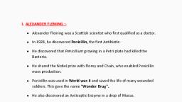
I B.SC. I SEMESTER, MICROBIOLOGY LABORATORY MANUAL, , , , , , , , , Sexual method of reproduction takes place by the Oogamous method. The species, are homothallic or heterothallic. Male sex organ is Antheridium and female sex organ, is Ascogonium., The Antheridium produce Antherozoids and Ascogonium an egg. The union of, Antherozoid with an egg results in Zygote formation. The Zygote undergo division, and develops new mycelium., Economic importance :, The Penicillium as an antibiotic from Penicillium notatum and Penicillium, chrysogenum , is a life saving drug against a number of bacterial disease.( Gr+ and, Gr-), It was discovered by Alexander Fleming, a nobel prize winner, , Department of Microbiology, Government Science College (Autonomous), Hassan, , 29
Page 30 :
I B.SC. I SEMESTER, MICROBIOLOGY LABORATORY MANUAL, , STUDY OF FUNGI - AGARICUS, Aim : Study of Agaricus ., Classification :, CLASS : BASIDIOMYCETES, ORDER : AGARICALES, FAMILY : AGARICACEAE, GENUS : AGARICUS, , , , , , , , , , , , , , , , Characteristics features :, It is commonly called Mushroom and it is Saprophytic fungus growing on damp, rotting roots, soil, dead and decaying organic matter, on cow dung hill etc., The vegetative plant body is Mycelium present bell the substratum, which is much, branched, septate, uninucleate in the beginning and later becomes dikaryotic. This, Mycelium gives rise to a fruiting body called Basidiocarp., Basidiocarp is an umbrella shaped, serial structure consisting of a stalk called Stipe., The terminal region of the Stipe has an expanded cap like structure called Pileus., When the Pileus is young, it is covered by a membrane called Velum., The Velum ruptures as the Pileus grows and leave a ring like structure called, Annulus., On the lower region of the Pileus, there are many vertically grown plates called Gills., On both sides of the Gills, Basidiospores are present., Structure (V/S) of the Gills :, A vertical section (V/S) through the Gills shows the following regions: Trama,, Subhymenium and Hymenium., Trama : It is the central interwoven mass of slender, elongated Hyphae which run, horizontally., Subhymenium : It is formed from Trama on both the sides and is made up of closely, packed tissue and is short., Hymenium : It is the fertile region of the Gill .It consists of club shaped structures, called Basidia .The Basidia are intermixed with sterile Hyphae called Paraphyses., Each Basidium produces four Basidiospores exogenously on a short Stalks called, Sterigmata., , Department of Microbiology, Government Science College (Autonomous), Hassan, , 30
Page 31 :
I B.SC. I SEMESTER, MICROBIOLOGY LABORATORY MANUAL, , , , , Economic importance :, The mushrooms are rich in vitamins, proteins, carbohydrates and hence it is used as, food and is used in the preparation of Biomass Proteins., It is cultivated in large scale as a commercial product. Button and Oyster Mushrooms, are edible for their rich nutrients., , Department of Microbiology, Government Science College (Autonomous), Hassan, , 31
Page 32 :
I B.SC. I SEMESTER, MICROBIOLOGY LABORATORY MANUAL, , EXPERIMENT No. 09, STUDY OF ALGAE - CHLORELLA, Aim : Mounting of Chlorella., Requirements : Chlorella sample, glass slide, coverslip, needle, forceps, safranin, stain, muslin cloth, microscope., Procedure :, A cleaned glass slide is taken., A small amount of Chlorella sample is taken on the slide with the help of needle, and forceps., The material is spread on the slide using needle and forceps. A drop of safranin, stain is added on the material., A drop of glycerin is added and then it is covered with a cover slip., The prepared slide is mounted on the stage of microscope and observed under low, power, high power and oil immersion objectives (a drop of Cedar wood oil or clove, oil is used)., Characteristics features :, CLASS, ORDER, FAMILY, GENUS, , : TREBOUXIOPHYCEAE, : CHLORELLALES, : CHLORELLACEAE, : CHLORELLA, , Occurrence :, It is a freshwater, microscopic, aquatic, green algae, found growing in freshwater, pools, moist soil, wet tree trunks, potted plants and also live as symbiotic, association with Lichens., Structure of thallus :, The thallus is unicellular or found in colonies., , Department of Microbiology, Government Science College (Autonomous), Hassan, , 32
Page 33 :
I B.SC. I SEMESTER, MICROBIOLOGY LABORATORY MANUAL, , Cell structure :, , , , , , , , , The cells are round or spherical in shape. The cell has an outer thick cell wall and, inner protoplast., The protoplast consists of a large shaped chloroplast parietal in position., There is a well developed nucleus, pyrenoid, air cavities, mitochondria and golgi, bodies are present., Starch is the reserve food material., Chlorella is used as food because it is rich in proteins, vitamins etc., It is also used in space because of the food value and it release more of oxygen., It is also used in sewage disposal and waste disposal., , Reproduction :, , , , It asexually reproduces by the means of cell division., Reproduction is also effected by the production of autospores which are developed, within a cell by the successive division of the protoplast. Two or even sixteen, autospores may be formed in each cell which are liberated out by the rupture of the, parent cell wall. These immobile spores form new individuals., , Department of Microbiology, Government Science College (Autonomous), Hassan, , 33
Page 34 :
I B.SC. I SEMESTER, MICROBIOLOGY LABORATORY MANUAL, , STUDY OF ALGAE - SPIROGYRA, Aim : Mounting of Spirogyra, Requirements : Spirogyra sample, glass slide, coverslip, Needle, forceps, safranin, stain, muslin cloth and microscope, Procedure :, A clean glass slide is taken, A small amount of Spirogyra sample is taken in the slide with the help needle and, forceps. The material is spread on the slide using needle and forceps. A drop of, safranin stain is added on the material., A drop of glycerin is added and then it is covered with the coverslip, The prepared slide is mounted on the stage of microscope and observed under the, low power, high power and oil immersion objectives (a drop of cedarwood oil or, clove oil is used), Characteristic features :, Class : Zygnematophyceae, Order : Zygnematales, Family : Zygnemataceae, Genus : Spirogyra, Occurrence:, It is a green algae growing in freshwater, ponds, pools, dishes etc.,, Structure of the thallus :, It is a branched, filamentous, green, thread like shining alga., The cells in the filament are long and cylindrical shaped., The characteristic ribbon or spiral shaped chloroplast is present throughout the cell,, hence the name Spirogyra., There is a central vacuole present in the cytoplasm near the cell wall., There is a single nucleus and number of pyrenoids are present along the length of, the chloroplast., Department of Microbiology, Government Science College (Autonomous), Hassan, , 34
Page 35 :
I B.SC. I SEMESTER, MICROBIOLOGY LABORATORY MANUAL, , , The reserved food is starch., , Reproduction :, It produces by means of sexual method by conjugation. It is of two types that is, lateral conjugation and scalariform conjugation., , STUDY OF ALGAE - DIATOMS, Aim : Mounting of Diatoms., Requirements : Diatom sample, glass slide, coverslip, needle, forceps, safranin stain,, muslin cloth and microscope., Procedure:, A clean glass slide is taken., A small amount of diatom sample is taken on the slide with the help of needle and, forceps., Material is spread on the slide using needle and forceps. A drop of safranin stain is, added on the material., A drop of glycerin is added and then it is covered with the cover slip., The prepared slide is mounted on the stage of microscope and observed at the low, power, high power and oil immersion objectives (a drop of cedarwood oil or clove, oil is used)., Characteristics features :, CLASS, : BACILLARIOPHYCEAE, ORDER, : PENNALES, FAMILY : PENNULARIACEAE, GENUS, : PINNULARIA(DIATOMS), Occurence :, The diatoms are of two types, namely pennales and centrales depends on the, symmetry., Pennales with bilateral symmetry and Centrales with radial symmetry., , Department of Microbiology, Government Science College (Autonomous), Hassan, , 35
Page 36 :
I B.SC. I SEMESTER, MICROBIOLOGY LABORATORY MANUAL, , , The diatoms occur in both freshwater and marine water as freely floating planktonic, forms., , Structure of thallus :, Pennate diatoms are with bilateral symmetry and cell is called Frustule., It consists of two overlapping halves or valves. The Upper half is called epitheca, and lower half is called hypotheca which fits into one another like a soap box. The, path of journey is the gridle., The cell consists of silicified and pectic wall and inner a number of static and, punctate, cytoplasm, two large chromophores and large nucleus .The, chromatophores consists in pigments like xanthophylls (golden brown) and, chlorophyll a,b etc., The cell shows two views, girdle view the girdle, striae and punctate are seen., Diatoms are also biological indicators of water pollution., Reproduction :, Diatoms reproduces by the method of cell division., Sexually by the formation of Auxospores., , STUDY OF ALGAE - GRACILARIA, Aim : Mounting of Gracilaria., Requirements : Gracilaria sample, glass slide, cover slip, needle, forceps, safranin, stain, muslin cloth and microscope., Procedure :, A clean glass slide is taken., A small amount of Gracilaria sample is taken on the slide using needle and forceps., The material is spread on the slide using needle and forceps. A drop of safranin, stain is added on the material., A drop of glycerin is added and then it is covered with the cover slip., The prepared slide is mounted on the stage of microscope and observed under low, power, high power and oil immersion objectives (A drop of Cedarwood oil or clove, oil is used.), , Department of Microbiology, Government Science College (Autonomous), Hassan, , 36
Page 37 :
I B.SC. I SEMESTER, MICROBIOLOGY LABORATORY MANUAL, , Characteristics features :, CLASS, : FLORIDEOPHYCEAE, ORDER, : GRACILARIALES, FAMILY : GRACILARIACEAE, GENUS, : GRACILARIA, Occurence :, It is microscopic red alga found in marine or sea water attached to the substratum, by the means of basal cushions like disc., Structure of thallus :, It consists of two parts namely Basal disc and upright leaf like structure called, Fronds., The thallus is multicellular that is multi axial (presence of rows of cells)., The transverse section of the thallus shows outer epidermis, inner cortex and central, Medulla ., Epidermis is compactly packed and whereas cortex and medulla are having loosely, arranged cells., The cortex shows the presence of chromatophores contains red pigments, Phycoerythrin and phycocyanin., Agar is produced by this plant which is used in microbiology lab experiments. Agar, is a polysaccharide having jellifying property. Hence it is used for the solidification, of the media., Agar is also used in the preparation of the sweet dishes., Reproduction :, It reproduces by the sexual method. There are three generations in the life history of, Gracilaria., Gametophytic phase-the plants are haploid producing female sex organCarpogonium, male sex organ- Spermatogonia in different plants., Tetrasporophytic phase- The plants are diploid bearing tetrasporophytes producing, haploid tetraspores., , Department of Microbiology, Government Science College (Autonomous), Hassan, , 37
Page 38 :
I B.SC. I SEMESTER, MICROBIOLOGY LABORATORY MANUAL, , EXPERIMENT No. 10, STUDY OF PROTOZOA - EUGLENA, Aim : Study of Euglena viridis., Requirements : Euglena viridis sample, glass slide, coverslip, needle, forceps,, safranin stain, muslin cloth and microscope., Procedure :, A clean glass slide is taken., A small amount of Euglena sample is taken on the slide with the help of needle and, forceps., The material is spread on the slide using the needle and forceps. A drop of safranin, stain is added on the material., A drop of glycerin is added and then it is covered with the cover slip., The prepared slide is mounted on the stage of the microscope and observed under, low power, high power and oil immersion objectives (A drop of Cedarwood oil or, Clove oil is used)., Classification :, CLASS : EUGLENOIDEA, ORDER : EUGLENALES, FAMILY : EUGLENACEAE, GENUS : EUGLENA, Characteristics features :, It is the microscopic, unicellular, freshwater protozoan found in pools, ponds and, ditches etc., It is a phytoflagellate because it posses both chloroplast and flagella., It is autotrophic, uninucleate, uniflagellate and heterotrophic in nature., The body of the organism is spindle shaped and has a blunt, narrow anterior end, and pointed posterior end., A long whiplash type of Flagella is present at anterior end and arises from the basal, granules or Blepharoplasts. Flagellum is meant for locomotion., The cell is covered by a plasma membrane called Pellicle which gives a definite, shape to the organism and gives protection., The Cytoplasm is differentiated into outer Ectoplasm and inner Endoplasm., Department of Microbiology, Government Science College (Autonomous), Hassan, , 38
Page 39 :
I B.SC. I SEMESTER, MICROBIOLOGY LABORATORY MANUAL, , , The Endoplasm has a large contractile vacuole helps in osmoregulation and, excretion., There is a definite nucleus and elongated chloroplast with Paramylum bodies., There is a Stigma or eye spot present anterolaterally, which determines the, direction., Reproduction :, Euglena reproduces by asexual method. It takes place by the means of Binary, Fission and Multiple Fission., , STUDY OF PARAMECIUM, Aim : Study of Paramecium., Requirements : Paramecium sample, glass slide, coverslip, needle, forceps, safranin, stain, muslin cloth and microscope., Procedure :, A clean glass slide is taken., A small amount of Paramecium sample is taken on the slide with the help of, needle and forceps., The material is spread on the slide using the needle and forceps. A drop of safranin, stain is added on the material., A drop of glycerin is added and then it is covered with the cover slip., The prepared slide is mounted on the stage of the microscope and observed under, low power, high power and oil immersion objectives (A drop of Cedarwood oil or, Clove oil is used)., Classification:, CLASS : OLIGOHYMENOPHOREA, ORDER : PENICULIDA, FAMILY : PARAMECIIDAE, GENUS : PARAMECIUM, Characteristics features :, It is commonly called as “Slipper animalcule” because of the shape of the body., It occurs abundantly in freshwater ponds rich in decaying animal and vegetable, matter., Department of Microbiology, Government Science College (Autonomous), Hassan, , 39
Page 40 :
I B.SC. I SEMESTER, MICROBIOLOGY LABORATORY MANUAL, , , , , , , , , , , , , , , , The body is made up of single cell 0.3 mm in length and is shaped like a sole of, slipper., One end is narrow called anterior and then the other broad end is called posterior, end., The organism has dorsal (arboral) and ventral (oral) surfaces., The oral groove is present in the oral surface near the anterior surface. The oral, groove leads to the mouth called Cytostome. The mouth opens into a gut, Externally body is covered by cell membrane to which are attached a number of, small hair like structure called Cilia, which are locomotory organs. They also help, to gather the food particles., Internally the body has Cytoplasm. Cytoplasm is differentiated into outer/peripheral, clear ectoplasm and inner/central dense endoplasm., The endoplasm consists of two contractile vacuoles meant for excretion, two nuclei, are present and large macronucleus involved in metabolic activities and a small, micronucleus involved in reproduction. A large number of food vacuoles are also, present., The ectoplasm contains defensive structures called Trichocysts are spindle shaped, filled with colourless fluid of high refractive index., The waste material is thrown out an opening called Cytophage which is, anterolaterally present., Reproduction takes place both by sexual and asexual methods., Asexual method is by Binary Fission., Sexual method is by Conjugation and Endomixis., , STUDY OF ENTAMOEBA, Aim : Study of Entamoeba hystolytica., Requirements : Entamoeba hystolytica sample, glass slide, coverslip, needle, forceps,, safranin stain, muslin cloth and microscope., Procedure :, A clean glass slide is taken., A small amount of Entamoeba hystolytica sample is taken on the slide with the help, of needle and forceps., The material is spread on the slide using the needle and forceps. A drop of safranin, stain is added on the material., A drop of glycerin is added and then it is covered with the cover slip., , Department of Microbiology, Government Science College (Autonomous), Hassan, , 40
Page 41 :
I B.SC. I SEMESTER, MICROBIOLOGY LABORATORY MANUAL, , , The prepared slide is mounted on the stage of the microscope and observed under, low power, high power and oil immersion objectives (A drop of Cedarwood oil or, Clove oil is used)., , Classification :, CLASS : LOBOSEA, ORDER : AMOEBIDA, GENUS : ENTAMOEBA, SPECIES : HISTOLYTICA, Characteristics features :, It is a parasitic protozoa occurring in the colon of the human beings. The parasite, has an active stage called Trophozoite resembles an Amoeba in its structure., It is a unicellular, microscopic organism occurred by a plasma lemma/plasma, membrane., The plasma membrane encloses the cytoplasm. The cytoplasm is differentiated into, outer/peripheral clear Ectoplasm and inner/central dense Endoplasm., The Endoplasm contains a single, large spherical nucleus, food vacuole,, RBC’s,WBC’s etc., A pseudopodium (false foot) is formed from the Cytoplasm and it helps for, locomotion and feeding., Reproduction is by Binary Fission and Multiple Fission. The life cycle includes two, stages-Trophozoit stage and Cyst stage., Encystment takes place to tide over the unfavorable conditions., It causes Amoebic dysentery in human beings in which the stools are acidic and, contain pure blood and mucus in which Trophozoites are present., Man is infected by the ingestion of contaminated food or water containing the Cysts, of the protozoa. It secretes an enzyme which dissolves the epithelial cells causing, Ulcer., , Department of Microbiology, Government Science College (Autonomous), Hassan, , 41
Page 42 :
I B.SC. I SEMESTER, MICROBIOLOGY LABORATORY MANUAL, , EXPERIMENT No. 11, DISPLAY OF PHOTOGRAPHS OF MICROSCOPES AND, SCIENTISTS MENTIONED IN THE THEORY, STUDY OF MICROSCOPES, STEREO MICROSCOPE, PRINCIPLE: This is based on the principle of phase contrast microscope. The, principle is to increase or decrease amplitude due to the interference between out, of phase light waves and in phase light is used to produce contrast in the image., In stereomicroscope beams of light are continued after passing through the, specimen. This produce interference pattern and have objectives of higher NA, and also use color to increase the contrast., The microscope consists of plane polarized light polarizer, a prism,, condenser, analyzer and eye piece. A beam of polarized light is split into two, beams at right angles to each other, which travels through the specimen which, once again combined and forced through an analyser. The resultant inference, pattern will produce spectro 3D image., Image seen through the stereo microscope are brilliantly colored because, of the phase changes of light wave that pass through the various components of, an object., APPLICATION:- It is used to observe 3D image of the specimens., , DARK FIELD MICROSCOPE, PRINCIPLE: When a field containing any particle such as dust or microbes is, placed on the side at the focal point of oblique rays each particle become visible, as a brightly illuminated speck because of the light reflected upwards from its, surface into the barrier of the microscope. The remainder of the field appears, dark hence the name is “dark field”., , Department of Microbiology, Government Science College (Autonomous), Hassan, , 42
Page 43 :
I B.SC. I SEMESTER, MICROBIOLOGY LABORATORY MANUAL, , WORKING: The ordinary compound microscope is fitted with a dark field, condenser that transmits a illumination of light as shown in figure., A high aperture dark field condenser is designed to prevent the entrance of the, central rays of light through the condenser on to the objective and the field is dark., The light rays are diffracted straight upwards into the tube of the microscope, all the peripheral rays are reflected obliquely to the center of the upper surface of, the microscope slide., The rays emerge from the upper surface of the slide as a hallow cone of light, apex down centered on the object. The oblique rays forming this inverted cone do, not reach the eye. Under some object is present to reflect or diffract them upwards., APPLICATION:- Dark field microscope is particularly valuable for the, examination of unstained microbes suspended in fluid wet mount and hanging, drop preparations., , FLUORESCENCE MICROSCOPE, PRINCIPLE: When a fluorescent dye such as auramine or archidine orange or, fluorescein etc is applied to the specimen to be observed under fluorescence, microscope, the dye absorbs short wave length of light and emit a longer wave, length of light rays from that of the incident rays when it emerges out of the, fluorescent compound., WORKING: The microscope consists of a mercury vapor lamp [Hg-lamp] a, condenser and 3 types of filters, namely heat filter, excitation filter and a barrier, filter. Mercury vapors lamp used as a light source and emit white light in the range, of 200-400nm [UV] and visible rays in the range of about 780nm, IR rays produced by the lamp is reduced by using heat filter without preventing, transmission of UV and visible rays. The cooled light passes through the, excitation filter which absorbed color of the light and allow blue light to pass, through, the barrier filter blocks out blue light and allow green light or others light, emitted by the fluorescing specimens to pass through and reach the eye., The eye perceives the stained portion of the specimen as glowing against a jet, black background. The unstained portion of the specimens are invisible., Department of Microbiology, Government Science College (Autonomous), Hassan, , 43
Page 44 :
I B.SC. I SEMESTER, MICROBIOLOGY LABORATORY MANUAL, , The excitation filter are dark and barrier filter are almost colorless. The, barrier filter are selected on the basic of the dye used. For the best results the dark, field condenser is used., APPLICATION: It is mainly found its application in labeled antibodies study and, in the study of Tuberculosis bacilli and also in the study of biological specimen., , PHASE CONTRAST MICROSCOPE, PRINCIPLE: The technique is based on the fact that when light passing through, the one material into another material of slightly different refractive index and, thickness, it will undergoes a change in phase. This retardation is called phase, shift. These difference in phase or wave front irregularities are translated into, variations in brightness of the structure and this is detectable by the eye., WORKING: The microscope has a phase contrast objective with diffraction plate, and a condenser with annular diaphragm. The annular diaphragm permits a ring, of light to pass through the condenser and objective. The objective has a phase, shifting element which makes the ring of light to retarded and advance., The phase shifting plate enhances the optical effect of difference. The object, appear either dimmer as brighter than the surrounding material depending upon, the phase ring. The contrast between the two is called phase contrast., APPLICATION: Mainly used in examination of wet mount hanging drop, preparation, to see transparent protoplasmic components without staining and, without killing. It is ideal instrument for observation of living protozoans. This, microscope was developed by Frederick Zernike in 1933., , TRANSMISSION ELECTRON MICROSCOPE, PRINCIPLE: In TEM beam of electron passes through the specimen forming an, image of the detailed structure of the specimen. This is similar to the light, microscope in contrast except for the source of illumination by invisible light, here, it is a beam of electrons. Instead of glass wares there are a series of electro, magnetic lenses for focusing the beam of electrons. The microscope was, constructed by Von Ferris and Kurea in berlin in 1938., Department of Microbiology, Government Science College (Autonomous), Hassan, , 44
Page 45 :
I B.SC. I SEMESTER, MICROBIOLOGY LABORATORY MANUAL, , WORKING: The modern electron microscope has an electron generator(Gun),, electromagnetic coils (lenses), three sets screen (for reviewing) and Vacuum, pump., The electron generator is a source of illumination called electron gun made from, tungsten filament. When the filament is heated to a high voltage, energy forced, through condenser lens and direct them forward the object by varying the, magnetic field strength, the beam can be properly focused on the object., The specimen to be observed must be extremely thin and must be supported, on a thin but strong film or metallic grid. The specimen is kept below the, condenser and an electron beam passes through it and is scattered depending on, the varying refractive index of the specimen. The electron beam is passes through, second and third sets of magnetic coils which finally produce the image., The final images are either projected on a fluorescent screen for viewing, or exposed on a photographic plate., The entire electron microscope must be kept in vacuum otherwise the, electrons gets scattered due to collisions with air molecule and fail to get focused., APPLICATION: For viewing ultra structure of the cell organelles, structural, details of large molecules and to observe minute details of the topographic, architecture of pollen grains., , SCANNING ELECTRON MICROSCOPE, PRINCIPLE: It reveals surface architecture of the specimen rather than the, internal details in as in TEM, the stream of electron is subjected to a narrow beam, which rapidly moves over the surface of the specimen and produce 3D image., Deflection between the primary and secondary electrons the resultant image is, detailed well., WORKING:- In SEM an accelerated beam of electron is produced from the, electron gun which is focused on the specimen by a condenser lens. The magnetic, lenses of a SEM are so constructed as to provide an extremely thin beam of, electrons., , Department of Microbiology, Government Science College (Autonomous), Hassan, , 45
Page 46 :
I B.SC. I SEMESTER, MICROBIOLOGY LABORATORY MANUAL, , The primary electron beam strikes the specimen, focus out electron from, the surface of specimen. These are the secondary electrons which are transmitted, to a collector. During this process, some of the primary electrons are also reflected, and transmitted to the collector but their number is far less the secondary, electrons., The secondary electrons are collected by a detector which generates an, electronic signals. The signals are then scanned in a manner of a TV system to, produce an image on a cathode rays tube [CRT]. The image in the CRT screen, reveals all the topographical details. The image on the CRT screen will be 3D., APPLICATION: It is primarily used to scan the surface architecture of both, biological and non biological specimens., SCIENTISTS WHO CONTRIBUTED TO THE FIELD OF, MICROBIOLOGY, ANTONIE VAN LEEUWENHOEK(1632-1723):, Antonie Van Leeuwenhoek is regarded as “father of Microbiology” as he was the, first one to observe the microorganisms under the microscope., He was a Dutch lens grinder and credited for inventing the microscope which is, a precursor of modern microscopes., During that period, he built microscopes with exceptionally high resolving power, with magnifying power of about 200-300X., He took a drop of pond water and observed a number of living organisms under, his microscope which included bacteria, algae, fungi, protozoans, etc., He also observed the swarming and zig zag movement of microorganisms. They, were tiny like molecules and move like animals he named them as “animalcules”, He also observed RBC, spermatozoa of animals using his microscopes., He published his observations and presented before the Royal Society of London, to become a member of that society., His work became the steps stone for a new branch of biology called, ‘Microbiology’., EDWARD JENNER (1749-1823):, Department of Microbiology, Government Science College (Autonomous), Hassan, , 46
Page 47 :
I B.SC. I SEMESTER, MICROBIOLOGY LABORATORY MANUAL, , He was an English physician, who developed the method of safe and efficient, method of immunization against small pox virus., He proved that when a person was inoculated with cow pox fluid, can be, protected against small pox. First he did this to a boy by name James Phipps., Jenner observed the Sarah Nelmes, a milkmaid who never contracted with small, pox since she was infected by cow pox which was a weak virus, He was the first person to introduce the vaccination against small pox. It is one, of the significant milestones in the History of microbiology., Jenner coined the term “Vaccination” (vacca means cow) which is the, introduction of attenuated cells into the body to trigger the immune response., Because of his pioneer work in the field of immunology, he is popularly known, as “The Father of Immunology”., He was interested in Ornithology, a study of birds and observed the cuckoo birds., LOUIS PASTEUR (1822-1895):, He was a French biologist, microbiologist and chemist and also regarded as father, of microbiology., He studied the optical isomer structure of tartaric acid, He disproved the theory of spontaneous generation and supported biogenesis by, conducting Swan Necked flask experiment., He demonstrated the fermentation of organic substances by the activity of micro, organisms., He found out the solution for spoilage of wine by studying the fermentation of, yeast and eliminating the contaminant lactic acid bacteria., He formulated vaccines for Anthrax, Chicken cholera, Rabies, etc., He developed pasteurization process which is used in dairies and other food, production units to prevent the microbial spoilage of products., He worked on Pebrine, a disease of silk worms and found the remedy that it can, be controlled by examining the egg laying by moths since the disease was transovarial., , Department of Microbiology, Government Science College (Autonomous), Hassan, , 47
Page 48 :
I B.SC. I SEMESTER, MICROBIOLOGY LABORATORY MANUAL, , , , , , , , , , , JOSEPH LISTER (1827-1912):, He was an English surgeon and regarded as “The Father of Antiseptic Surgery”, He used carbolic acid (now known as Phenol) as the anti-microbial agent while, operation, dressings wounds, which kills the microbes and prevent the wounds, from infection and inflammations., He developed standard method of sterilization of surgical instruments and, antimicrobial agent before and after operation and is known as “Antiseptic, Surgery’., His technique saved the countless number of lives from the jaws of death due to, wound infection., Lister also developed a method of repairing kneecaps with metal wire and, improved the technique of mastectomy., Lister studied the mechanism of coagulation of blood and role of blood vessels, in the first stages of inflammation., , ROBERT KOCH (1843-1910):, A German physician regarded as the father of ‘Bacteriology’ due to his, contributions in the field of microbiology., He was responsible for the advancement of hypothesis popularly known as, Koch’s Postulates to prove the causal relationship between a specific disease and, specific microorganisms., He introduced the method of obtaining pure cultures of microorganisms by ‘pour, plate method’., He discovered the bacteria that cause Anthrax, TB, and Cholera etc., He introduced the technique of staining of bacteria using aniline dye for, observation., As suggested by Walther and Fanny Hesse, Koch started to use Agar to grow and, isolate pure cultures of bacteria, Koch observed the phenomenon of acquired immunity., Koch’s assistant Julius Richard Petri, developed the petri dish, which made the, observations of bacteria easier., , Department of Microbiology, Government Science College (Autonomous), Hassan, , 48
Page 49 :
I B.SC. I SEMESTER, MICROBIOLOGY LABORATORY MANUAL, , EXPERIMENT No. 12, STUDY OF THE PRINCIPLE AND APPLICATIONS OF, IMPORTANT INSTRUMENTS USED IN THE MICROBIOLOGY, LABORATORY, AUTOCLAVE:, Autoclave is an instrument which is used in microbiology laboratory to sterilize, the media and other related materials. The autoclave is used to sterilized the glass, wares, media, discarded cultures, metallic instruments, etc. There are different kind, of Autoclaves namely, horizontal and vertical. Depending upon the requirement, these can be employed for sterilization., Principle:, Autoclave works on the principle of “latent heat”. The killing action of heat on, the organisms can be done by using increase in the steam in a closed system. The, water molecules become aggregated resulting in increase in their penetration., General temperature employed for proper sterilization is 121.8 0C for 20 minutes, with the pressure of 15lb/inch2., Construction:, The Autoclave is usually of pressure cooker type made up of gun metal sheets, which is supported in an iron case. It is closed by swing door which is fastened by, radical bolts tightly. The steam passes from below at the base. The side walls are, heated by the steam jacket. It has a provision to record the temperature and pressure., It also consists of safety valve that guards against the accidents., Operation:, Sufficient water is poured in the autoclave to the required level. All materials, which has to be sterilized are kept inside in the basket provided. Door is locked, and, the exhaust valve is opened. Power cord is connected to heat up the coils. Water, boils and start to produce steam which attains the temperature around 100 0C. Now, the exhaust valve is closed. When the pressure of 15lb/inch 2 reached time is, recorded. After 20 minutes Autoclave is switched off and exhaust valve is opened., , Department of Microbiology, Government Science College (Autonomous), Hassan, , 49
Page 50 :
I B.SC. I SEMESTER, MICROBIOLOGY LABORATORY MANUAL, , After complete evacuation of steam, the door is opened. With the help of a hand, glove the materials are taken out., Care should be taken not to open the autoclave soon after completing sterilization., Because the outside air enters into the autoclave and possibility of and then open the, lid and taken out the materials to the working table., PRESSURE COOKER:, It is a miniature and alternative to Autoclave, very commonly used equipment in, the lab., Principle: It is working on the principle of moist heat sterilization. The high, temperature of the steam under the pressure kills the microorganisms by inactivating, the enzymes and coagulating the proteins., Construction and Operation: It consists of a body and a lid, made of gun metal or, stainless steel or aluminum. The body has a heavy base for durability. It is provided, with a handle for grip, body has a stand inside and a container. The lid is provided, with a lid handle, vent valve, weight, safety valve and a rubber gasket made air tight, and prevent the escape of the steam., Sufficient amount of water is added into the pressure cooker, and the cleaned, glass wares, media etc. to be sterilized are kept in the bowl or basket. The lid is, closed tightly., The pressure cooker is operated at 15lbs/sq inch of pressure at 121Oc temperature, for 15-20minutes., The pressure cooker is used to sterilized the glass wares, media, discarded, cultures, metallic instruments, etc., Care should be taken not to open the pressure cooker soon after completing, sterilization because the outside air enters into the pressure cooker and possibility of, cracking the glass wares. Allow to release the pressure completely and then open the, lid and taken out the materials to the working tables., HOT AIR OVEN:, It is important microbiology lab equipment, which sterilizes the material by dry, heat., PRINCIPLE: The high temperature of the dry heat causes dehydration of bacterial, cells and kills the microorganisms by inactivating the enzymes., , Department of Microbiology, Government Science College (Autonomous), Hassan, , 50
Page 51 :
I B.SC. I SEMESTER, MICROBIOLOGY LABORATORY MANUAL, , CONSTRUCTION AND OPERATION: It is consisting of an insulated cabinet, which is held at a constant temperature by means of electricity and regulated by a, thermostat., The inner surface of the steel cabinet wall is insulated by thick asbestos lining to, reduce the radiation of heat. There are a number of adjustable perforated racks or, shells to keep the materials to be sterilized., The electrical connection is made at the bottom. There is a thermostat, pilot, lamps, indicator on the front side. There is an air circulating fan placed inside the, unit. There is a thermometer inside the cabinet to note the temperature. At the sides, there are vents to escape excess heat., The hot air oven is used to sterilize the glass wares, metallic instruments, Petri, plate, test tubes, pipettes, etc. the instrument is operated at a temperature of 1601800c for about 1-2 hours allow it to cool., Care should be taken not to open the hot air oven soon after completing, sterilization. Because the outside the air enters into the hot air oven and possibility, of cracking the glass wares. Take out the materials to the working tables., INCUBATOR:, It is important microbiology laboratory equipment used to incubate the plated, microbial cultures at appropriate temperature for particular days. The bacterial, growth takes 24-48 hours at 370c and fungal growth requires room temperature 250c, for 4-5 days., Microbiological incubators maintain constant temperature and operates from room, temperature to 2000C. To sterilize, the 1600C temperature is maintained for 1 hr. It, consists of a steel cabinet or box plated with a door with a glass panel to see the, objects inside. Inner chamber lining of door are insulated with asbestos which, prevents the escape air outside. The inner chamber is having 4-5 adjustable, perforated racks to keep the culture plates, flasks, test tubes etc. for incubation., The incubator is provided with a thermometer, temperature adjustment indicator, lamp, power button, and heating coil with electrical connection., , INOCULATION NEEDLE OR LOOP:, These are important instruments widely used in microbiology experiments to, transfer the cultures into liquid or solid media., Department of Microbiology, Government Science College (Autonomous), Hassan, , 51
Page 52 :
I B.SC. I SEMESTER, MICROBIOLOGY LABORATORY MANUAL, , It consists of steel or Bakelite or glass handle, long steel rod and screw holder,, which holds a long or sharped nichrome or platinum wire. The wire is straight, because of inoculation needle or it is made into a loop in case of inoculation loop., Before inoculation the needle or loop are dipped into the bottle of alcohol is, flamed to red hot. Then it is cooled and used for inoculation. This process is called, incineration., The inoculation loop is used to transfer or inoculate the cultures of bacteria, fungi, to petri plates, test tubes. The loop is also used in streak plate method, broth culture,, spot inoculation etc., MEMBRANE FILTER:, It is important microbiology lab equipment used to sterilized some thermolabile, liquids which cannot be sterilized by heat. The sterilization of liquids using, membrane filter is called Cold Sterilization., Membrane filter is used to remove bacteria and other contaminated organisms, from the liquid or air., It consists of a filter assembly containing a membrane filter disc, a rubber corks, a filter funnel and a filter flask. The filter funnel is made up of either glass or steel, with a screw cap and an air nozzle. At the bottom of the funnel, there is a screw, assembly to keep the filter disc., Membrane filter is a cellulose paper or plastic disk with a pore size of 0.45micro, meter in diameter. The membrane filter can be used only once. The liquid to be, sterilized is taken in the funnel and suction is applied to draw the liquid through the, membrane disc leaving the contaminating organisms., The materials like sugar solutions, blood serum to separate exotoxins from cells, and to separate bacteria from viruses, the membrane filter is used., QUEBEC OR DIGITAL COLONY COUNTER:, It is a microbiological laboratory instrument used to count the number of, microbial colonies developed on agar plates after inoculation and incubation., The colony counter consists of a metallic box provided with a circular graduated, glass plate at the center in a slanted position., On the glass plate there are a number of squares marked on which incubated, plate is placed for counting the colonies., A magnifying lens is fixed at the top of the box which helps to magnify the fully, grown colonies., Department of Microbiology, Government Science College (Autonomous), Hassan, , 52
Page 53 :
I B.SC. I SEMESTER, MICROBIOLOGY LABORATORY MANUAL, , The colony counter provides an adequate diffused illumination which helps to, see the colonies., A digital colony counter is also used for this purpose, which shows the exact, number of colonies present is digitally recorded with the help of a digital marker, pen., , LAMINAR AIR FLOW SYSTEM, A laminar flow cabinet is a carefully enclosed bench designed to prevent, contamination of biological samples, or any particle sensitive materials. Air is, drawn through a HEPA (High Effiency Particulate Air) filter and blown in a very, smooth, laminar flow towards the user. Due to the direction of air flow, the sample, is protected from the user but the user is not protected from the sample. The, cabinet is usually made of stainless steel with no gaps or joints where spores might, collect., Such hoods exist in both horizontal and vertical configurations, and there are, many different types of cabinets with a variety of airflow patterns and acceptable, uses., Laminar flow cabinets may have a UV-C germicidal lamp to sterilize the, interior and contents before usage to prevent contamination of experiment., Germicidal lamps are usually kept on for 15 minutes to sterilize the interior and, no contact is to be made with a laminar flow hood during this time. During this, time, scientists normally prepare other materials to maximize efficiency. (It is, important to switch this light off during use, to limit exposure to skin and eyes as, stray ultraviolet light emissions can cause cancer and cataracts.), BIOLOGICAL SAFETY SYSTEM, A biosafety, cabinet (BSC), also, called, a biological, safety, cabinet or microbiological, safety, cabinet, is, an, enclosed,, ventilated laboratory workspace for safely working with materials contaminated, with (or potentially contaminated with) pathogens requiring a defined biosafety, level. Several different types of BSC exist, differentiated by the degree, of biocontainment required. BSCs first became commercially available in 1950., The primary purpose of a BSC is to serve as a means to protect the laboratory, worker and the surrounding environment from pathogens. All exhaust air is HEPADepartment of Microbiology, Government Science College (Autonomous), Hassan, , 53
Page 54 :
I B.SC. I SEMESTER, MICROBIOLOGY LABORATORY MANUAL, , filtered as it exits the biosafety cabinet, removing harmful bacteria and viruses. This, is in contrast to a laminar flow clean bench, which blows unfiltered exhaust air, towards the user and is not safe for work with pathogenic agents. Neither are most, BSCs safe for use as fume hoods. Likewise, a fume hood fails to provide the, environmental protection that HEPA filtration in a BSC would provide. However,, most classes of BSCs have a secondary purpose to maintain the sterility of materials, inside., BOD INCUBATOR:, BOD incubator is the most versatile and reliable low temperature incubator, which is designed to maintain at 20°C, necessary for Biological Oxygen, Demand/Biochemical Oxygen Demand (BOD) determination. BOD incubators, provide controlled temperature conditions for accelerated tests and exposures. The, biological oxygen demand (BOD) is an empirical test in which standardized, laboratory procedures are used to determine the relative oxygen requirements of, microbes in wastewaters, effluents, and polluted waters and in simple words, It is a, chemical process that determines how fast biological organisms use up oxygen in a, body of water or it measures the oxygen required for the biochemical degradation of, organic material and the oxygen used to oxidize inorganic materials, such as sulfides, and ferrous iron The seeding and dilution procedures provide an estimate of the, BOD at pH 6.5 to 7.5., , pH METER:, A pH meter is a scientific instrument that measures the hydrogenion activity in water-based solutions, indicating its acidity or alkalinity expressed, as pH. The pH meter measures the difference in electrical potential between a pH, electrode and a reference electrode, and so the pH meter is sometimes referred to as, a "potentiometric pH meter". The difference in electrical potential relates to the, acidity or pH of the solution. The pH meter is used in many applications ranging, from laboratory experimentation to quality control., , Department of Microbiology, Government Science College (Autonomous), Hassan, , 54