Page 1 :
MODERN’S ZOOLOGY - VOL. I : SEMESTER .., , CIRCULATORY SYSTEM, , The circulatory or blood-vascular system of earthworm is closed typeie., the blood flows;, channels with definite walls. The circulatory system consists of four parts — (i) blood (8 blog,, glands (iii) blood vessels and (iv) hearts., , & Blood : The blood is composed of a fluid plasma and colourless corpuscles. The blood isry, ‘ due to the presence ofa respiratory pigment, haemoglobinorerythrocrunin, dissolved inthe plasm,, itqivesa ed colour to blood and aids in the transportation of oxygen for respiration. The corpus, are amoeboid, colourless and nueleated, like leucocytes of the vertebrates., , (ii) Blood glands : The blood glands are reddish, rounded bodies lying on the alimentary can, in the segments 4th, 5th & 6th (Fig, 12). They probably formed blood corpuscles and haemoglobi, , ii) Blood vessels: The number, nature and arrangement of blood vessels in earthworm ay, very different in the first thirteen segments from that in the rest of the body. It is therefore, necessay, to describe the system in two regions separately as follows :, (A) Arrangement of blood-vessels behind the 13th segment, , In this region there are three main vessels : dorsal, ventral and subneural., , @ Dorsal vessel : Located above the intestine in mid-dorsal line, it is the largest blood vest, of earthworm (Fig. 18). It is visible form outside through the thin and semitransparent skin ai, blackish tinge. It is thick, muscular and rhythmically contractile wall. ;, , , , , , , , , , , , , , , , , , , , , , , si a ee SET =, PETITE TT Tc, erated ddl, , CE Sa :, , AN, , BR
Page 2 :
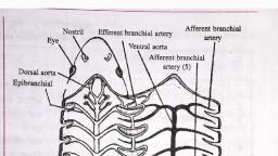
RTE LESTE, , ANNELIDA : PHERETIMA, POSTHUMA (EARTHWORM) i,, F SSTERIOR FACECOMMISSURAL DORSO ANTERIOR, , OF SEPTUM VESSEL INTESTINAL, FACE OF, VESSELS | SEPTUM, , , , , DORSAL VESSEL, , SUPRA-INTESTINAL, EXCRETORY, DUCTS, , , , Fig. 19. Pheretima: Blood vascular system behind 13 segments., , Gi) Ventral vessel : It is a large vessel that runs mid-ventrally between the alimentary canal and, nerve cord. It also extends throughout the length of the body. It has a thin wall without muscles., Internal valves are also absent, because the blood flows in it from in front backwards. Ventral vessel, isa distributing channel. In each segment, three branches arise from it-a pair of ventro tegumentary, vessels and an unpaired ventro-intestinal vessel (Fig. 20). Each ventro-tegumentary vessel runs, , DORSAL BLOOD VESSEL, DORSO-INTESTINAL COMMISSURAL, , b nr |, , INTESTINAL, sus-NEURAL, VESSEL VESSEL, , Fig. 20. Phereti T.S. body through the septum (right) and Ventrotegumentary vessel (left) segments., , , , backwards, pierces throu the intersegmental septum, to reach the succeeding segment. It, Supplies blood, by tect branches to body wall and integumentary nephridan. Before, , behind it gives offasmall septonephridial branch, supplying theseptal nephridia., The ventro-intestinal branches of ventral vessel supply blood to the intestinal wall., , Subneural : It is a slender vessel which runs beneath the nerve cord in mid-ventral, Wn tase spe cent up to the posterior end. It is a delicate vessel without valves, and muscles in its wall. Flow of blood is from in front backwards. It is a collecting vessel and receives, , Blood from ventral nerve cord and ventral body wall by a pair of small branches. It pour its blood into, _« the dorsal vessel by a pair of commissural vessels in each segment., () Arrangement of blood vessels in first 13 segments :, Dorsal and ventral vessels extends as such in this region of body, but the subneural vesse, bifurcated, in the 14th segment, into two lateral longitudinal vessels. Besides this, an i, | “pra oesophageal vessel is also present in this region of body., ‘cl, , ‘
Page 3 :
) MODERN’S ZOOLOGY = VOL. I + SEMESTER... 4, , (i) Dorsal vessel : It retains its muscular nature and valves but becomes a distributing Vesse),, his region. Some blood is supplied to buccal cavity, gizzard, oesophagus and pharyngeal Nephrig, yy means of small branches, one pair in each of the 3rd, 4th, 5th, 6th and 8th segments. Most of, slood is, however transferred to ventral vessel by 4 pairs of inflated, loop-like branches, locate,, round the gut, one pair in each of the segments, 7th, 9th, 12th and 13th. These branches have thig, muscular and rhythmically contractile walls, called hearts. They pump blood from dorsal to venj,,, vessel, while flow in opposite direction is prevented by internal valves. Each ‘heart’ of segment {._, and 13th connected with the supraoesophageal vessel also by means of a small branch. Hence, the., hearts are called latero-oesophageal hearts (Fig. 21 B), while those of the 7th and 9th are simp), called “lateral hearts” (Fig. 21A). The latter have 2 pairs of additional valves in their middle regio,, , , , , , , , SUPRA, OESOPHAGEAL, VESSEL, , VENTRAL, VESSEL, , , , VALVES, mi 8B, , Fig. 21. Pheretima: Hearts in section. A. Lateral heart, B. Latero-oesophageal heart, , (ii) Ventral vessel : The ventral vessel is a continuation of the ventral io, region and extends up to the second segment. It retains its ibe inwecrtc. the oe, also. However, it does not supply the gut in this region. It supplies bl a,, , a pair of ventro-tegumentary vessels to tegumentary nephridia, lood, has eco, (iii) Lateral oesophageal vessel : The two lateral, , os sc cronreag the subneural vessel. eae ia i. formed in the _, , the pre-intestinal part of the gutup tothe 2nd 2gment s ane oe ovat ;, , wall, septa, nephridia and genital organsby means ofa aie nent, these lect blood from, , it sends blood to the supra oesophageal, also through a pair of arog een emtary vessel. Morea’ Which, , loops in each of the 10th and 11th . e = Pair of thin walled, non-pulsatile, ante'# Rly, , Gy) oesophageal vessel
Page 5 :
x r MODERN'S ZOOLOGY — VOL. I: SEMESTER _,, , =kidney). In Pheretima, these are relatively more numerous, smaller and located inall except the, two segments. They are segmentally arranged, hence also called segmental organs. These are typ,, cally unbranched and their inner ends open into coelom by a ciliated funnel, called nephrostom,, Such a nephridium opening by a ciliated funnel, is termed a metanephridium. They are small siz, or micronephridia, as compared to large sized metanephridia of Nereis and leech. According to th,, location and structure, the nephridia of Pheretima are of three types - (A) septal (B) integument,, and (C) pharyngeal., , (A) Septal nephridia :, @) Position. These are the largest nephridia of Pheretima, and are situated to both the faces, , each intersegmental septum behind the 15th segment (Fig. 23). There are 20 to 25 nephridia in ead, row. Thus, there are 80 to 100 septal nephridia in each segment., , 2 DUCTS OF