Notes of S. Y. BSc, Zoology Study of Scoliodon.pdf - Study Material
Page 1 :
1, , STUDY OF SCOLIODON, 3.1, , SCOLIODON-THE INDIAN SHARK (DOGFISH), Systematic position, Habit and habitat, , 3.2, , External characters, , 3.3, , Digestive system, food, feeding and physiology of digestion, , 3.4, , Respiratory system, , 3.5, , Blood vascular system, , 3.6, , Nervous system and sense organs, , 3.7, , Male urinogenital system and female reproductive system, , Fishes occupy a unique place in the animal kingdom, since they are the forerunners of all the higher, forms of life, and all the essential characters of higher vertebrates make their first appearance amongst them., Fishes are distinguished by the presence of paired and median fins supported by fin-rays and by the fact that, they breathe throughout life by means of gills borne on visceral arches. Fishes living at the present time may, be divided into two groups: (1) Cartilaginous fish or Chondrichthyes (2) Bony fish or Osteichthyes., Chondrichthyes are characteristic by the absence of true bone. The genus Scoliodon is a cartilaginous fish., The genus Scoliodon was first described and named by Miiller and Henle in 1837. The commonest and most, abundant species of sharks found along the coasts of India belong to the genera Scoliodon., 3.1 Systematic Position:, Phylum, Chordata, Sub-phylum, Vertebrata or Craniata, Section, Gnathostomata, Class, Chondrichthyes, Order, Pleurotremata, Family, Carcharinidae, Genus, Scoliodon, Species, sorrakowah, Habits and Habitat:, The natural home of sharks is the Indians sea. It is carnivorous fish and voracious feeder. The, Scoliodon is active swimmer and aquatic breather like other fishes. Scoliodon exhibits sexual dimorphism, i.e. sexes are separate and distinguishable externally. Fertilization is internal. The Scoliodon is, viviparous i.e. gives birth to the young ones. It has a wide distribution in the Indian Ocean, Bay of, Bengal, Eastern Pacific Ocean and in the Atlantic Ocean along the coast of South America., Economic Importance:, Scoliodon has great educational and experimental value because of its availability and size., Sharks are used as human food in many countries. Scoliodon fins are dried and then boiled to yield a, gelatinous substance favoured for soups. The dried skin of shark is called shagreen is used for covering, carcasses, jewel boxes, sword handles and ornamental works. The dried skin of shark also used for, smoothing and polishing furniture., 3.2 External Characters:, The Scoliodon has a long, laterally compressed spindle-shaped body tapering at the
Page 2 :
2, , both ends. The full grown specimen is about 60 cm long. The dorsal and lateral sides of body are, pigmented dark grey while the ventral side is white. The body surface is very rough to touch because, of presence of placoid scales. The entire body is divisible into head, trunk and tail, though there are no, distinct boundaries between these three regions. The body supports several fins that help the Scoliodon, in locomotion and in maintaining balance.', , Head: The head is flattened and dorso-ventrally compressed. Head is prolonged in front into thin,, blunt, wedge-shaped snout. The mouth is wide crescentic in shape, found on the ventral surface and, is guarded by the upper and lower jaws which contain one or two rows of backwardly directed conical, teeth. The large circular eyes are situated at the sides of the head and each eye is provided with a, movable nictitating membrane. The skin forms upper and lower eye lids. The nostrils are obliquely, placed on the ventral surface of snout but they are for olfactory function instead of respiratory function., Several amputlary pores are present on the upper and lower surface of head. There are five vertical slits, on each side are' present called the branchial or gill clefts.
Page 3 :
3, , Fig. 3.4: Scoliodon V. S. of skin showing Development of a Placoid scale, Trunk: This region constitutes the major part of body extends from just behind the last gill cleft up, to the cloacal aperture. Trunk is thickest part of the body and is laterally compressed. Trunk contains, median unpaired fins and paired lateral fins. A median unpaired fin includes first dorsal fin, second, dorsal fin and ventral fin. The first dorsal fin found anterior to middle part of the body. The second, dorsal fin is found a little behind the first dorsal fin. The ventral fin is found on ventral side, little, distance to the position of second dorsal fin. There are 2 pairs of lateral or paired fins on trunk. The, fins of the anterior pair are called as pectoral fins and those of the posterior pair as the pelvic fins. The, pectoral fins are large and triangular expansions, originate from the ventro-lateral margins of the trunk, just behind the gill slit. The pelvic fins are much smaller than the pectoral fins and arise close together, from the ventral surface of the junction of the trunk and tail and enclose the cloacal aperture. In female,, these fins are simple and sub-triangular appendages but in male, each pelvic fin bears along its inner, edge a stiff, rod-like copulatory organ called the clasper. A pair of claspers is found attached to the, pelvic fins in male only. Each clasper has a groove on its dorsal surface which leads into a cavity, the, siphon at the base of clasper. The_cloacal aperture is an.elongated, opening at the base of the tail on, the ventral side between the two pelvic fins. It is an outlet of a common chamber, the cloaca into, which rectum, urinary and genital ducts open. There is a pair of openings situated on elevated papillae, on either side of the cloaca; these are the abdominal pores, through which the coelom communicates, with the exterior., Tail: The portion of the body behind the cloaca is called tail. The tail is laterally compressed and is, bent up-wards at a small angle. Such tail is known as heterocercal tail. The caudal fin extends along
Page 4 :
4, , the dorsal and ventral surfaces of the tail in the the median line and forms a dorsal epicaudal lobes, and ventral well developed hypocaudal lobes. At the junction of caudal fin and tail is a caudal pit., Histological structure of skin, The skin or integument consists of two distinct layers, an outer epidermis and the, inner dermis., 1. Epidermis: The epidermis composed of many layers of epithelial cells. The stratum germinativum, or Malpighian layer is the innermost layer rests on basement membrane. The cells are cuboidal and, they divide mitotically forming new cells externally. Numerous unicellullar mucous glands are, present, in, the, epidermis, and, they secrete mucus. The mucus is helpful for reducing the friction in water and preventing the settling, of foreign organisms on the skin. It is ectodermal in origin., 2. Dermis: It is mesodermal in origin and it consists of dense connective tissue mixed with smooth, muscle fibres, blood capillaries, pigment cells and nerves. The pigment cells lie just below the, epidermis. This rough skin is due to the presence of closely lying minute dermal denticles called, placoid scales which are arranded in regular oblique rows and from the exoskeleton of the shark, covering the entire surface of the body., Structure of Placoid scales:, Placoid scales are derived from both epidermis and dermis of skin. The placoid scales or dermal, denticles are characteristics of elasmobranch or chondricthyes fishes like sharks, rays, skates, chimeras, etc., Placoid scales or odontoids are embedded in the skin in regular oblique rows, forming the exoskeleton The, placoid scales are minute dermal denticles of dentine embedded in the dermis. A typical placoid scale is, distinguished into the basal plate and spine., Basal plate: It is a diamond-shaped or rhomboidal plate formed of cement like material. It lies embedded in, the dermis and is firmly attached to it by strong fibres of connective tissue. The basal plate is perforated by a, small opening which leads into the pulp cavity of the spine. In live condition the blood vessels, nerve fibres, and lymph channels enter the pulp cavity through this opening. The pulp cavity is filled with numerous, odontoblast, cells, connective tissue, fibres, nerve cells and blood vessels which collectively form the pulp., , The Spine: It is a flat, backwardly directed trident structure projecting out of the skin and giving it
Page 5 :
5, , roughness. The surface of spines is not smooth but presents a stratified appearance under the, microscope. The spine composed of a hard calcareous substance, the dentine which is external coated, with a hard dense enamel like substance, the vitrodentine. The vitrodentine is secreted by the, epidermal cells., , Importance of Scales in Fishes:, , I., 2., 3., 4., , The main function of scales is protection and for that purpose only they are differently modified into, spines, knobs or bony plates., The structure and arrangement of scales' has a taxonomic value., Scales are also useful in identification of fishes., Scales form an important tool in the palaeontological study of fishes, as they are well preserved and, get embossed clearly on the surrounding clay., , Coelom and Viscera:, In Scoliodon, the coelom is spacious and is divided into two unequal cavities, the pericardial and the, abdominal cavities, separated from each other by a membranous partition, the septum transversun. The, pericardial cavity is comparatively smaller in size and triangular in shape and lies beneath the Pharynx, and surrounds the heart. It is enclosed between tightly fitting smooth layers of peritoneum lining the, outer wall of the cavity and the inner pericardial layer which adheres closely to the heart itself. The, pericardial cavity contains a clear colorless fluid, the pericardial fluid. It communicates wit h the, abdominal cavity through aperture in the septum transversum called the pericardio-peritoneal canal., The abdominal cavity is very large surrounding the viscera and communicating with the exterior, through a pair of abdominal pores situated on papillae, one on either side of the cloacal aperture., The abdominal cavity is lined with the peritoneum and is also filled with a colorless coelomic fluid ., The alimentary canal is suspended in the body cavity by a double fold of peritoneum called, mesentery. The mesentery is incomplete, only the anterior and posterior portions of the mesentery, are well developed. The anterior portion of the mesentery forms a large flap, the mesogaster which, suspends the stomach, while the posterior portion, the mesorectum suspends the hinder part of the, alimentary canal. There are other membranes which attach different organs to one another and are, called, omenta (singular omentum). The membrane which connects the stomach with the liver is known as, gastro-hepatic omentum, while the membrane connecting the spleen with the stomach is the gastrosplenic omentum. In Scoliodon, the viscera comprise the various organs of the body lying in the, body cavity. The important organs are heart, liver, stomach, spleen, pancreas, gonads and kidneys.
Page 6 :
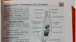
6, , 3.3 Digestive system of Scoliodon: The digestive system can be described under the following parts:, a) Alimentary canal,, b) Glands of alimentary canal and, c) Physiology of digestion, a) Alimentary canal: The alimentary canal of Scoliodon comprises the mouth, buccal cavity, pharynx,, oesophagus, stomach, intestine and rectum and cloaca.
Page 7 :
7, , Mouth: It is a wide ventral cresentic openings which leads into a spacious dorso-ventrally compressed, buccal cavity. The buccal cavity is lined with a thick mucous membrane raised ventrally into a thick, fold to form the so-called tongue which is non muscular and non glandular.The mucous membrane is, rough due to the presence of dermal denticles. The teeth are oblique and have sharp more or less, compressed cusps, the edges of which are smooth and non-serrated. The teeth are alike in shape,, homodont, and are borne in several parallel rows on the inner margin of the upper and lower jaws., The teeth are used to catch the prey and prevent its escape but not to crush or masticate it. Through, there are several rows of teeth yet only one row function at a time (polyphyodont) and the old row, is replaced by the new one. There are no glands in the buccal cavity comparable to the salivary, glands of higher vertebrates., , Buccal Cavity: It is a large, dorsoventrally compressed cavity opens into the pharynx, on either, side of which lie the internal openings of the spiracle and five gill pouches. The spiracle is vestigial
Page 8 :
8, , represented by an inconspicuous oval pit and the gill-pouches are large. The cavity of pharynx is, lined with mucous membrane containing numerous dermal denticles., Pharynx: The pharynx narrows posteriorly to form the short oesophagus. The oesophagus has, thick muscular walls with an internal lining of mucous membrane raised into longitudinal folds., Oesophagus: The pharynx becomes narrow posteriorly and merges into the oesophagus. It is, short, dorso-ventrally compressed and lined with mucous which is highly folded. The mucous is, composed of ciliated cells and nuicellar mucous glands. The oesophagus posteriorly widens to, form a large muscular stomach., Stomach: The stomach consists of two chambers, cardiac and pyloric stomach. The stomach is, bent on itself and forms a J -shaped organ, the long proximal limb of which is called the cardiac, stomach, while the short distal limb is called the pyloric stomach. At the junction of cardiac and, pyloric limbs there is a blind outgrowth, the blind sac. The inner mucous lining of the cardiac, stomach is also thrown into prominent longitudinal folds that end in the depression of the blind, sac. The lining of the pyloric stomach is quite smooth proximally but is slightly folded distally. At, the end of pyloric stomach there is a muscular bursa entiana. The opening of pyloric stomach into, the bursa entiana is guarded by a circular band of muscle fibers called the pyloric valve. The bursa, entiana continues into the intestine., Intestine: The intestine is a wide tube running straight backwards into the abdominal cavity and, opening positively into the rectum. The internal surface of the intenstine is increased by a, characteristics fold of the mucous membrane, the scroll valve, having one edge attached. to the, inner wall of the intestine and the other rolled up longitudinally on itself into a scroll, making an, anti-clockwise spiral of about two and half turns. In a transverse section the scroll valve looks like, a watch spring. The scroll valve serves not only to increase the extent of the absorptive surface of, the intestine but also prevents the rapid flow of food through the intestine., Rectum: The rectum is the last part of the alimentary canal. The tubular rectal (caecal) gland opens, dorsally into the rectum. The rectum leads into the cloaca into the alimentary canal as well as the, urinogentical ducts open. The rectal gland is small, tubular, thick diverticulum that arises the dorsal, side of the rectum. It secretes a fluid which poured into the intestine but its action is unknown.
Page 9 :
9, , Digestive Glands:, 1. Liver: It is a large, massive, elongated, bilobed, yellowish gland, occupies gteater part of the abdominal, cavity. The right and left lobes are leaf shaped and attached to the septum transversum by a median suspensory, ligament. They are united anteriorly but lie apart posteriorly and extend backwards freely in the abdominal, cavity. The right lobe of the liver possesses a thin walled, V shaped sac anteriorly called the gall bladder. It, stores the bile. The bile from the gall bladder is carried by long bile duct which' on its way receives branches, from the right and left lobes of liver and forms the common bile duct. The common bile duct opens in the, duodenum., Liver produces bile, stores glycogen and fat and destroys the worn out erythrocytes., 2. Pancreas: It is a compact, whitish, ribbon like bilobed gland situated between the cardiac and pyloric, stomach. It shows a longer dorsal lobe running parallel to the posterior part of the cardiac stomach and smaller, ventral lobe lying close to the pyloric stomach. The pancreatic duct runs across the complete length of the, gland and opens into the intestine opposite to the opening of bile duct. The interior of pancreas contains, exocrine as well as endocrine cells. The exocrine cells are arranged in cords or their groups form acini. They, are serous and secrete the pancreatic juice. The endocrine tissue can be observed outside the epithelial cells of, smaller ducts or as single cells at the base of larger ducts., 3. Rectal gland: It is short thick diverticum arising from the dorsal wall of the rectum. It secretes a fluid, which poured into the intestine but its action is unknown., 4. Gastric glands: These glands occur in the wall of the stomach which secret gastric juice as well as, hydrochloric acid., 5. Intestinal glands: These glands occur in the wall of the intestine which secrete intestinal juice for digestion, of food., Physiology of digestion:, Food: The new born babies and young ones are bottom feeders and they feed exclusively on prawns. As, growth proceeds they change their diet to crabs. small soles etc. The full grown Scoliodon mainly feeds on, fishes such as mackerel, oil sardine, silver bellies etc. and also crabs, lobsters and worms. Sharks feed at one, or two days intervals.
Page 10 :
10, , Ingestion: The prey is held with the help of teeth and jaws and engulfed, entirely without mastication., Digestion: By the process of digestion, the complex and nonabsorbable food is transformed to simple, absorbable form. The digestion mainly occurs in the cardiac stomach. As compared to the intestinal digestion, it is quite fast. The cells of the musous membrane of the cardiac stomach secrete, gastric juice which contains hydrochloric acid and pepsin. The gastric juice digests proteins but not chitin., Action of gastric juice:, (1), , Pepsinogen HCl, HCl Pepsin, (Inactive), (Active), (2) Proteins + Pepsin, ~ Syntonin, peptones, proteoses., Digestion in Intestine: The semi-digested food from the stomach enters the intestine and 'mixes with bile and, pancreatic juice. The intestinal digestion and absorption needs a period of three to five days which, depends on the temperature of the surrounding water. Warm water accelerates the process., Action of Bile:, (1) It makes the acidic food alkaline which is necessary for the action of pancreatic juice., (2) Trypsinogen, (Inactive), (3) Fats, , . Bile, Enterokinase, , Bile, , Trypsin, (Active), , Emulsification, , Action of pancreatic juice:, Pancreatic juice contains trypsin in the form of inactive trypsinogen, amylopsin and lipase. It acts as, follows, , (1), , Chymotrypsinogen Enterokinase, (Inactive), , Chymotrypsin, (Active), , Trypsin, (2) Proteins, , Polypeptides, peptones, proteoses., Chymotrypsin, (3) Polypeptides, peptones, proteoses Erepsin, Amino-acids, (4) Polysaccharides, (5) Maltose, , Amylases, , Maltase, , (6) Emulsified fats, , Disaccharides (Maltose), , Glucose (Monosaccharide), , Lipase, , fatty acids, glycerol., , Absorption: The digested food is absorped into the blood over the surface of intestine and scroll valve., 3.4 The Respiratory system:, Scoliodon is adapted to respiration in an aquatic medium and breathes by means of gills, borne in a, series of gill-pouches on either side of the pharynx. Water enters the buccal cavity and pharynx through the, mouth and passes out through the gill-slits, bathing the gills on its way through the branchial pouches. There, are five pairs of gill pouches in Scolioclon. Each pouch is compressed antero-posteriorly and communicates,, on, the one hand with the cavity of the pharynx by a large internal branchial aperture, and on the other, with, the exterior by a narrow external branchial aperture. The mucous membrane lining the gill-pouches is raised, into a series of horizontal folds, the branchial lamellae which are richly supplied with blood capillaries. Each
Page 11 :
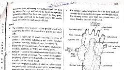
11, , gill-pouch has two sets of gill-lamellae one on its anterior wall and another on its posterior wall. The posterior, set of lamellae being longer than the anterior set. Successive gill pouches are separated from one another by, stout fibro-muscular partitions called the interbranchial septa, in the inner or pharyngeal border of which are, imbedded the visceral arches, with their branchial rays., These arches thus alternate with the gill pouches, each being related to the posterior set of lamellae of, one pouch and the anterior set of the next. These two sets of lamellae attached to a visceral arch constitute a, complete gill or holobranch, while a single set makes hemibranch or a half-gill. A gill-pouch thus contains the, posterior hemibranch of one gill and the anterior hemibranch of the succeeding gill. The hyoid arch bears gilllamellae on its posterior surface only and, therefore, has only a half-gill or hemibranch; the first four branchial, arches bear gill-lamellae on both surfaces called as holobranch; while the fifth branchial arch is, entirely gillless called as hemibranch., , Mechanism of Respiration:, The fish depresses the floor of the buccal cavity by the contraction of the hypobranchial muscles, and, consequently, the buccal cavity is enlarged and its volume capacity is increased. Simultaneously with the, lowering of the floor of the buccal cavity, the mouth is opened and the water rushes in to fill the enlarged, buccal cavity. The cavity of the pharynx is next enlarged by the raising of the branchial arches, while the
Page 12 :
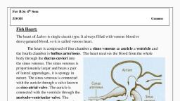
12, , mouth is closed by the action of the adductor muscles. Following the line of least resistance, the water enters, the branchial pouches, and is forced out through the external gill-slits. In the gill pouches, the water comes in, contact with the capillaries of the gill-lamellae, where the non-oxygenated blood is separated from the oxygen, dissolved in water only by the very thin capillary-wall, so that the oxygen passes into the blood, while the, carbon dioxide in the blood diffuses out into the water. Thus an efficient exchange of oxygen (O2) and carbon, dioxide (CO2) take place between the blood and sea water by a process of endosmosis and exosmosis., , 3.5 The Blood Vascular System:, The blood vascular system includes the heart, arterial system and venous system., Heart:, The heart of Scoliodon is a bent muscular tube, it lies in the pericardial cavity below pharynx. . It is reddish, brown and conical organ. The heart consisting of the following chambers:, 1. The sinus venosus, 2. The auricle, 3. The ventricle and, 4. The conus arteriosus., 1. The sinus venosus: It is a thin-walled triangular chamber placed transversely along the base of the, pericardial cavity. Two large veins, the ducti Cuvieri, enter the sinus laterally, while the two hepatic sinuses, open into it in the postero-median line. Anteriorly, the sinus venosus opens into the auricle through the sinuauricular aperture, guarded by a pair of membranous valves which prevent the backward flow of the blood., 2. The auricle: The auricle is a large triangular sac with walls thicker than those of the sinus venosus. It is, situated in front of the sinus venosus and dorsal to the ventricle. The sides of the auricle projecting on either, side of the ventricle give it the characteristic ear-like or auricular appearance. It communicates with the, ventricle by the auriculo-ventricular aperture guarded by a bilabiate valve., 3. The ventricle: It is the most prominent part of the heart and is supported ventrally by the coracoids cartilage., It has very thick muscular walls, the inner lining of which is traversed numerous muscular strands that give, ventricle and spongy texture. The ventricle opens in the conus arteiosus and opening is guarded by cuspid, valves. The valves are provided by chordate tendinae ( elastic threads), 4. The conus arteriosus: The conus arteriosus is a stout; muscular tube which extends anteriorly from the, ventricle to the front of the pericardial cavity. The interior of the conus arteriosus is provided with two, transverse rows of semi-lunar valves, each row containing three valves a dorsal and two ventro-laterals. In, addition to these, there is always a small accessory valve on either side of the dorsal. Fine tendinous threads, from the free ends of the valves are attached anteriorly and posteriorly to the muscular processes of the wan
Page 13 :
13, , to hold the valves in place. The conus arteriosus is continued forwards through the wall of the pericardium as, the ventral or cardiac aorta., , Working of Heart: The function of the heart is to pump the blood to the different parts of the body. Since the, blood passes through heart only once in its the circuit, it is obvious that there should be sufficient pressure in, the heart to force the blood through the different organs of the body. This is effected by the rhythmic, contraction of the different parts of the heart in a definite Succession and at regular intervals. The heart-beat, or contraction starts in the sinus venosus, and the blood brought to it from the different parts of the body by, the veins is forced into the atrium or auricle through the sinu-auricular aperture. The membranous valves, guarding this aperture prevent the blood from flowing back into the sinus venosus. The muscular contraction, next spreads over the auricle and drives the blood into the ventricle. Here again, the return of blood into the, auricle is prevented by the bilabiate valves guarding the auriculo-ventricular aperture. Then wave of, contraction passes to ventricle and the contraction of the ventricle forcing the blood into the conus arteriosus., As the wave of contraction passes from the ventricle to the conus and thence into the ventral aorta, any return, of the blood into the ventricle is prevented by the two rows of semi-lunar valves present in the conus arteriosus., Then the blood passes into the afferent arteries which break up into the capillaries of the gills where the blood, is oxygenated. Thus, only the venous blood (Deoxygenated) passes through the heart in Scoliodon., This is known as the single type of circulation and the heart is called venous or branchial heart. A, cardiac branch of the vagus nerve forms a nerve plexus in a patch of muscles in the sinus venosus, to form a sinuauricular node where the heart-beat starts.
Page 14 :
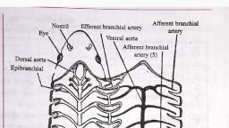
14, , Arterial System: The arterial system comprises of the following vessels., 1. The ventral aorta and afferent branchial arteries: Ventral aorta arises from the conus arteriosus and, upturned. Ventral aorta extends along the ventral surface of the pharynx right up to the Posterior border of the, hyoid arch, where it bifurcates into two branches, each of which again divides into two forming the first and, second afferent branchial arteries. The first afferent branchial artery runs along the posterior border of the, hyoid arch supplying numerous branches to the gill-lamellae of the hyoidean hemibranch. The second afferent, branchial follows a similar course along the first branchial arch supplying branches to the anterior and, posterior gill-lamellae along its whole length. The third, fourth and fifth afferent arteries arise equal distant, from each other from the ventral aorta and run along the ventral surface of the second. third and fourth visceral
Page 15 :
15, , arches. All the afferent arteries arise by their own separate openings from the ventral aorta which supply, deoxygenated blood to gills for oxygenation., 2. The efferent branchial and epibranchial arteries: The blood from the capillaries of the gills is collected, by a series of blood-vessels called the efferent branchial arteries which form loops round the gil1-cIefts. There, are nine efferent branchial vessels running- along the anterior and posterior borders of the five gill-clefts. The, posterior efferent artery of each loop is much larger than the anterior and receives the major portion of the, blood from each gill; while the anterior and posterior efferents of adjoining clefts communicate with one, another by short horizontal connecting vessels, placed about the middle of each gill, across the interbranchial, septum. Each of the four efferent branchial loops is continued into an epibranchial artery which runs, backwards and inwards to the mid-dorsal line. These four pairs of epibranchials unite to form the median, dorsal aorta., 3. Arteries of dorsal aorta: The dorsal aorta is formed by union of four pairs of epibranchial arteries. The, dorsal aorta runs back below the vertebral column and enters the tail as a caudal artery which passes, through the haemal canals of the caudal vertebrae and goes to the tail. The dorsal aorta gives off a, number of paired and median arteries. These include buccal arteries arises from anterior end and give, blood to roof of the buccal cavity, the radices also rise from anterior end and, join the common carotid., Pair of slender arteries called subclavians arises in front of the last epibranchials going to the pectoral, fins. Each subclavian gives off a lateral artery, and dorso lateral artery. It then enters the pelvic fin as, the branchial artery. The coeliaco mesenteric artery is arising from little behind the last epibranchial, arteries. It is again divided into coeliac and anterior mesenteric artery. The coeliac gives off gastric, and hepatic supplying blood to oesophagus, stomach and liver. The anterior mesenteric arteries and, their branches supplying blood to scroll valve. The pancreatic artery carrying blood to the pancreas., The gastro-splenic is also a median artery and it sends genital to gonads, posterior gastric supplying, the pyloric stomach and spleen and dorso intestinal supplying blood to the dorsal wall of the intestine., The posterior rmesenteric artery arises near the end of the trunk and supplies blood to the rectal gland., Iliac arteries are pair of slender arteries arising from the aorta at the level of pelvic fins and they, supply blood to pelvic fins. There are number of paired segmental or
Page 16 :
16, , parietal arteries given off from the dorsal aorta. The parietal divides into dorsal and ventral parietal., The dorsal parietal supplies the dorsal and lateral body-wall muscles, vertebral column, dorsal fins,, while ventral parietal supplies ventral body wall muscles and sends renal branches to kidney.
Page 17 :
17, , 4. The Arteries of the head: The head receives its blood-supply direct from the efferent branchials unlike the, rest of the body which is supplied by the branches of the dorsal aorta. All the arteries of the head originate, from the first hyoidean efferent and consist of three arteries arise from the first efferent branchial of, each side, they are an external carotid going to head and lower jaw, an afferent spiracular artery which, passes around the spiracle and is then known as the spiracular epibranchial artery supply blood to, the brain. The external carotid sends orbital artery to eye muscle,abuccal artery to the muscles of the, lower jaw, a maxilla-nasal artery, to the olfaclory sacand muscles of the upper jaw, and then enters, the rostrum as the rostral artery. The hyoidean artery arises from the first efferent branchial artery. It, sends an ophthalmic artery to the eye-ball and enters the cranium. Here it joins the internal carotid, forming cerebral artery. It again divides into anterior and posterior cerebral arteries which supply, blood to brain and form basilaries artery which runs backwards all along the ventral side of the spinal, cord as a spinal or myelonal artery. The mandibular artery arises from the ventral end of the first, pretrematic efferent branchial artery. It forms submental artery and provides blood to muscles of, lower jaw., 5. The hypobranchial blood plexus: It is present in the ventral wall of pharynx includes four, longitudinal vessels, a pair of median hypobranchial arteries and a pair of lateral hypobranchial arteries., The two median hypobranchial arteries are interconnected by transverse vessels and meet posteriorly, to an unpaired artery and gives off pair of coronary artery to the heart wall and pericardial artery to, the pericardium., Venous System:, In Scoliodon the blood distributed to different parts of the body by the arteries and their branches and, returned to the heart by the veins. The venous system consists of wide, thin walled, non-contractile, blood spaces called sinuses, instead of narrow, tubular muscular veins. The system can be divided into, following parts:, 1. Anterior cardinal system, 2. Posterior cardinal system, 3. Lateral abdominal system, 4. Portal system, 5. Cutaneous system, 1. Anterior cardinal system: This system collects blood from anterior part of body and, it contains two large sinuses called anterior cardinal and inferior jugular sinus., (a) Anterior cardinal sinus: They are large, ventral and collect blood from head, and branchial regiondorsally and finally open into the ducti Cuvieri. The number, of small veins from rostrum joins to form orbito- nasal or anterior facial vein. This vein passes, backwards and forms nasal or olfactory sinus. Nasal and orbito-nasal sinuses open into large, orbital sinus located in orbit below eye-ball. The right and left orbital sinuses are interconnected, by an inter-orbital vein. The orbital sinuses pour their blood into a large anterior cardinal sinus, through a narrow post- orbital sinus. In addition the anterior cardinal receives blood from five, dorsal nutrient sinuses from the gills., (b) Inferior jugular sinus: It is small ventral present below the gill clefts and receives blood, from the floor of the buccal cavity and the pharynx. It also receives five ventral nutrient sinuses, from the ventral region of the gills. The anterior cardinal and inferior jugular sinuses and finally, open in ducti Cuvieri., 2. Posterior cardinal system:
Page 18 :
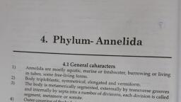
18, , The posterior cardinal ·system consists of caudal vein, two renal vein and two large posterior cardinal sinuses., The caudal vein collect blood from tail and bifurcate into right and left renal portal vein in kidney. Small, Parietal veins from body wall join the renal portal veins to form posterior cardinal sinuses. The blood from, gonads collects in a pair of genital sinus. All along their course, they receive segmental veins from the bodywall. Finally each sinus opens into the ductus Cuvieri., 3. Lateral abdominal system:, It, is, formed, by, a, pair, of, large, vessels, running, along, the, lateral, sides., They, are, present, outside, the, peritoneum, in, the, trunk, region., It, starts at the level of the pelvic girdle and collects blood from inner lateral, body, wall,, paired, fins, and, cloacal, region., Posterior,, they, are, connected, with each other by a commissural vein. Each lateral abdominal is formed by:, a).Cloacal, vein:, Small,, draining, blood, from, the, wall, of, the, cloaca., b). Femoral vein: Dorsolateral and drains blood from the pelvic fin., c). Brachial vein: Anterior from the pectoral fin., The lateral abdominal joins anterior with brachial vein and forms the subclavin vein which opens into the, ductus Cuvieri., 4. Portal system: There are two types of portal systems namely hepatic and renal., (a), Hepatic, portal, system:, The, blood, from, alimentary, canal, and, digestive glands carries to the liver is called as hepatic portal system. It consists of a large hepatic, portal vein which receives number of branches namely, anterior gastric vein from cardiac stomach,, anterior lieno- gastric vein from pyloric stomach and spleen, ventral intestinal vein from the ventral wall, of the intestine and intra intestinal vein from the ventral wall of the intestine and intra intestinal vein from, scroll valve Blood from the liver is returned by a pair of large hepatic sinuses which open into sinus, venosus., (b) The renal portal system: The blood collects from tail and distributes into the kidneys by, caudal vein. In the abdominal cavity, caudal vein bifurcates into right and left renal portal veins. The, blood from kidneys is discharged into posterior cardinal sinsues., 5. Cutaneous system: It consists of a dorsal, a ventral, and two lateral veins. These are best seen in transverse, sections of the body and tail regions. The dorsal cutaneous vein lies below the skin along the mid-dorsal line, and collects blood from the skin of the dorsal side. The ventral cutaneous vein lies in the mid-ventral line, beneath the skin, while the lateral cutaneous vein runs along the sides’ ventral to the lateral line canal.
Page 19 :
19, , 3.6 Nervous system of Scoliodon: The nervous system of Scoliodon consists of three parts:, (1) Central nervous system: It includes brain and spinal cord., (2) Peripheral nervous system: It includes cranial and spinal nerves., (3) Automatic nervous system:, (1) Central nervous system:, (A) Brain: The brain is divided into three parts: fore-brain, mid-brain and hind-brain., a).Fore-brain: The fore-brain includes olfactory lobes, cerebrum and the diencephalon., The olfactory lobe consists of pair of stout stalks, the olfactory peduncles extending forwards, and outwards from the antero lateral angle of the cerebrum. Olfactory peduncles, end in a bilobed mass called olfactory bulb or lobe, closley applied to the olfactory sac of its, own side. The olfactory tract and bulbs enclose narrow cavities, the olfactory ventricles or, rhinocoels.
Page 20 :
20, , The cerebrum is undivided massive structure with no median groove to separate it into right and, left cerebral hemispheres. It contains pair of cavities called lateral ventricles or paracoels separated by, median partition and they are continued with foramen of Monro. The dorsal surface of cerebrum is, quite smooth but on mid ventral surface, there is small opening, called neuropore through which emerge, a pair of delicate nerves, the terminal or pre-olfactory nerves. Each of these nerves bears a ganglion, along its course and runs along side the olfactory tract of its own side to innervate the mucous membrane, of, the, olfactory sac., The cerebrum is continued behind into the narrow diencephalon which is very short and is, completely hidden by the forward prolongation of the cerebellum. The roof of the diencephalon is, extremely, thin, and, membranous,, being, non-nervous, in, character,, but, it, contains numerous blood vessels forming the anterior choroid plexus. Ventrally its anterior margin bears, optic chiasma of two optic nerves. Just behind the chiasma a hollow projection called infundibulum is situated, on the floor. Attached posteriorly to infundibulum is a sac like hypophysis and both make up the pituitary, body. Close to the lateral sides of infundibulum lie two thick walled oval sacs called the lobi inferiors, the, distalend which are continued into a pair of glandular sacs, the sacci vasculosi with thin walls., (b) Mid brain:, It is not a prominent part and remains closely concealed dorsally by cerebellum and ventrally by the, infundibullar outgrowths. It consists mainly of a pair of large, rounded dorsal swellings the optic lobes or, carpora bigemina, which are the centres of sight and hearing. Each optic lobe has a cavity called optocoel. Ill, and IV cranial nerves arise from midbrain., (c) Hind brain: It consists of two parts cerebellum and medulla oblongata., Cerebellum is large, elongated and rhomboidal structure overhanging the optic lobes in front and part, of medulla behind. Its dorsal surface is irregularly folded and divided into three lobes by two transverse, furrows. It has a cavity called metacoel. Anterior end of hind brain arises, a pair of hollow outgrowths, the, carpora restiformia or auricular lobes., Medulla oblongata, the last part of the brain is a triangular structure and continues posteriorly into the, spinal cord. Medulla is roofed over by a thin, non-glandular and vascular membrane, the posterior choroid, plexus. It has a wide cavity called fourth ventricle or myelocoel., Functions of Brain:, 1. Olfactory lobes and cerebrum are useful for sense of smell, 2. The cerebellum is the seat of regulation of balance and muscular control., 3. The lobi inferiors and sacci vasculosi are centres for smell and taste., 4. Optic lobe has optic, olfactory, gustatory and acoustic-lateral sensory centres, 5. The medulla oblongata contains the respiratory centres.
Page 21 :
21, , (B) Spinal Cord: It is the extension of medulla oblongata up to the end of tail, within the neural canals of, vertebral column. It encloses a central canal and inner grey matter covered by outer white matter. It has shallow, dorsal fissure and well marked ventral fissure. It has at its centre, a narrow cavity called central canal filled, with cerebrospinal fluid., Function: It conducts the sensory and motor impulses to and from the brain. It is also a centr for local and, spontaneous reflex action.
Page 22 :
22, , 3.7 Urinogenital System:, The excretory and reproductive organs are closely related to each otherand are deals with together, hence it called as urinogenital System. In Scoliodon Sexes are separate and the urinogenital system, includes different organs in the male and female fishes., Male Urinogenital System:
Page 23 :
23, , The kidneys are a pair of long ribbon-like glandular structures, lying dorsal to the peritoneum and extending, anteriorly to the base of the liver and posteriorly to the side of the cloaca. The posterior portion of the kidney, is greatly thickened and laterally compressed and forms the chief organ of excretion. In this portion, uriniferous tubules are present The anterior part is comparatively narrow and non-renal part, which results, from the fact that this portion cones into the service of the genital system. In this portion uriniferous tubules, are absent. . The posterior broader part of each kidney is excretory and contains masses of coiled, uriniferous tubules with peritoneal funnels, Malphighian bodies and collecting tubules. The, collecting tubules of the anterior region are very small and open into Wolffian duct, while the, collecting tubules of the posterior region open into a common duct, the ureter. The ureters finally, open into a wide chamber called urinogenital sinus and it opens into the cloaca.The urineis hypotonic, to blood. The genital part of the system includes a pair of testes, vasa efferentia, a pair of Wolffian, ducts, a pair of sperm, sacs, a pair of siphons and a pair of claspers., Testes: There are two elongated testes, each running from the liver to the rectal gland. They are, attached to the abdominal wall by a double fold of peritoneum called mesorchium. Each testis is, composed of numerous seminiferous tubules lined with germinal epithelium. Each testis is made, up to numerous lobules with median central canal, Vasa efferentia: From each testis arise several thin tubular vasa efferantia which open into the, anterior and of that vas deferens. These ducts are convoluted called as epididymis. The vas, deferens which passes back to form a broad seminal vesicle. The seminal vesicle of both sides, open behind into a large triangular chamber called urinogenital sinus., Spem sac: On either side of the urinogenital sinus lies a club shaped sperm sac. The function, sperm sac is unknown., Siphons: A pair of elongated siphons lies on the ventral side of the body between the skin and, muscles., Claspers (Myxopterygium): These are paired stiff, rod like copulatory organs which are modified, pelvic fin. Each claspers is a tube partially open on the dorsal side forming a triangular groove, which open into cloaca.
Page 24 :
24, , Female Urinogenital System:, There is no direct connection between kidneys and genital organs in the female. Therefore anterior part of, kidneys is extremely reduced. The posterior part of kidney is thick and massive and which is functional renal, part. It also extends to the posterior limit of the cloaca. From each kidney arises a ureter and two ureter unite, to form a common ureter which opens into a urinary sinus. The female genital part of the system includes, paired ovaries, oviducts, shell glands and uteri., Ovary: These are large, yellowish, lobulated, saccular bodies and vary in form, and size as per the age of, shark., They, are, located, in, the, abdominal, cavity-and, attached, to, the, anterior, abdominal, wall, middorsally, by, a, fold, of, peritoneum, called, mesovarium., The, surface, shows, a, number, of, rounded, projections, which, contain, the, developing, ova., They, extend, back, from the base of liver and merge into the epigonal organ., Oviducts:, They are large tubes extending along the complete length of the, body, and, are, also, known, as, mullerian, ducts., Anteriorly, they, are, narrow, and, unite, mid, ventrally, below, the, oesophagus., They, open, into, the, coelom, by, zigzag, slit, like, aperture,, the, ostium, or, oviducal, funnel, lying, ventral, to the oesophagus and posterior' to transverse septum
Page 25 :
25, , Shell gland: The gland is also known as oviducal or nidamental gland which is, heart, shaped, and, shows, narrow, middle, mucus, secreting, zone, and, posterior large shell secreting zone, Uterus: Towards the posterior region, the oviduct dilates to form a uterus. The two uteri unite to form a short, vagina, which opens into cloaca., Copulation and Fertilization, For copulation, one or both claspers of the male are inserted into the cloaca of the, female. The forwardly directed spines of the scales on the claspers facilitate their, insertion and check their slipping out during copulation. The seminal fluid with sperms is passed into, the grooves of the claspers. The apopyles are closed by tilting the claspers., The muscular walls of the siphons contract forcing the sea-water present in them into the, grooves of the claspers. With this water, the seminal fluid is flushed into the cloaca of the female., The sperms swim up to the oviducts, reaching in front of the shell glands where fertilization occurs., Mature eggs reach in the shell glands from oviducal aperture and oviducal funnel.
Page 26 :
26, , Development, After fertilization, the development of the embryos takes place in the uteri. Three to, seven embryos develop in each uterus. During the breeding season, two uteri occupy, greater part of the abdominal cavity. The mucous membrane of the uteri forms as many, chambers as there are embryos. The chambers are full of a fluid that protects the embryos. In the, early stages, the embryo gets nourishment from the yolk of the egg contained in a reservoir called, yolk sac and conveyed to the embryo by a tubular yolk stalk, which is connected to the gut of the, embryo., When the yolk is used up, the yolk sac becomes folded and embedded in the uterine, wall. This connection between embryonic and maternal tissues is called the yolk sac, placenta. In the mean time, the blood vessels extend into the yolk sac by way of the yolk, stalk, which thus becomes placental or umbilical cord. Attached to the umbilical cord are, numerous tubular appendicula which also absorb nourishment from the uterine wall., When embryos are fully formed, the mother gives birth to them. Thus Scoliodon is, viviparous but some sharks are oviparous and ovoviviparous, Excretion and Osmoregulation:, Scoliodon is ureotelic i.e. the end product of nitrogen metabolism is urea. A large, quantity of urea is retained in the body as an adaptation to marine life. Retention of urea is brought, about in two ways:, 1. Synthesis of urea in all tissues, and, 2.Absorption of urea from the glomerular filtrate by the special urea absorbing, segments of the uriniferous tubules of the kidney., The urine is, thus, hypotonic to sea-water and consists mainly of water and salts The high urea, content makes the osmotic pressure of elasmobranchs higher than that of, sea-water, this would mean a constant flow of water, but this is prevented by the reduced, permeability of the gills. Thus, urea is not lost and water is constantly removed by the, kidneys enabling sharks to live in sea-water. Excess of urea is excreted chiefly through, gills.
Page 27 :
27
 Learn better on this topic