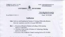
Page 1 :
Department of Zoology Government First Grade College. K R Nagar., , LABORATORY MANUAL, FOR B.Sc., IN, , 2. RANJITHA J, Faculty of Zoology, , 3. VAISHNAVI S, M.Sc., , 1
Page 2 :
Department of Zoology Government First Grade College. K R Nagar., , 1., , MICROSCOPE- Simple and Compound, , 2., , Study of different cell types Using Methylene blue stain, , 3., , Study of different stages of mitosis in root tip of Allium cepa, , 4., , Study of various stages of meiosis in grasshopper testis., , 5., , Study of permeability of cells using salt solutions of different, concentration., , 6., , Study of parasites in humans., , 7., , Procedures for preparation of temporary and permanent slides, , 8., , Identification of different types of mutants in Drosophila – white, eye, bar eye, sepia eye, vestigial wing and yellow body, , 9., , Squash preparation of salivary gland/ polytene chromosomes of, Drosophila larva., , 10., , Preparation of Human karyotype and study of Chromosomal, abnormalities in Humans – Turner’s, Klinefelter’s and Down’s, syndromes, Cri-du chat and Jacobsen syndrome, compliments and photos), , 11., , Preparation of family pedigree, , 12., , Additional information, , (Chromosomal, , 2
Page 3 :
Department of Zoology Government First Grade College. K R Nagar., , 1. MICROSCOPE-Study of different parts of Simple and, Compound Microscope., Aim: To study different parts of Simple and compound microscope, 1. SIMPLE OR DISSECTING MICROSCOPE, The simple or dissecting microscope has a single lens system. It is, composed of a single convex lens through which an inverted image of the object, is seen. In this microscope, in a sense, the magnifying lens is a simple, microscope., Simple, , microscope, , has, , the, , following parts:, (i) Foot,, (ii) Stand,, (iii) Vertical limb,, (iv) folded arm,, (v) A simple convex lens,, (vi) Glass stage,, (vii) Reflecting mirror,, (vii) Clips, and, (ix) Adjustment screw., Operation of Simple Microscope: To see any object, clean and dry the stage,, place the object on it and place the eye close to one side of the lens and adjust, the mirror. Turn the focussing screw up and down as the need may be to get a, sharp and distinct image of the object., , 3
Page 4 :
Department of Zoology Government First Grade College. K R Nagar., , 2. COMPOUND MICROSCOPE:, Parts of Compound Microscope: The compound microscope comprises of the, following part:, 1. Foot. The foot gives support to the microscope and bears its weight. The foot, comprises two parts the tripod foot and horse-shoe-shaped foot., 2. Mirror: The mirror is fitted below the stage having concave and plane, surfaces. It reflects light upward through the diaphragm and the hole in the, stage., 3. Stage: The stage is a rectangular, platform with the sides attached to, the arm by means of a broad rigid, fork or bracket. There is a hole in, the stage which admits light from, the mirror through the condenser., There is a pair of clips fitted on the, stage, which keep the slide in a, fixed position over the stage., 4., , Body-tube.:, , It, , carries, , the, , objectives at the lower end and the, eye-piece at the upper end. It also, holds the eye-piece and objectives, and the proper working distance, from each other. It possesses a rack, and pinion arrangement to bring the, object in the proper view., 5. Nose-piece. In the nose-piece low power and high power lenses are fitted., 6. Arm. It supports the body-tube and coarse adjustment., , 4
Page 5 :
Department of Zoology Government First Grade College. K R Nagar., , 7. Condenser. It is present in between the mirror and stage. It is used for, focussing 1light and adjusting the intensity of illumination., 8. Coarse adjustment: The coarse adjustment is very important for the rapid, and precise focussing of the object. It is attached to the body-tube. It moves the, body-tube up and down approximately to the correct distance from the, specimen. The coarse adjustment has a large head. By turning the head of, coarse adjustment in the clock-wise direction, the body-tube moves downward,, while by turning it in anti-clock-wise direction, the body-tube moves upwards., 9. Fine adjustment: The fine adjustment is very essential for the perfect image, of the object. It is attached with the arm on either side in the form of horizontal, small heads parallel to the coarse adjustment heads. It also moves the body-tube, up and down, like the coarse adjustment. A slight revolution of fine adjustment, is required for the exact and sharp focussing of the object., 10. Eye-piece or ocular: The eye-piece or ocular lies at the top of the bodytube. The eye-piece may be replaced with another of lower or higher, magnification. The eye-pieces are generally 5x, 6x, &x, 10x, 12x, and 15x., 11. Objectives. The objectives are attached with the nose-piece. These contain, lenses of different magnifications, 5x, 10x, 40x, and oil immersion., Focussing of Microscope, 1. Place the prepared slide to be examined on the stage of the microscope, and try to keep the, object in the centre of the hole of stage., 2. Lower the body-tube with the help of coarse adjustment until the low power objective, comes close to the cover glass or until it cannot be moved down., 3. Look through the eye-piece and raise the objective with coarse adjustment until the object, comes in view. 4. Make the focus sharp with the help of fine adjustment., 4. If the object is to be examined under high power, first focus it under low power and then, change to high power objective by using the fine adjustment, For sharp focussing. Do not try to focus directly under high power., 6. While focussing under high power never use coarse adjustment., , 5
Page 6 :
Department of Zoology Government First Grade College. K R Nagar., , 2. STUDY OF DIFFERENT CELL TYPES USING, METHYLENE BLUE STAIN, Aim: To study different cell types using Methylene blue stain., Materials:, i., , Glass microscope slides, , ii., , Plastic cover slips, , iii., , Paper towels or tissue, , iv., , Methylene Blue solution 0.5% to 1%, , v., , (Mix approximately 1 part stock solution with 4 parts of water), , vi., , Plastic pipette or dropper, , vii., , Sterile, individually packed cotton swabs, , Method:, 1. Take a clean cotton swab and gently scrape the inside of your mouth., 2. Smear the cotton swab on the centre of, the microscope slide for 2 to 3 seconds., 3. Add a drop of methylene blue solution, and place a coverslip on top4. Remove, any excess solution by allowing a paper, towel to touch one side of the coverslip., 5. Place the slide on the microscope, with 4, x or 10 x objective in position and find a, cell. Then view at higher magnification., , Discussion:, , Methylene, , blue, , stains, , negatively charged molecules in the cell,, including DNA and RNA. The cells seen, are squamous epithelial cells from the, outer epithelial layer of the mouth. The small blue dots are bacteria from our, teeth and mouth., , 6
Page 7 :
Department of Zoology Government First Grade College. K R Nagar., , 7, , Note:, 1. Concentrated methylene blue is toxic if ingested., 2. This dye is toxic when ingested and it causes irritation when in contact with, the skin and eyes., 3. Wear gloves while using stain., , SKELETAL MUSCLE CELL:, Each muscle is made of many long, cylindrical fibers, arranged in parallel arrays., Because skeletal muscle cells are long and cylindrical,, they are commonly referred to as muscle fibers (or, myofiber)., Skeletal muscle fibers can be quite large compared to, other cells, with diameters up to 100 μm. and lengths, up to 30 cm., Each myofiber is multinucleated cell, Fibers are unbranched with dark and light striations., These fibers are composed of numerous fine fibrils,, , Skeletal muscle fiber under microscope, , called myofibrils composed of Actin and Myosin filaments., , NEURON:, , 1., , Neurons are the cells, , that are characteristic of, nervous tissue., 2., , The main part of a, , neuron is the cell body,, which is also known as the, soma (soma = ―body‖)., 3., , The cell body contains
Page 8 :
Department of Zoology Government First Grade College. K R Nagar., , the nucleus and most of the major organelles., 4. Neurons are usually described as having one, axon—a fiber that emerges, from the cell body and projects to target cells., 5. The other processes of the neuron are dendrites, which receive, information from other neurons at specialized areas of contact called, synapses., 6. Where the axon emerges from, the cell body, there is a special, region referred to as the axon, hillock., 7. Within the axon hillock, the, cytoplasm changes to a solution, of limited components called, axoplasm., , Neuron under microscope, , 8. Many axons are wrapped by an insulating substance called myelin, which, is actually made from glial cells., 9. There are gaps in the myelin covering of an axon; each gap is called a, node of Ranvier., 10.At the end of the axon is the axon terminal, there are usually several, branches each of which ends in an enlargement called a synaptic end bulb., , 8
Page 9 :
Department of Zoology Government First Grade College. K R Nagar., , 3. STUDY OF PERMANENT SLIDES OF DIFFERENT, STAGES OF MITOSIS IN ONION ROOT TIP, Introduction, Mitosis is a process of cell division in which chromosomes replicate and, get equally distributed into two daughter cells so that these cells have the, same number and type of chromosomes as are present in the parent cell so it, is otherwise called equational division., 1. PROPHASE: - ( pro- first ; phasis – stage ), a) The already duplicated chromosome, becomes progressively shorter and, thicker due to condensation., b) As, , a, , result, , of, , condensation,, , chromosomes, , become, , Chromosomes, , consist, , strands, , called, , distinct., of, , two, , chromatids., , Chromatids attached to centromeres., c) The chromosomes start moving towards nuclear membrane. As a result, the, central space of nucleus becomes empty., d) The duplicated centrioles move towards the opposite sides. Both the, centrioles pairs radiate out fine micro tubular fibrils called aster rays (aster)., The spindle fibers appear between the centrioles present at the poles. The, whole structure consisting of two centrioles with their aster rays and spindle, fibers is called astral figure., e) The nucleolus diminishes in size and finally disappears., f) The nuclear envelop is partly disintegrated., 2. METAPHASE: - (meta – after or second; phasis – stage), a) Spindle fibers are completely formed., b) The spindle fibers that are attached to chromosomes are called chromosomal, fibers., , 9
Page 10 :
Department of Zoology Government First Grade College. K R Nagar., , c) Here they are attached at the region of the, centromere. Those which extend between one, pole to the other are called continuous fibers., d) The chromosomes aggregate in the center of, the cell. They come to lie in the equatorial, places to form an equatorial plate., e) Nucleolus and nuclear membrane disappears., 3. ANAPHASE:- ( Ana – up ; phasis – stage ), a) The two chromatids of each chromosome separate completely to become, daughter chromosomes. The two daughter, chromosomes move away from the equator, to the opposite poles., b) The movement of the chromosomes takes, place, , by, , the, , chromosomal, , shortening, , fibers., , During, , process, , of, , anaphasic, , movement of chromosomes, the centromeres lead the path and the arms trail, behind. As a result, the anaphasic movement chromosomes appear V, L, J, and I shaped., 4. TELOPHASE:- ( telos – end ; phasis –, stage), a) In telophase daughter chromosomes move, and reach the opposite poles where they, become thin and thread like again. These, threads overlap one another to form a fine, chromatin network., b) The spindle fibers disintegrate and disappear., c) A new nuclear membrane is formed around each chromatin reticulum., d) Nucleolus reappears., , 10
Page 11 :
Department of Zoology Government First Grade College. K R Nagar., , 4. STUDY OF PERMANENT SLIDES OF VARIOUS, STAGES OF MEIOSIS IN GRASSHOPPERS TESTIS., Introduction: Meiosis is a division which occurs in a diploid cell and gives, rise to four haploid cells, each having half the number of chromosomes as, compared to the parent cell. It is known as reductional division., MEIOSIS I: Sequential phases of meiosis I seen during karyokinesis are;, Prophase I,, Metaphase I,, Anaphase I and, Telophase I., , Prophase I: The longest phase of meiosis and is further divided into 5 stages, namely leptotene, zygotene, pachytene, diplotene and diakinesis., a) Leptotene (leptose-slender, tene-thread), 1. It is a first stage of prophase I., 2. Chromosomes appear thin, long and, uncoiled. Each is a double chromosomes, consists, , of, , two, , chromatid., , These, , chromosomes held firmly., 3. Each chromosome appears as a string of, beads, the beads are the chromomeres., 4. The telomeric ends of chromosomes, converge towards once side of the nuclear membrane forming a loop like a, bouquet., 5. Nuclear membrane and nucleolus are intact., 6. The centrioles start moving to opposite poles to form asters., a) Zygotene (zygon-paired), 1. It is the second stage of prophase I., 2. Homologous chromosomes undergo pairing (or synapsis) and each paired, unit is called a bivalent., , 11
Page 12 :
Department of Zoology Government First Grade College. K R Nagar., , 3. Each bivalent consists of four chromatids and is therefore called a tetrad., 4. The two chromatids of the same, chromosome, , are, , called, , sister, , chromatids and those belonging to two, different, , chromosomes, , of, , a, , homologous pair are termed as nonsister chromatids., 5. The paired homologous chromosomes, are joined by a roughly thick protein containing framework called a, synaptonemal complex., 6. This complex extends along the whole length of the paired chromosomes and, is usually anchored at either end to the nuclear envelop., 7. Synaptonemal complex helps to stabilize the pairing of homologous, chromosomes and to facilitate the cytogenetical activity called recombination, or crossing over., 8. Chromosomes undergo condensation., 9. Nucleoli tend to disappear., 10., , Asters move apart towards the poles., , b) Pachytene (pachus – thick ; tene - thread), 1. It is the third stage of prophase I., 2. Here the pairing is complete. The paired, chromosomes or bivalent become short, and thick., 3. Each bivalent chromosome shows two, sister chromatids and hence is called, tetrad stage., 4. Coiling takes place between non sister chromatids of tetrad and segments are, exchanged. This process is called genetic crossing over. It results in, recombination of genes. After crossing over, the two chromatids of a, chromosome become dissimilar., 5. The points of interchange are X shaped are called chiasmata., , 12
Page 13 :
Department of Zoology Government First Grade College. K R Nagar., , 6. Centrioles start moving towards poles., 7. Nucleus and nuclear membrane starts disappearing., c) Diplotene ( diplo – double ; tene - thread), 1. It is the fourth stage of prophase I., 2. Here chromosomes show a high degree of, condensation., 3. The, , attractions, , between, , homologous, , chromosomes disappear., 4. Repulsion, , between, , homologous, , chromosomes begins., 5. Each tetrad now appears in different shapes i.e., X – shaped, 8 shaped or O, shaped., 6. Repulsion results in terminalization of chiasmata, finally they disappear., 7. Nucleolus disappears., d) Diakinesis ( dia – across ; kinesis- separation), 1. It is the last stage of prophase I., 2. In this stage the bivalent chromosomes, become more condensed and evenly, distributed in the nucleus., 3. The, , nucleolus, , breaks, , down, , and, , disappears., 4. Spindle fibers appear., 5. Nuclear membrane breaks down., 6. Chromosomes, , release, , in, , to, , the, , cytoplasm., 7. During diakinesis the chiasma moves from the centromere towards the end of, chromosomes and intermediate chiasmata diminish., 8. This type of movement of the chiasmata is known as terminalization., , Metaphase I, 1. After prophase I cell enters to metaphase I., 2. It is characterized by the completion of spindle formation., , 13
Page 14 :
Department of Zoology Government First Grade College. K R Nagar., , 3. Asters are formed around the centrioles at two poles., 4. The paired chromosomes are arranged at the equatorial plate of the spindle, fibers., 5. The spindle fiber becomes attached to the chromosome at the region of, centromere., 6. In this stage arms are towards the equator and centromere towards the pole., , Anaphase I, 1. It is characterized by the movement of chromosomes., 2. The two chromosomes in each bivalent, segregate and move to the opposite poles., 3. There is no splitting of centromere., 4. Segregation of homologous chromosomes, and their migration towards opposite poles, cause reduction in chromosome number from, diploid to haploid., , Telophase I, 1. Segregated chromosomes reach opposite poles., 2. Spindle fibers disintegrate., , 14
Page 15 :
Department of Zoology Government First Grade College. K R Nagar., , 3. Nuclear membrane and nucleoli are formed around the chromosomes at each, pole., 4. Chromosomes decondense to become a chromatin network., , (Cytokinesis: - cleavage furrow appears around the equatorial plate which, gradually deepens and divides the cytoplasm into two daughter cells)., , MEIOSIS II: In this division, the two chromatids of each chromosome separate, from each other and go to separate daughter cells. As a result the number of, chromosomes remains the same as produced by meiosis I. Therefore meiosis II, is also called equatorial division. The sequential phases seen during, karyokinesis of meiosis II are Prophase II, Metaphase II, Anaphase II and, Telophase II., , Prophase II, 1. It is the first stage of meiosis II., 2. The, , separated, , chromosomes, , or univalent, , chromosomes condense again., 3. Nuclear membrane breaks down and nucleoli, disappear., 4. Centrioles migrate to opposite poles., , 15
Page 16 :
Department of Zoology Government First Grade College. K R Nagar., , 5. Spindle fiber appears., , Metaphase II, 1. It is the second stage of meiosis II., 2. Here, spindle apparatus is fully formed, with the continuous and discontinues, fibers. Spindle fibers become attached to, the centromeres., 3. Chromosomes, , are, , arranged, , on, , an, , equatorial or metaphase plate., 4. Chromosomes are highly condensed., 5. Centrioles occupy opposite poles., Anaphase II, 1. The two chromatids of each chromosome separate completely. The, chromatids, , are, , now, , called, , daughter, , chromosomes., 2. The chromosomes move towards the poles, along the spindle fibers., 3. At the end of anaphase II, each cell has, two groups of chromosomes, each group, having haploid number., Telophase II, 1. Daughter chromosomes reach opposite poles., 2. Spindle fibers disintegrate., , 16
Page 17 :
Department of Zoology Government First Grade College. K R Nagar., , 3. Nuclear membrane and nucleoli are formed around the chromosomes at each, pole., 4. Chromosomes decondense to become chromatin network., (Cytokinesis: cleavage furrow appears around the equatorial plate which, gradually deepens and divides the cytoplasm into daughter cells. Thus from two, meiotic divisions four haploid daughter cells are formed)., , 17
Page 18 :
Department of Zoology Government First Grade College. K R Nagar., , 5. PERMIABILITY OF CELLS USING SALT, SOLUTION OF DIFFERENT CONCENTRATION, Aim: To check the permeability of cells using salt solutions of different, concentration, Materials:, i., , Glass slides and cover slip, , ii., , Beetroot, , iii., , Salt solution of different concentration, , iv., , Microscope, , Introduction:, The cell membrane is a semipermeable membrane that separates the, interior of all cells from the surrounding environment. The semipermeable, membrane allows some particles, ions, or water molecules across the, membrane, but blocks others. Cell membrane consists of a phospholipid bilayer, which makes the membrane partially permeable. Permeability can be altered by, various different variables (e.g. temperature, solvent concentration like ethanol)., Cell membrane permeability can be measured by utilising beetroot cells,, containing betalain (a purple pigment). At higher permeability of the cell, membrane, more pigment leaks out of cells. The permeability can therefore be, measured by the amount of pigment leaked from beetroot cells into an aqueous, solution., , Procedure:, 1. Two stock solutions of NaCl and CaCl2 (anhydrous) of conc. 1.88%, (which is more or less isotonic to cell sap) are prepared., 2. Six petridishes are taken and the solutions are added in the following, pro-portions:, [P.T.O], , 18
Page 19 :
Department of Zoology Government First Grade College. K R Nagar., , 3. Equal slices of beet root of uniform thickness are cut with the help of a, cork borer and these are thoroughly washed in distilled water to remove, the adhering anthocyanin pigment from the wounded cells., 4. Four such slices are then transferred to each petridish containing the above, solutions in the aforementioned proportions., 5. Observations are made at 5 minutes interval for half an hour to note, whether any red colour diffuses into the external solution., Observation:, Sl., , Petridish, , 5, , 10, , 15, , 20, , 25, , 30, , No., , number/, , min, , min., , min., , min., , min., , min, , Concentration, , Diffusion of red colour appears first in petridish (b), then in (c) and (d), lastly in (a)., No colour diffusion takes place in case of petridishes, (e) and (f)., , 19
Page 20 :
Department of Zoology Government First Grade College. K R Nagar., , Inference:, NaCl increases the permeability of the plasma-membrane to a point, which is injurious to it but CaCl2 acts in a reverse way, i.e., it decreases the, permeability to a point which is injurious. When these two salt solutions are, mixed in a proper ratio, their combined effect on the plasma-membrane, neutralizes the individual effect and the permeability remains unaltered., Since Ca is a divalent cation (having two positive charges) and Na being, a monovalent cation (having one positive charge), the effect of Na ions, (increasing permeability) is counterbalanced by the effect of Ca ions, (decreasing permeability) at 2 : 1 ratio of Na : Ca. Thus one Ca ion antagonises, two Na ions., , 20
Page 21 :
Department of Zoology Government First Grade College. K R Nagar., , 6. STUDY OF PARASITES IN HUMAN, 1. GAIRDIAClassification:, Phylum: Sarcomastigophora, Class: Zoomastigophora, Order: Diplomanodida, Causes Giardiasis, Exists in two morphological form- trophozoite and cyst, Trophozoite:, It is the active feeding stage of parasite which is responsible for colonization in, intestine., i., , The shape of trophozoite is, pear shape or tennis racket, shape with broad round, anterior end and a tapering, posterior end., , ii., , It measures 9-21 µm in, length, , and, , 5-5µm, , in, , breadth., iii., , The dorsal surface is convex while, ventral surface is concave with a, sucking disc (adhesive disc) which acts, as an organ for attachment., , iv., , Behind the adhesive disc lies a pair of, large curved and transverse median, bodies., , v., , It is bilaterally symmetrical and all, organs of body are paired., , vi., , Giardia under microscope, , They have two median bodies, two axostyle, two nuclei and four pairs of, , 21
Page 22 :
Department of Zoology Government First Grade College. K R Nagar., , flagella., vii., , Each nucleus consists of large central karyosome giving a characteristic, face like appearance to the parasite in stained preparation., , viii., , Cytoplasm is uniform and finely granulated., , Cyst:, i., , It is an infective stage of parasite., , ii., , A fully mature cyst is oval or ellipsoidal in shape and measures 8-12µm, in length and 7-10µm in breadth., , iii., , Cyst is surrounded by a thick cyst wall. Cytoplasm is granulated and is, separated from the cyst wall by clear space., , iv., , The axostyle lies more or less diagonally., , v., , A cyst contains 4 nuclei., , vi., , The remaining of flagella and the margins of sucking disc may be seen, inside the cytoplasm., , 2. TRYPANOSOMA, Phylum: Sarcomastigophora, Class: Zoomastigophora, Order: Kinetoplastida, Causes: Sleeping sickness, Structure:i., , Trypanosome has colourless, elongated and flattened leaf like body., , ii., , It is spindle shaped about 10 to 40 long and 1 to 5 broad., , iii., , A, , firm, , but, , elastic, , pellicle, supported by, fine microtubules covers, the body and maintain its, shape., , 22
Page 23 :
Department of Zoology Government First Grade College. K R Nagar., , iv., , A long thread like flagellum project from the front end of the body., , v., , All along its length over the body, the, flagellum is connected by a fin-like, undulating, , membrane, , formed, , of, , cytoplasm and folded pellicle., vi., , There is a large oval nucleus and a, long, , oval, , narrow, , band, , Trypanosoma in blood, , like, , mitochondrion in the cytoplasm., vii., , A conspicuous mass of DNA called Kinetoplast is embedded in the, mitochondrion near the gullet., , viii., , In between the reservoir and the nucleus, Golgi apparatus is found. In, trypanosomes large deeply-staining volutin granules are found scattered, in the cytoplasm., , ix., , Endoplasmic reticulum with ribosomes is also present, , 3. WUCHERERIA BANCROFTI, Phylum: Nematoda, Class: Phasmida/Secernetea, Order: Filaroidea/Spirurida, Causes: Filariasis, i., , Adult Wuchereria bancrofti worms are, elongated,, , cylindrical,, , hair-like, , and, , slender worms., ii., , The body is slightly curved with rounded, ends., , iii., , They are often creamy-white in colour,, though occasionally their body looks, transparent., , iv., , The body has numerous nuclei dispersed, throughout their body cavity., , 23
Page 24 :
Department of Zoology Government First Grade College. K R Nagar., , v., , The mouth is very small and is devoid of buccal capsule., , vi., , Male and female Wuchereria, bancrofti are separate and, exhibit, , distinct, , sexual, , dimorphism, vii., , Head End: The head or, cephalic end of both males, and, , females, , is, , slightly, , enlarged and terminates in a, slightly round swelling. It, has two circles of distinct, papillae and is connected to, the main body by a short and, constricted neck., viii., , Micrograph: Wuchereria sp., , Tail End: The tail end of the, worms lacks any nucleus. Male adults have a ventrally curved tail caudal, papillae and post-anal papillae. The tail end of female worms gradually, tapers, is rounded at the tip and lack any papilla., , 24
Page 25 :
Department of Zoology Government First Grade College. K R Nagar., , 7. TEMPORARY AND PERMANENT SLIDE, PREPARATION OF AVAILABLE MATERIAL, a) Temporary slide preparation of onion root tip to study stages of, mitosis, Aim: To prepare a temporary squash of onion root tip to observe different, stages of mitosis., Requirements: onion bulb, tray filled with sand or glass tubes with water,, glacial acetic acid, ethyl alcohol, 2% acetocarmine stain or aceto-orcein stain,, 1N HCL, 45% acetic acid, spirit lamp, watch glass, glass slides, cover slips,, blotting sheet, molten wax or nail polish and compound microscope., Procedure, Preparation of slide, 1. Take onion root tips into a watch, glass., 2. Add 1N HCL and gently warm on a, spirit lamp., 3. Transfer the treated onion root tips on, to a glass slide and stain with few, drops of stain acetocarmine or acetoorcein for about 30 minutes., 4. Remove excess of stain by using, blotting sheet., 5. Add 45% acetic acid and cover the material with cover, slip., 6. Place the blotting paper on the top of cover slip tightly., 7. Gently squash the material by tapping with the blunt end of a pencil to, spread the material into a thin film., 8. Seal the margins of cover slip using molten paraffin wax or nail polish., 9. Observe under the microscope to identify different stages., , 25
Page 26 :
Department of Zoology Government First Grade College. K R Nagar., , b) Permanent slide preparation of available material, Aim: To prepare permanent slides of the paraffin sections of different organs., Procedure:, Staining of paraffin sections, 1. The slides are at first deparaffinised by keeping them in the xylene for nearly, 30 minutes to one hour, (Preferably by giving a change in xylene after 15 to 30 minutes)., 2. The deparaffinised slides are now passed through a downgraded series of, ethyl alcohol, a process often termed as running down slide to water (or, hydration), because a series of alcohols of decreasing strengths are used., (During this procedure, never at any time the slides should be allowed to get, dried.), The entire procedure of hematoxylin-eosin staining is carried out in the, following order., 1. Hydrate deparaffinised slides by passing through a graded series of ethyl, alcohol in descending order of, 100%, 90%, 80%, 70%, 50%, and 30% and water., 2. After hydration, stain the slide in hematoxylin by keeping it for nearly 2 to 5, minutes in the stain., 3. Wash the slide in water and observe it behind any white back ground; if over, stained (sections turn reddish brown on washing with water) then destain by, giving one or two drops of acid water (a coupling jar of distilled water + 1 drop, of HCl.)., 4. Immediately transfer the slides under running tab water for about dipping 3 to, 5 minutes, then the sections turn blue in colour., , 26
Page 27 :
Department of Zoology Government First Grade College. K R Nagar., , 5. Now dehydrate the slides by passing through a graded series of alcohol in, ascending order up to 70% alcohol., 6. Counter stain in eosin by giving one or two quick dips is the stain solution,, and then washes it in a fresh grade of 70% alcohol., 7. Dehydrate further by passing through ascending grades of ethyl alcohol, i.e., 80% 90% rectified spirit, absolute alcohol I and absolute alcohol II., 8. Allow the slides to remain in the absolute alcohol for about 5 to 10 minutes,, so that they can be completely dehydrated., 9. Now transfer slides to Xylene I for clearing, where by, Xylene penetrates the, tissue and replaces the tissue alcohol., 10. Give one or two changes of Xylene and mount in DPX mountant or Canada, balsam., , T.S of mammalian duodenum (micrograph), , 27
Page 28 :
Department of Zoology Government First Grade College. K R Nagar., , 28, , 8. IDENTIFICATION OF DIFFERENT TYPES OF, MUTANTS IN DROSOPHILA – (white eye, bar eye, sepia, eye, vestigial wing and yellow body), 1) White eye mutant, a) White eyed Drosophila was one of the first, mutations known in Drosophila., b) Normal eye color in Drosophila is red. The, white eye is due to sex linked recessive, mutation present in the X-chromosome., c) This mutant was first discovered by T. H,, Morghan. In Drosophila melanogaster,, white eye color is recessive to normal red, eye color., d) There are no drosopterinoid pigments in the eye., , 2) Bar eye mutant, a) Instead of normal oval shaped eye, bar eye flies, have narrow, slit like eyes., b) This phenotype is inherited in the same way as, dominant X-linked mutation., c) It is discovered by T. H. Morgan., , Bar eyed mutant, , d) The normal wild type females have about 800, facets in each eye. Heterozygous females have 350 facets, while homozygous, females average about 70 facets., e) Females were occasionally recovered with even facet and were designated as, bar., 3) Sepia eye mutant, a) In sepia eye mutant the color of the eye is coffee, brown., b) It is result in the recessive gene mutation only occur, Sepia eyed mutant
Page 29 :
Department of Zoology Government First Grade College. K R Nagar., , when two sepia eyed flies mate., c) It is an autosomal mutation of 3rd chromosome., , 4) Vestigial wing mutant, a) These wings are reduced and are held at right, angles to the body., b) Wing veins are still visible., , c) Vestigial wing is a recessive mutation and, the gene located in the 2nd chromosome., d) The flies cannot fly., e) The vestigial wing mutant was discovered by, T. H. Morghan., , 5) Yellow body mutant, a) It is a body mutant of Drosophila melanogaster., b) Genotype is y/y., c) It is sex-linked recessive mutant., d) The gene y is located on the X-chromosome at a, distance of 1.0 locus., e) Yellow, , body, , mutant, , is, , phenotypically, , characterized by the appearance of yellow colour, body instead of grey body, wing veins and bristles are, yellow. The body hair tips and bristle tips are also yellow., , Yellow body mutant, , 29
Page 30 :
Department of Zoology Government First Grade College. K R Nagar., , 9. SQUASH PREPARATION OF SALIVARY GLAND, CHROMOSOMES OF DROSOPHILA LARVA, Aim: Preparation of salivary gland chromosomes of Drosophila larva., Introduction: Salivary gland chromosome is one of the giant chromosomes, found in animals and plants. In animals it is found in the salivary glands,, malpighian tubules, the epithelial cell lining of the gut and in the fatty cells of, the larvae of certain diptera. The polytene chromosomes of salivary glands in, Drosophila larvae can be demonstrated easily in laboratory., Procedure, a) Dissection of 3rd instar larva for salivary gland:-, , Dissection of 3rd instar Drosophila larva for salivary gland, , 1. Take a few drops of saline on a clean slide and put the 3rd instar larva in it., 2. Locate the junction of thorax and abdomen., 3. Take two needles, one in each hand. Press the first needle firmly on the, posterior end of thorax and other needle at the junction of thorax and, abdomen., 4. Pull the second needle so that abdomen is separated from head and thorax., 5. Then press the thorax with the needle and observe that the salivary glands are, seen floating in the saline water on the slide., b) Preparation of slide:1. Take a clean slide; put a drop of acetocarmine on it., 2. Transfer the salivary glands in acetocarmine on slide and cover it with a, cover-slip., , 30
Page 31 :
Department of Zoology Government First Grade College. K R Nagar., , 3. Leave it for 10 minutes and then warm it gently., 4. Gently squash the material by tapping with the blunt end of a pencil to spread, the material into a thin film., 5. Seal the margins of cover slip using molten paraffin wax or nail polish., 6. Observe the slide under microscope and for details observe under high power, of microscope., Observation:, 1. These are large sized, hence, called giant chromosomes., 2. These chromosomes present, alternate pattern of dark bands, and light inter bands., 3. The dark bands contain rich, amount of DNA and RNA and, composed of much coiled, , Polytene chromosome, , chromonemal thread., 4. The light bands contain large amount of proteins and little amount of DNA, and RNA., 5. A polytene chromosome is multistranded, it is formed of large number of, chromonemal threads or strands., 6. A polytene chromosome exhibits puffs and Balbiani rings at certain points., The puffs are made of lateral extensions of bands of chromonemal strands, into side loops., 7. The puffs and Balbiani rings are related with metabolic activities of the, chromosomes., 8. These chromosomes help in the synthesis of proteins, nucleic acids and, formation of nuclear material., 9. These were discovered by Balbiani in 1881., , 31
Page 32 :
Department of Zoology Government First Grade College. K R Nagar., , 10., , PREPARATION OF HUMAN KARYOTYPE, AND STUDY OF CHROMOSOMAL, ABNORMALITIES, , a) Human Karyotype, Aim: To prepare karyotype of human and study chromosomal abnormalities., Introduction:, Karyotype is a composite picture of a set of chromosomes in the mitotic, metaphase of an individual made by taking a photo micrograph of specially, prepared cells and then cutting out the chromosomes, matching them and, arranging them in the descending order of length (largest to smallest)., The characteristics which are described by a karyotype are:a), , The chromosome number., , b), , Relative size of different chromosomes., , c), , Position of centromere and length of chromosomal arms., , d), , Presence of secondary constrictions and satellites., , e), , Banding pattern of the chromosomes., , f), , Features of sex chromosomes., , Materials:, i., , Micrograph of metaphase plate, , ii., , Scissor, , iii., , Glue, , Preparation:, Karyotype is prepared from microphotographs of metaphase chromosomes. The, metaphase chromosome is selected because at this stage the chromosome will, have maximum condensation (maximum thickness)., , 32
Page 33 :
Department of Zoology Government First Grade College. K R Nagar., , The individual chromosomes are cut out from the microphotographs and then, they are lined up by size with their respective pairs to form the karyogram., A uniformly accepted pattern is used for the arrangement of chromosomes in, preparation of karyogram. The chromosomes of the organism are ordered in a, series of its decreasing size (largest chromosome at first and smallest at last). In, humans, autosomes are numbered from 1 to 22. Sex chromosomes are arranged, after the autosomes. Chromosomes in the karyogram are aligned along a, horizontal axis shared by their centromeres., Karyotyping is the process of preparation of the karyotype of a species. It is, now most commonly used in clinical diagnosis and clinical genetics., , Observation:, When the chromosomes are, morphologically paired and are, arranged based on their size, and shape 7 distinct groups are, reorganized. The groups have, been denoted by letters A, B,, C, D, E, F and G., Characters, , of, , different, , groups of chromosomes in human karyotypes, Group A: It includes 3 pairs of chromosomes. They are the longest pairs of, chromosomes. The 1st pair is metacentric having secondary constriction. 2nd, pair is sub metacentric and the 3rd pair is metacentric. They are closely, distinguished from one another on the basis of the length and position of, centromere., Group B: It includes 4th and 5th pair of chromosomes. They are long but, shorter than group A chromosomes. The centromeres are sub medium. They are, , 33
Page 34 :
Department of Zoology Government First Grade College. K R Nagar., , distinguished from one another the chromosome 4 is slightly longer than, chromosome 5th., Group C: This group has more number of chromosomes. It includes 6 to 12, pairs of chromosomes. The chromosomes of this group are submetacentnic. The, 10th chromosome has a secondary constriction., Group D: The chromosomes 13 to 15 are included in this group. The, chromosomes are acrocentric because the position of centromere is terminal., All are denoted as SAT chromosomes. Since satellite is present at the shorter of, all the chromosomes. The 13th chromosome has a secondary constriction also., Group E: This group includes chromosomes 16 to18. 16th is metacentric, 17th, and 18th chromosomes are submetacentric. The 16th pair has a secondary, constriction. These chromosomes are shorter than group D., Group F: This group includes the chromosomes 19 to 20. They are shorter., The centromeres are nearly terminal and the chromosomes are metacentric. A, satellite is present at the short arm of each chromosome. They have nucleolar, organizer also., Group G: X and 'Y' chromosomes are included in this group and can be, distinguished from 21 and 22 due to its greater variability in appearance., , 34
Page 35 :
Department of Zoology Government First Grade College. K R Nagar., , A human karyotype shows metacentric sub metacentric and acrocentric, chromosomes. When they are arranged descending order, the normal human, karyotype includes 22 matching pairs of autosome and 2 X chromosomes in, female or one X and one Y chromosome in male., Significance of karyotype, 1., , Karyotypes of different species can be easily compared., , 2., , Similarities in the karyotypes represent the evolutionary relationship., , 3., , Karyotypes can be used to solve taxonomic disputes., , 4., , It can indicate primitive and advanced features of an organism., , 5., , It reveals the numerical anomalies and structural anomalies of the, , chromosomes., 6., , Karyotype analysis gives important diagnostic information in sex, , determination, detection of birth defects, genetic disorders and detection of, some cancers., , b) Chromosomal abnormalities in human, Chromosome abnormalities can be numerical or structural. A numerical, abnormality mean an individual is either missing one of the chromosomes from, a pair or has more than two chromosomes instead of a pair. A structural, abnormality means the chromosome's structure has been altered in one of, several ways., , Numerical abnormalities, 1) Turner’s syndrome, i., , The Turner's syndrome is a sex chromosomal abnormality in female., , ii., , It was first noticed by Dr. Hendry and H. Turner (1938)., , iii., , Woman with Turner’s syndrome have rudimentary ovaries and under, development anatomically and physiologically they are female although, unable to menstruate and ovulate., , 35
Page 36 :
Department of Zoology Government First Grade College. K R Nagar., , iv., , Instead of normal ovaries only ridges of whitish tissue occur (streak, gonad) i.e. why many authors use the term ―gonadal disgenesis‖ in place, of Turner’s syndrome., , v., , The affected women are short., , vi., , They are subnormal intelligence (mentally defective)., , vii., , The cells of female with Turner’s syndrome have 45 chromosomes, instead of 46. Here there is only one X chromosome instead of two., , viii., , The absence of one X is due to non-disjunction of the sex chromosome, during the formation of egg by the mother. The mother produces either, an XX egg on an egg devoid of X chromosomes. If the egg which has no, chromosomes is fertilized by normal X bearing sperm an individual with, Turner’s syndrome is produced., , ix., , This single X chromosome woman is sterile and she is an under, developed female., , x., , The incidence of Turner's syndrome is one in 25.000 births., , 2n=44+X=45, , 36
Page 37 :
Department of Zoology Government First Grade College. K R Nagar., , 2) Klinefelter’s' s syndrome, i., , Klinefelter’s syndrome is due to sex chromosomal abnormality in male., , ii., , It occurs as high as one in 506 births., , iii., , It is manned by Hardy F. Klinefelter’s in 1941., , iv., , The individual with this syndrome is phenotypically male. But their, testes are under developed., , v., , The breasts are larger and legs are also longer than the average, , vi., , They have highly pitched voice., , vii., , Most patients are sterile mentally defective., , viii., , The cells of males with Klinefelter’s syndrome have 47 chromosomes, instead of 46. Thus this sterile male possesses XXY sex chromosomes, constitution. Despite of two X chromosome the possession of Y enables, the patient to have masculine characters. As in mongolism affected one, is born more after to older women., , ix., , This abnormality is due to non-disjunction under abnormal condition, older women produce an XX egg and an egg devoid of X chromosome., If the XX egg is fertilized by Y bearing sperm Klinefelter’s- syndrome, results., , 2n=44+XXY=47, , 37
Page 38 :
Department of Zoology Government First Grade College. K R Nagar., , 3. Down’s syndrome- 2n=45+XX or XY, i., , Down syndrome is a genetic disorder, caused when abnormal cell division, results in an extra full or partial copy of, chromosome 21., , ii., , Children, , and, , adults, , with, , Down, , syndrome have distinct facial features., Though not all people with Down, syndrome have the same features, some, of the more common features include:, a. flattened face,, b. small head,, c. short neck,, d. protruding tongue,, e. upward slanting eye lids (palpebral fissures),, f. unusually shaped or small ears, and, g. poor muscle tone., , iii., , Broad, short hands with a single crease in the palm, relatively short, fingers and small hands and feet., , 38
Page 39 :
Department of Zoology Government First Grade College. K R Nagar., , iv., , Tiny white spots on the colored part (iris) of the eye called Brushfield's, spots., , v., , Short height, , vi., , Down syndrome may also be associated with other health conditions,, including endocrine problems, dental problems, seizures, ear infections,, and hearing and vision problems., , STRUCTURAL ABNORMALITIES, Cri-du-chat or "cat's cry syndrome", i., , Cri-du-chat or "cat's cry syndrome" is caused by a deletion of, chromosome 5p, which is written "5p-.", , ii., , Babies with Cri-du-chat have a high-pitched cry, poor muscle tone, a, small head size, and low birth weight., , iii., , They also have problems with language and may express themselves by, using a small number of words or sign language., , iv., , Other health problems include delays in walking, problems with feeding,, hyperactivity, scoliosis, and severe intellectual disability., , v., , Most people with Cri-du-chat may have a normal life span, unless they, are born with other serious organ defects., , 39
Page 40 :
Department of Zoology Government First Grade College. K R Nagar., , Jacobsen syndrome, Jacobsen syndrome is a condition caused by a loss, of genetic material from chromosome 11. Because, this deletion occurs at the end (terminus) of the, long (q) arm of chromosome 11, Jacobsen, syndrome is also known as 11q terminal deletion, disorder., The, , signs, , and, , symptoms, , of, , Jacobsen, , syndrome:, i. Most affected individuals have delayed development, ii. people with Jacobsen syndrome have been diagnosed with attentiondeficit/hyperactivity disorder (ADHD), iii. Jacobsen syndrome is also characterized by distinctive facial features., Including, iv. Small and low-set ears ,Widely set eyes (hypertelorism) with droopy eyelids, v. Skin folds covering the inner corner of the eyes (epicanthal folds), vi. A broad nasal bridge downturned corner of the mouth, vii. A thin upper lip and a small lower jaw Affected individuals often have a large, head size (macrocephaly) and a skull abnormality called trigonocephaly, which gives the forehead a pointed appearance., , 40
Page 41 :
Department of Zoology Government First Grade College. K R Nagar., , 11., , PREPARATION OF FAMILY PEDIGREE, , Introduction:, One method that is extensively used by human geneticists and genetic, counsellors is the construction and analysis of pedigree charts. The pedigree, charts consist of a set of symbols which convey the details regarding the', transmission of a trait over a number of successive generations. It is possible,, after a careful study of pedigree chart to conclude whether a trait is dominant or, recessive and whether it is an autosomal trait or a sex linked one., Aim: To prepare and analyse the pedigree charts., Requirements, Information about traits in a family for more than one generation., Procedure, Select a family with anyone of the monogenic traits like rolling of tongue, blood, groups, ear lobes, widow’s peak and colour blindness., Ask the person exhibiting the trait as to who in his/ her family has the trait in, question., Prepare a pedigree chart on the basis of the information collected, using, appropriate symbols. (Refer page: 49), Examine the pedigree chart carefully to find out whether the disease is, autosomal recessive, autosomal dominant, X-linked dominant or recessive and, Y-linked dominant or recessive., Explanation, Autosomal Dominant Trait- Blood Groups, Free hanging earlobes, Widow’s, Peak, Rolling of tongue. The encoding gene for these genes is present on any of, the autosomes. In these traits, the mutant allele is dominant., Such types of traits exhibit the following features:, , 41
Page 42 :
Department of Zoology Government First Grade College. K R Nagar., , The traits get transmitted from the parents to either gender., It affects males and females equally., The trait is present in each of the generations, i.e., the pedigree is vertical., Some common traits of this type include blood groups, polydactyly,, brachydactyly, the dimple in cheeks, etc., Autosomal Recessive Trait, The mutant allele of such traits is recessive., Salient features of such type of traits include:, It is found equally in multiple male and female siblings whose parents are, carriers., Homozygous siblings for defective alleles, but parents are heterozygous., If men and women who are genetically related are married to each other,, they might exhibit this trait., X-Linked Dominant Traits, The encoding gene for such traits is located on the X chromosome. The mutant, allele is dominant in this trait., The features of such type of traits are:, Inheritance is vertical and is found in all the generations., If the female is affected, half of her sons are also affected., If the male is affected, all the daughters will be affected but no sons will, be affected, i.e., there is no male-to-male transmission., X-Linked Recessive Traits- Colour Blindness, In such type of traits, the mutant allele is recessive to the wild type allele. The, features of X-linked recessive traits include:, This is expressed only by homozygous females but homozygous and, hemizygous males., , 42
Page 43 :
Department of Zoology Government First Grade College. K R Nagar., , If the female is the carrier, about half the sons are affected. If the female is, homozygous, 50% of the daughters and 100% of the sons can be affected. That, is why the male population is the most affected., Pedigree analysis Problems:, 1. Below is given the pedigree of a family, certain individuals of which are, affected by an inherited metabolic disorder alkaptonuria. The disease is, caused by a defect in the metabolism of the amino acid: phenylalanine,, Answer the questions given below after a careful analysis of the pedigree., 1) Does the above pedigree suggest an autosomal or sex chromosomal, Inheritance?, 2) Does the inheritance pattern suggest the involvement of a dominant allele?, Or a recessive allele?, 3) How would you explain the marriage between III-3 and a IV-I individuals?, 4) Assuming the dominant and recessive alleles are designated as A and a what, are the genotype of I-2, II-3, II-4, III-3 and IV-1 individuals?, , 43
Page 44 :
Department of Zoology Government First Grade College. K R Nagar., , 2. Analyze the following pedigree and answer the questions as directed., , a) Is the trait an autosomal or a sex chromosomal one?, b) Is the gene that causes the trait a dominant or a recessive one?, c) What could be the genotypes of I-1, II-2, II-1, II-6, II-7, III-5 and III-7, Individuals, assuming the dominant allele is S and recessive allele are s., , 44
Page 45 :
Department of Zoology Government First Grade College. K R Nagar., , ADDITIONAL INFORMATIONS:, Ex: 1, , Simple microscope:, Parts:, The foot is also called a supporting stand. It bears the weight of the, microscope. The vertical limb on the stand is fitted with an adjustment screw for, focussing. The simple convex lens is mounted on a vertical limb through a, folded arm. The glass stage is a sort of platform. The slide with the specimen or, the entire specimen is kept over the stage. The clips hold the slide firmly. The, reflecting mirror is used for getting the light. The magnification of simple, microscope ranges from 4 to 40 times, depending upon the magnification of the, lens. Simple microscope is used to magnify small animals like, bed bug, ant,, louse, etc., or to see large sections, or to perform dissections of small animals., Compound microscope:, The compound microscope consists of three types of lens systems which, are as follows: (i) Condenser lens system occurs beneath the specimen and its, function is the collection and focussing of the light rays on the object or, specimen which is placed on the stage of the microscope. (ii) Objective lens, system remains near and above the specimen. It produces and magnifies the, images of the specimen. (iii) Eye-piece lens system or ocular lens system, remains near the eyes of the observer and it magnifies and forms the image, (secondary) of the (primary) image previously produced by the objective., Setting the Microscope:, 1. Take out the microscope from its case and place it gently on the working, table, keeping the arm towards yourself and stage away., 2. The base of microscope should be kept several centimetres away from the, edge of the table., , 45
Page 46 :
Department of Zoology Government First Grade College. K R Nagar., , 3. Clean the body and stage of the microscope with neat cloth and the lenses, with lens paper., 4. Rotate the nose-piece so as to make the low-power objective in line with the, body-tube., 5. By using the coarse adjustment raise the body-tube about 2.5 cm above the, stage., 6. Open the diaphragm of the condenser for passing the light on the stage., 7. Look through the eye-piece with your left eye. Hold the edge of the mirror, and tilt and turn it towards the light source; adjust its position so that it reflects, light upwards through the hole in the stage. Plane mirror should be used and, avoid direct sun-light., 8. Do not use coarse adjustment when viewing through high power objective., 9. The slide should not be dirty or wet when it is to be observed under the, microscope., 10. Microscope should be kept usually in the up-right position., 11 .When not in use the microscope should be kept in its case, Focussing of Microscope, 1. Place the prepared slide to be examined on the stage of the microscope, and, try to keep the object in the centre of the hole of stage., 2. Lower the body-tube with the help of coarse adjustment until the low power, objective comes close to the cover glass or until it cannot be moved down., 3. Look through the eye-piece and raise the objective with coarse adjustment, until the object comes in view. 4. Make the focus sharp with the help of fine, adjustment., 4. If the object is to be examined under high power, first focus it under low, power and then change to high power objective by using the fine adjustment, , 46
Page 47 :
Department of Zoology Government First Grade College. K R Nagar., , for sharp focussing. Do not try to focus directly under high power., 6. While focussing under high power never use coarse adjustment., , EX: 3, Growing root tips of onion, 1. Take a medium sized onion bulb and carefully remove the old roots using a, sharp blade., 2. Place the onion bulb on a suitably sized glass bottle filled with water. Care, should be taken so that the basal part of the bulb is always in contact with the, water. Keep it there for about 5 days for new roots to grow., 3. Cut a few root tips and transfer to a vial containing freshly prepared fixative, solution (1 part glacial acetic acid and 3 parts of ethyl alcohol) for about 24, hours. This process is called fixation., 4. Preserve the onion root tips in 70% alcohol., Note: cutting of root tips should be done in the morning between 7-8 a.m., during the summer and between 9-11 a.m. during the winter., , 47
Page 48 :
Department of Zoology Government First Grade College. K R Nagar., , EX: 4, , 48
Page 49 :
Department of Zoology Government First Grade College. K R Nagar., , Ex: 10, According to the position of the centromere, three distinct types of, chromosomes are observed. They are given below., Metacentric chromosome: The position of the centromere is medium. This, gives the chromosomes a V shaped configuration., Sub metacentric chromosome: The position of the centromere is terminal and is, very close to one end of the chromosome. So that one arm is minute and the, other is much a longer., Human karyotyping:, A normal human karyotype includes 22 matching pairs of chromosomes and is, called as autosomes. The autosomes are numbered from 1 to 22 in the, descending order of length., , Apart from autosome the female has 2 X, , chromosome of equal size whereas male has one X chromosome and unequal, sized 'Y' chromosome. These sex chromosomes are not numbered. The haploid, set found in sperm and egg cell consists of 22 autosomes plus one of the sex, chromosomes (a total of 23)., Down’s syndrome, i., , Varies in severity among individuals, causing lifelong intellectual, disability and developmental delays., , ii., , It's the most common genetic chromosomal disorder and cause of, learning disabilities in children., , iii., , It also commonly causes other medical abnormalities, including heart and, gastrointestinal disorders., , iv., , Infants with Down syndrome may be average size, but typically they, grow slowly and remain shorter than other children the same age., , v., , A woman's risk of conceiving a child with Down syndrome increases, after 35 years of age., , 49
Page 50 :
Department of Zoology Government First Grade College. K R Nagar., , Ex: 11, , 50