Page 1 :
rach different organs to one, ind are called omenta (singular pyloric stomach bursa entiana, |, The membrane which connects 4, with the liver is knows as pyloric valve, spatic omentum, while the, , ‘connecting the spleen with the, , is the gastro-splenic omentum eee, , ESTIVE SYSTEM mucous membrane, , of scroll vaive, , , , , , , , , , , , , , , , — bile duct, , , , openings of bile duct, , intra-intestinal, , Migestive system of Scoliodon, artery, , ibed under the following, , tumen of intestine scroll valve in T.S., , entary canal,, ds of alimentary canal and, jology of digestion., , Canal, tary canal of Scoliodon, the mouth, buccal cavity,, “oesophagus, stomach, intestine, popening in the cloaca through openings of uterus x 7, , rectal gland, J scroll vaive in L.S., , valve at rectal opening, opening of rectal gland, urinary papilla, , ath. It is a ventral crescentic Fig. 14.20. Scoliodon. Intestine cut open to show scroll, warded by upper and lower lips valve!, , folds of integument., , ecal cavity. The mouth leads into a spacio, ed with jaws. The buccal cavity is, , ha thick mucous membrane. It is raised, , nto a thick fold to form the so-called, , ch is non-muscular, non-glandular, otrusible. It is internally supported, cartilage. The mucous membrane is, , € to the presence of dermal denticles, , teeth are oblique and have sharp, , the edges of, , us dorso-ventrally compressed buccal cavity., , , , , ventral lobe of pancreas, , pyloric end of stomach, , large intestine, , 21. Small intestine of Squalus acanthias, (spiny dogfish) cut open to show, , spiral valve., avity comparable to the, , Fig. 14., , glands in the buccal ¢, , , , Scanned with CamScanner
Page 2 :
3. Pharynx- The buccal, cavity merges posteriorly Wh, the large pharynx lined ry spinal cord, endoderm. The pharynx on either, lateral side bear the internal j, , openings of the spiracles and five, vertical internal gill-slits of gillpouches. The spiracle is vestigial, represented by an inconspicuous, oval pit having 00 gill lamellae, and external opening and the, gill-pouches are large. The cavity, of pharynx is lined with mucous, membrane containing numerous, dermal denticles., , 4. Oesophagus. The, pharynx narrows posteriorly, oe pad ie cartilage of pectoral fin stomach cutaneous jhe,, oesophagus. The oesophagus ein, a coy poe Fig. 14.22. T.S. of Scoliodon passing through stomach andiive:, membrane raised into longitudinal folds., , 5. Stomach. The oesophagus, widens posteriorly to form the large, muscular stomach. The stomach is bent, on itself and forms a J-shaped organ, the, long, wider distensible proximal limb, which is called the cardiac stomach,, while the short narrow distal limb is, called the pyloric stomach. At the, junction of cardiac and pyloric limbs, there is a blind outgrowth, the blind sac lateral, and sphincter valve. The oesophageal CUtaneous vein, opening into the cardiac stomach is, guarded by an oesophageal valve, developed from a circular fold of mucous, membrane. Like oesophagus, the inner, mucous lining of the cardiac stomach js, also thrown into prominent longitudinal, folds that end in the depression of the, blind sac. The lining of the lori, stomach is quite smooth a, ne e proximally but, is slightly folded distally. At the end of, pyloric stomach there is a muscular bursa, on oe Opening of pyloric stomach, , © Oursa entiana is guarded bya, , circular band of mu:, pyloric valve. scle-fibres called the, , =, , , , , , Scanned with CamScanner
Page 3 :
-Scoliodon : A Cartilaginous Fish (The Dogfish), , _ Intestine. The bursa entiana continues into the intestine. The intestine is a wide tube, straight backwards into abdominal cavity and its middle region is like cardiac stomach, eter. Its narrow anterior part receives the bile and pancreatic ducts. The internal mucous, Fthe intestine is folded anti-clockwise into a characteristic fold the scroll valve. Its one, tached to the inner wall of the intestine and the other rolled up longitudinally on itself, oll, making an anti-clockwise spiral of about two and a half turns (Fig. 14.20). In a, se section (Fig. 14.23) the scroll (spiral) valve looks like a watch-spring. The scroll valve, not only to increase the extent of the absorptive surface of the intestine but also prevents, nid flow of food through the intestine. In Scylliorhinus and Squalus, etc., the scroll valve, bout 14 to 15 tums arranged like a spiral-staircase of a tower (Fig. 14.21)., , 7. Rectum. The short and narrow rectum is the last part of the alimentary canal. The, rectal (caecal) gland opens dorsally into the rectum. Its function is not known, The, | opens into the cloaca through anus. Into the cloaca also open the urinogenital ducts., , lands of the Alimentary Canal, glands of alimentary canal (digestive glands) are liver and pancreas., , . Liver, rectal gland, and spleen. The liver is an elongated yellowish gland, consisting of, , bes which extend back along the greater part of the abdominal cavity. The two lobes are, anteriorly and attached to septum transversum by a ligament, but remain apart posteriorly., ight lobe of the liver carries a V-shaped thin-walled sac, the gall bladder, which stores the, reted by the liver. The gall bladder communicates with the anterior end of the intestine, bile-duct. In a full grown specimen the bile duct is about 3 cm long. The liver produces, stores glycogen and fat. It also destroys wornout red blood corpuscles since Kupffer cells, 2. Pancreas. The pancreas is a compact bilobed gland situated in the angle between the, ac and pyloric stomach. It comprises a longer dorsal lobe running parallel to the posterior, of the cardiac stomach and a smaller ventral lobe lying closely applied to the pyloric stomach,, pancreatic duct opens into the intestine just opposite to the opening of the bile-duct., 3. Rectal gland. The rectal or caecal gland is a short thick hollow diverticulum arising, the dorsal wall of the rectum. It is richly vascularised and formed of lymphoid tissue. It, harges a fluid into the intestinal lumen but the physiological effect of the fluid is unknown., 4, Spleen. It is a large lymphoid organ attached with the cardiac and pyloric stomach. It, ced lymphocytes and, thus, has no physiological relation with the alimentary canal., , Food and Physiology of Digestion, Scoliodon is carnivorous and feeds mainly on other fishes, but the diet may also include, k-crabs, lobsters and spider-crabs. Food is swallowed without mastication. The teeth of, liodon only prevent the escape of prey from the mouth and do not perform the function of, tication. Probably the teeth may be used for tearing the food. Since there are no salivary, nds in the mouth there is no digestion in the buccal cavity. The wall of the pharynx possesses, Merous mucous glands. The secretion of these glands has no digestive function but simply, ps in the passage of food. The oesophagus also has no digestive function. The digestion starts, ly in the cardiac stomach. The mucous membrane of cardiac stomach secretes the gastric juice, ich contains pepsin and hydrochloric acid that converts proteins into syntonin, proteoses and, ptones. The gastric juice is not able to digest chitin. The pyloric stomach and scroll-valve have, digestive activity of their own but they activate the pancreas. The pancreas secretes trypsin,, nylopsin, and lipase. As the semi-digested food enters the intestine it is acted upon by the, e and the pancreatic juice. The bile makes the food alkaline and, thus, helps the action of, creatic juice. The trypsin ‘acts on the remaining proteins, the amylopsin converts starch into, , , , Scanned with CamScanner
Page 4 :
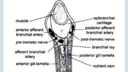
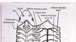
acids. The digested food is absorbed into the, , and lipase turns fats into fatty, tine i d scroll-valve., , the extensive surfaces of the intestine ani, , RESPIRATORY SYSTEM, , bloog, , In Scoliodon, the respiration, is aquatic, since the animal, resides in water. It breathes by, means of gills borne in a series of, gill-pouches on either lateral side, of the pharynx. Water enters the, mouth and after passing through, the buccal cavity, pharynx, gillpouches bearing gill-lamallae,, goes out through the external, gill-slits after bathing the gills., , , , (i) Respiratory Organs, There are five pairs of gillpouches bearing gills, arranged, in a series behind the hyoid, arch in the lateral walls of the, pharynx. Each gill-pouch is, compressed antero-posteriorly, and communicates with the cavity, of the pharynx through a large, internal branchial aperture, and with the exterior through, a narrow external branchial, aperture (commonly called gill- we, slit). The endodermal mucous :, membrane of gill-pouches or, interbranchial septa is raised Fig. 14.24. Scoliodon. Pharynx exposed to show gill-pouche>, into a series of horizontal folds 7, to form lamelliforms branchial lamellae or gill-filaments. The branchial lamellae have arid, blood supply, and they have a very thin covering membrane through which blood is ¢P™, to sea water for an exchange of gases. Each gill-pouch has two sets of gill-lamellae, one of, anterior wall and the other on the posterior, Each set of lamellae is a half gill or hemi, so that gill-pouch has two hemibranches. The gill-pouches are separated from each 0, fibro-muscular partition called the inter-branchial septa (they should more correctly al, intra-branchial septa because each lies between two successive gill-pouches). The interbrane, , septa extend well beyond the branchial lamellae, then each bends posteriorly to form 4 1 i, s, 150, , y |, , { | Spirack, , $ le, OP NS= |, , , , , , aM, A, i inh, , , , fh, Tat, yt, , , , ee the lamellae as well as external gill-slit. The inner part of each interbranchial, , a Es ea visceral arch with slender gill-rays. Visceral arches 4 ‘, gic comb-like gill-rakers which project i 5 i anchial 2p, , from entering the food, project inwards to protect the internal branchi, , E: a] isceral a }, e ach visceral arch supports the Posterior branchial lamellae or hemibranch of Por, gill-pouch and the anterior branchial lamellae ;, demibranchs with their j, , Scanned with CamScanner