Notes of Final B.Sc CBZ - 5 Sem, Zoology CHICK DEVELOPMENT - Study Material
Page 1 :
UNIT IV, 7 hr., • Development of chick: Structure of hen’s egg, cleavage,, blastula, gastrulation – origin and development of, primitive streak., • Fetal Membranes: Development, structure and functions, of amnion, horion, yolk sac and allantois., • Placenta: Histological and morphological classification, with examples. Placental hormones., hormones
Page 3 :
STRUCTURE OF HEN’S EGG, • Ovum is surrounded by a membrane called vitelline mebrane., • Mass of white yolk at the centre – latebra, • The latebra is surrounded by a many concentric layers of yellow and white yolk.
Page 4 :
•, , A strand white yolk runs upwards from the latebra. It is called the neck of latebra., , •, , It spreads on the upper surface of the ovum as a plate called the nucleus of, Pander., , •, , On the nucleus of Pander rests a mass of disc- shaped cytoplasm with a nucleus., This is called the blastodisc.
Page 5 :
•, •, •, •, , Blastodisc alone develop into the embryo proper – animal pole., As this passes down in to the oviduct, fertilization takes place and additional egg, membranes are added., The vitelline membrane is surrounded by the albumin or the white of the egg., The albumen is formed of three layers, namely an inner less dense layer, a middle dense, layer and an less dense layer.
Page 6 :
•, , Between the albumen and the vitelline membrane there is another membrane called, chalaziferous membrane., , •, , At each end of the egg, this membrane is twisted into yards called chalazae., , •, , The chalazae keep the blastodisc up always in whatever position the egg is turned.
Page 7 :
• The albumen is surrounded by a, double-shell membrane., • The two membrane is separated by, an air space at the broad end of the, egg., • Since hen’s egg is covered be a, calcareous shell, it is called cleidoic, egg., • The egg contains large amount of, yolk, it is called mesolecithal or, macrolecithal or polylecithal egg.., • The yolk is oriented towards one, pole, the egg is called telolecithal, egg.
Page 8 :
FERTILIZATION, • In birds, fertilization is internal., • Polyspermy
Page 12 :
Structure of hen’s egg
Page 13 :
Development of chickchick cleavage, • Cleavage is the successive mitotic division of the zygote into continuously, diminishing sized cells and result in blastula., blastula, • Cleavage is restricted to blastodisc and the yolk remains uncleaved. Such, cleavage is called meroblastic or discoidal cleavage., • The central part of blastodisc is whitish and circular. It is surrounded by a, darker marginal zone known as the periblast, which merges with the, underlying white yolk.
Page 14 :
Chick CleavageCleavage I, • After five hours of fertilization the first cleavage will appear. It is confined to, the centre of blastodisc., •, , It is meridional in plane., , • It cannot completely divide the blastodisc., blastodisc, • Blastomeres are not formed
Page 15 :
Chick CleavageCleavage II, • Second cleavage plane is also meridional., meridional, • It takes place at right angles to first cleavage., cleavage, • Even because of second cleavage clear blastomeres are not formed.
Page 16 :
Chick CleavageCleavage III, •, , The third cleavage consists of two furrows lying at right angle to the seemed one., , •, , It is vertical and parallel to the first division., division, , •, , It is in the two sides of first division., , •, , As a result of this division eight blastomeres are formed., , •, , But they do not show boundaries., boundaries
Page 17 :
Chick CleavageCleavage IV, • The fourth cleavage takes place in such a way that eight central, blastomeres and eight peripheral blastomeres (marginal blastomeres) will, form., • Only at this stage of division definite cells are formed., • The central eight cells are completely separated from yolk.
Page 19 :
Primary blastocoel, • A horizontal cleft appears below the central cells., • This cleft cuts off a single superficial layer of central cells from the, undivided cytoplasm beneath., • A fluid then begins to collect between this layer of cells and the, cytoplasm, , Establishes a shallow space called primary blastocoel or, , segmentation cavity or sub-germinal, germinal cavity.
Page 20 :
Horizontal cleavage, •, , As the primary blastocoel is being formed, a single layer of central cells is formed, above the cavity., , •, , Then a horizontal cleavage furrow appears among the central cells., , •, , This cleavage furrow is parallel to the surface., surface, , As a result, the single layer of, , central cells is converted into two layer above the primary blastocoel.
Page 21 :
Periblast tissue, •, , As cleavage proceeds, some of the nuclei from the dividing blastoderm area enter the, uncleaved portion of the cytoplasm located around and below the enlarging primary, blastocoel., , •, , These nuclei arrange themselves here and there in the cytoplasm. This mass of cytoplasm is, converted into a syncytial mass., , •, , This syncytial cytoplasm is called periblastic tissue. It is made up of two general areas,, namely peripheral periblast and central periblast., periblast, , •, , The peripheral periblast lies around the margin of the blastderm and central periblast lies, beneath the rim of the primary blastocoel.
Page 22 :
Chick development - BLASTULATION, •, , Blastula is an embryonic stage formed by cleavage., cleavage The blastula of chick is discoidal in, Discoblastula., , •, , Discoblastula consists central mass of cells called blastoderm. Below the blastoderm,, there is a cavity called primary blastocoel or segmentation cavity., , •, , The primary blastocoel is located in the central part of the blastula. As a result, the, central part of the blastoderm is free from the yolk and the peripheral part is closely, adhering to the yolk.
Page 23 :
•, , The central transparent part of the blastoderm is called Area Pellucida and peripheral, opaque region is called Area opaca., , •, , The Area Pellucida gives rise to embryonic tissue and Area Opaca gives rise to extra, embryonic tissue., , •, , Between the area pellucida and area opaca is a thin layer of cells which is known as, marginal zone. The embryo is now called the blastula.
Page 24 :
•, , At blastula stage the embryo reaches the uterus., , •, , The egg is laid by the female about the time the blastula is formed. At the time of, laying, the blastoderm is composed of 20,000, 20, to 60,000cells. Most of the cells of the, area pellucida remain at the surface, forming an “upper layer” called the epiblast., , •, , Shortly before the egg is laid some cells delaminate from the epiblast and ingress, into the subgerminal cavity in clusters forming the primary hypoblast.
Page 25 :
•, , At the same time a sheet of cells begins to delaminate and migrates from the posterior edge of, the area pellucida under the surface., , •, , These delaminated cells at the posterior edge of area pellucida gradually link up with each, other and with the primary hypoblast, to form a continuous layer of flattened cells, which lie, over the yolk on the floor of the subgerminal cavity. This layer is called as secondary, hypoblast or endoblast.
Page 27 :
Development of chickchick Gastrulation, • Gastrulation is a prolonged process initiated, , soon after the beginning of, , incubation after laying of the egg and is completed in about 4 days., • The gastrulation is the conversion of blastula into gastrula by morphogenetic, movement., • It is preceded by some pre-gastrular movements of certain cells resulting in their, separation from the blastoderm and formation of a lower layer called the, hypoblast., • Cells that remains at the upper layers of the blastoderm constitute the epiblast.
Page 28 :
• Morphogenetic movement results in the formation of the germ layers, namely, ectoderm, mesoderm and endoderm., • Gastrulation takes place by convergence, involution, divergence and, elongation., • The process of gastrulation is a complicated process and is expressed, as primitive streak., • The formation of primitive streak takes place at about 18 hours.
Page 29 :
Development of chick - origin and development of, primitive streak, • The primitive streak formed on the epiblast surface during the first, 10-18 hours of incubation at 37.5--385°C., • The primitive streak can be considered as the equivalent of an, elongated blastopore lip of amphibian embryos., • It forms as a result of convergence of epiblast cells to the dorsal, midline of the blastoderm.
Page 30 :
• There is a great tendency to form blastopore, due to heavy yolk it is not possible., • This tendency makes the prospective mesoderm, material to push along the mid anterior side., • This results in the formation of primitive streak., streak, Material from the posterior lateral side reaches, the streak, undergo involution and diverge on, the same side.
Page 31 :
• Primitive streak is a structural expression of gastrulation., • Fully formed primitive streak consists of a primitive groove and primitive, folds on its either side, • Primitive streak is homologous to the blastopore lip then the primitive, groove is homologous to the amphibian blastopore., • Continued convergence of more and more lateral mesoderm results in the, anterior elongation of the primitive streak., streak
Page 32 :
• In addition to convergence another kind of movement viz., forward, streaming and stretching takes place., place, • Circular area pellucid now assumes pear shape. In this way short, primitive streak becomes definitive primitive streak.
Page 33 :
• The primitive streak extends anteriorly upon about three fourth the length of, area pellucida where it ends in a deep pit called Hensen's Node with thick, borders., • Hensen’s node is the functional equivalent of the dorsal lip of the amphibian, organizer., • Posterior end of the primitive streak is comparable to the ventral region of, the blastopore which forms future anus., anus
Page 34 :
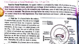
FOETAL MEMBRANES, • The embryos are covered and protected by a set of membranes called foetal, membranes., • These membranes are developed from the tissue lying outside the embryo., Hence they are also called extra-embryonic, embryonic membranes., • The main function of the foetal membranes are to provide protection,, nutrition, respiration and excretion to the embryo., • All the foetal membranes disappear before or immediately after hatching, hatching., • Types of foetal membranes, CHORION, AMNION, ALLANTOIS, YOLK SAC
Page 35 :
DEVELOPMENT OF FOETAL MEMBRANES, •, •, •, •, , These membranes will develop from blastoderm cells., The central part of blastoderm will give embryo proper, The marginal blastoderm will give extra embryonic membranes., Amnion and chorion will develop from somatopleurae, yolk sac and, allontois, will develop from splanchnopleurae., splanchnopleurae, , AMNION, • Amnion is a foetal membrane or extra embryonic membrane that covers, foetus., • The animals developing an amnion are called amniota., Eg: Reptiles, birds and mammals, • The animals which do not develop an amnion are called anamniota. Eg:, Fishes, amphians etc.
Page 36 :
• Composed of two layer, an outer somatic mesoderm and inner, ectoderm., • It encloses the a cavity filled with amniotic fluid., • The amnion is connected to the embryo on the ventral side by a stalk, called somatic umblicus.
Page 37 :
DEVELOPMENT OF AMNION, • Amnion develops from somatopleure., somatopleure During development, the, somatopleure develops certain folding called amniotic folds., • The amniotic folds develop into amnion., • Amnion ruptures at the time of hatching., hatching, FUNCTION OF AMNION:, 1. The amniotic fluid provides a liquid medium for the embryo., 2. It is called the artificial swimming pool of the embryo., 3. The amniotic fluid functions as a shock absorber., 4. It prevents the adhesion of the embryo to the shell.
Page 38 :
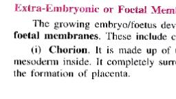
CHORION, • It is also called serosa., • It surrounds the entire embryo and lies outside. It lies close to the shell., • It s made up of two layers, namely an outer ectoderm and inner somatic, mesoderm., • The cavity enclosed by the chorion is called extra-embryonic, extra, coelom., • The chorion develops from somatopleure containing an outer ectoderm and, inner somatic mesoderm., • Chorion ruptures at the time of hatching., hatching, • Function: Helps in respiration and protection
Page 39 :
ALLANTOIS, •, , It develops from splanchnopleure., , •, , It is attached to the hindgut by a narrow stalk called allantoic stalk., , •, , Allantois is supplied by a allantoic arteries and a single allantoic vein., , •, , In later stage splanchnic mesoderm of allantois and somatic mesoderm of chorion, fuse together to form a chorio-allantoic membrane., membrane, , •, , Allantois ruptures at the time of hatching.., , Function:, collects the excretory products from the embryo., , •, , It is excretory in function, , •, , It helps in respiration., , •, , It absorbs calcium from the shell. This helps to rupture of the shell at the time of, hatching.
Page 40 :
YOLK SAC, • Yolk sac encloses the yolk., • It is made up of an inner endoderm and outer splanchano mesoderm. It, develops from splanchnopleure., • It is attached to the midgut by a narrow stalk called yolk stalk., • It opens into the midgut by an yolk duct., duct, • The endoderm of yolk sac has many finger-like, finger, folds called yolk sac septa., • The yolk sac gradually decreases in size as the yolk is consumed., The yolk sac provides nutrition for the embryo.
Page 41 :
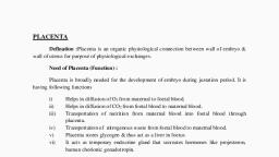
PLACENTA, • It is a special kind of tissue that connects between the mother and the foetus,, formed by the inner lining of uterus and the foetal membranes, , Found in, , mammals, • It is formed by the contributions of embryonic and maternal tissue. The, process of formation of placenta is called placentation, • It serves mainly for the transport of nutrients and oxygen from the mother to, the foetus., • Forms a placental barrier and prevents the direct mixing of foetal and, maternal blood cells.
Page 42 :
CLASSIFICATION OF PLACENTA
Page 44 :
CLASSIFICATION OF PLACENTA BASED ON THE, TYPE OF FOETAL MEMBRANES INVOLVED, YOLK SAC PLACENTA, • Yolk sac placenta is formed by yolk sac and, chorion. It is also called chorio-vitelline, placenta or yolk sac placenta., • Ex – Marsupials like Opossum
Page 46 :
CLASSIFICATION OF PLACENTA BASED ON THE, DISTRIBUTION OF VILLI, • The chorio- allantoic placenta, is classified into six types based on, the distribution of villi on the surface of chorion., Diffuse placenta – pig & horse, Cotyledonary placenta – sheep, cow & deer, Intermediate placenta- Camel & giraffe, Zonary placenta- Elephant, dog cat etc., Discoidal placenta – Rat &Rabbit, Metadiscoidal placenta, 1. Monodiscoidal – Man, 2. Bidiscoidal - Monkey
Page 48 :
Classification of placenta based on the type, of tissues involved - Histology, • Based on the tissue layer involved in the placenta.
Page 51 :
EPITHELIOCHORIAL PLACENTA, • In epitheliochorial placenta, the uterine epithelium of the mother makes, contact with the chorion of the embryo., embryo, • It is the simplest type of placenta. The villi of the chorion dip into the, chorio allantoic placenta., crypts of the uterine wall. It is chorio• This type of placenta is provided with 6 types of tissues., tissues, –, –, –, –, –, –, , Maternal endothelium, Maternal connective tissue, Maternal uterine epithelium, Chorion, Foetal connective tissue, Foetal endothelium, , Egg; Pig, Horse etc
Page 54 :
ENDOTHELIOCHORIAL PLACENTA, • In endotheliochorial placenta, the endothelium of the mother makes, direct contact with the chorion., • The uterine epithelium and maternal connective tissue are, eroded., Eg: Dogs and cats etc.,
Page 55 :
HAEMOCHORIAL PLACENTA, • In haemochorial placenta, the chorion of the embryo directly dip into the, maternal blood sinuses., • The uterine epithelium, maternal connective tissue and the maternal, endothelium are eroded., • Thus the chorionic villi directly dip into the maternal blood sinuses., , Eg; Man, Monkey and bats etc.
Page 56 :
HAEMOENDOTHELIAL PLACENTA, • Foetal blood vessels dip into maternal blood pools., • The uterine epithelium, the maternal connective tissue, maternal, endothelium and the chorion are eroded., eroded, • The maternal blood has only one barriers, the foetal endothelium to reach, the embryo., Eg; Rabbit and Rat etc,
Page 58 :
Placental hormones, • Placenta of mammals acts temporarily as an endocrine organ., •, , Secretes a various of hormones, , • Example: In rat on twelfth day of the pregnancy, placenta secretes the, hormone, rat chorionic mammoluteotrophin - which is responsible for the, maintenance of the corpora lutea and later progesterone (which is, responsible for pregnancy to continue to term)., • Horse placenta secretes the pregnant mare serum gonadotrophin (PMSG), a, luteotrophic hormone, in addition to progesterones and oestrogens., • In some animals, such as rabbit, the placenta does not seem to secrete any, hormone.
Page 60 :
Human Chorionic Gonadotropin, • hCG is a glycoprotein that contains galactose and hexosamine produced by the, syncytiotrophoblast, • hCG is primarily luteinizing and luteotropic and has little FSH activity, • It maintenance of corpus luteum till 6 week of pregnancy., • It stimulate the leyding cells of the male fetus to produce testosterone., Human Chorionic Somatotropin (hCS), Secreted by the placenta at about the fifth week of pregnancy., • Decreases insulin sensitivity, decreased utilization of glucose in the mother, making larger quantities of glucose available to the fetus., • Promotes the release of free fatty acids from the fat stores of the mother,, provides alternative source of energy for the mother’s metabolism during, pregnancy.
Page 61 :
Relaxin, • Relaxin hormone is produced by the placenta in very small amounts., •, , It causes the relaxation of the symphysis and sacroiliac joints during pregnancy, and also reduces the tension of cervix., , Estrogen, • Causes enlargement of the mother’s uterus, • Causes enlargement of the mother’s breasts and growth of the breast ductal, structure, • Causes enlargement of the mother’s female external genitalia., • The estrogens also relax the pelvic ligaments of the mother, so that the sacroiliac, joints become relatively limber and the symphysis pubis becomes elastic during, parturition.
Page 62 :
Progesterone, • Progesterone helps in the maintenance of pregnancy and prevents, premature parturition., • In fact, progesterone stops the menstruation and the release of, ovum from the ovary until the pregnancy., pregnancy
 Learn better on this topic