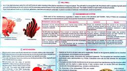
Page 1 :
AAAE, , , , call Biology r, 22's" o., , [PAPER-l SSS EMESTTERR = 1], , CHAPTER — 4., , , , , , , , , , , CELL BIOLOGY _, , Cell biology (formert:, discipline that studies cel], they contain, interactions, , Y cytology, from the Greek kytos means contain") is a scientific, ls, their physiological properties, their structure, the organelles ., ei one Gol ions with their environment, their life cycle, division and death. This, is done both on a microscopic and molecular level. Cell biology research encompasses, poth the great diversity of single-celled organisms like bacteria and protozoa, as well as, the many specialized cells in multicellular organisms such as humans, plants, and, , sponges. Cell biology revolves around the concept that the cell is the fundamental unit of, life. Thus, cell biology is in fact study of life., , 7 CELL, The cell (from Latin cella, meaning "small room") is the basic structural, functional, , and biological unit of all known living organisms. Cells are the smallest unit of life that, can replicate independently, and are often called the "building blocks of life”., , , , , , , , , , , , , , , , , , , , , , [Cell Wall, + Cytoplasm, t Chloroplast, 1 Mitochondria, S— I, (aa Nucieolus Sheath__ = yj, vall J, Chromatia geet, prs asngphore, : hycobilisome :, [> Vacuoles Naps OR i, Gas vacuoles., cyan ;ophycin, 6-Granales, o-Granules, , , , , , , , Fig. 4.1: (A) Eukaryotic and (B) Prokaryotic plant cells, , On the basis of the type of nucleus and structural organization, cells are categorized, into two basic types, namely, the prokaryotic type and the eukaryotic type., , In prokaryotic cells, the nucleus is not true as it does not contain nuclear membrane,, nucleoplasm or nucleoli. The hereditary material or the deoxyribonucleic acid (DNA), molecules lie in contact with the cytoplasm as the nuclear envelope is absent. These cells, also lack certain cell organelles such as endoplasmic reticulum, mitochondria, Golgi, complex, lysosomes, etc. In other words a prokaryotic cell is characterized by a primitive, or an incipient. nucleus and exhibits structural simplicity, e.g., bacterial cell, cells of blue, green algae. 2, , ’ A typical eukaryotic cell differs from the prokaryotic cell in having a well defined, nucleus having a definite nuclear envelope separating the hereditary material from the, cytoplasm The nucleus also contains nucleoplasm and nucleoli. These cells are further, , 4/F.Y. B.Sc. — A New Course in Botany (Sem-I & II)
Page 3 :
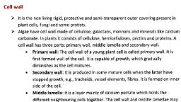
Cell Biology, , , , P's", A typical eukaryotic cell can best be studied under the following heads :, Cell 4, , , , , , Cell wall Protoplasm, , (only in plant cell) |, , [Rte oe oe Mel a aah sR, Plasma membrane Cytoplasm Nucleus, , , , , , , , , , , , Hyaloplasm Non-living cell Living cell, contents: contents or organelles, , , , r, , Endoplasmic, , oe -——, , Mitochondria, , , , Ribosomes Centrosome, , , , , , , , reticulum (in animal cell), Plastids Golgi Lysosomes, (in plants) complex, Fs ah i>) pele eh ol aa aes), Nuclear Nucleoplams Niclestng Chromatin, membrane reticulum, ULTRA-STRUCTURE AND FUNCTIONS OF CELL WALL a, , , , The external non-protoplasmic covering of a plant cell is called cell wall. It is, , composed of cellulose, hemicellulose and pectic compounds and distinguished into three, layers, viz.,, , , , i) middle lamella,, ii) primary cell wall and, iii) secondary cell wall., , i) Middle lamella : It is viscous, jelly-like, intercellular substance cementing the, adjacent cells. It is formed during cytokinesis and is made up of calcium and, magnesium pectates., , ii) Primary cell wall Yiejs the first wall secreted by protoplast during early stages of, growth and development) Since it is first to develop, it forms the most peripheral, part of the cell wall. It ‘consists of a loose network of cellulose microfibrils, embedded in an amorphous gel like matrix or ground substance of, polysaccharides such as pectin, lignin and hemicellulose. Following substances, are involved in the formation of cell wall :, , a) Cellulose microfibrils which forms a dense network. The adjacent molecules, are joined through hydrogen bonds forming a continuous lattice which is, , , , , , , , , , , , , , embedded in the, Bee Manors kt Middle lamella, b) Pectic : \ Primary, , . polysaccharides cell wall, constitute the Plasmadesmata r, second network, *. Pit field, rich in, galactouronic Secondary., acid residue. It Ce cell wall, forms alata ‘ Cytoplasm, , ciu:, , ou calcium” (A) Cell Wall (B) Plasmodesmata, bridges and a, ae a ig. 4.2 : Structure of cell wall, , interactions.
Page 4 :
P potany (F.Y.B.Se.) (Sem, = 1,, , ANew Course in, , eeoeg® oe, . eh to third network which, e to, , , , , , , , , terweaves through, , , , , , , , , , , , , , , , , , , , , , , , , , teins give TiS a Di eee 3 Cell, - Be ie ears. Jy packed and cross-linking * compl ch, «fibres are loosely PAC’ 1 cell is able to Brow Fumie, In young cells the cellulose fibre “linking of fibres, ai] expansion by additig,, However, in mature cells, due to cr0-e wail Joosening and © nal ab, The protein expansion is responsible . *, canoes ee iaeiles to the cellulose microfibrils. number of glucose mee ee if, Cellulose is polysaccharide made uP O' 7 Ts chains join '0 sleet 3 st, glycosidic bonds to form a chain. Many ce featies join 10 21) rf ible Bl M ac, micelles or elementary fibrils. Several mit acrofibrils run in all possil le planes pi, microfibrils together form macrofibrils. Be mi ache compounds to form primary q eA, form a meshwork joined by organic matrix like ane through which runs number 9 ree, wall. At intervals it shows a number of perforats cc, delicate cytoplasmic strands called plasmodesmata. apy Pe, Cellulose chains 7 m, Primary cell eer TI, Microfibrils pt, er, Meshwork |, fibrils q, al, molecules, Micelles Macrofibril as, pl, | Fig. 4.3 : Ultr ep 1 wall (Diagrammatic), ‘ig. 4.3 : Ultrastructure of primary cell wi Bi “ % al, Plasmodesmata: The cell wall is not uniform in thickness. During deposition of cell] P, wall some areas are left unthickened at intervals. These are called primary pit fields. They) oO, are perforated by small pores through which cytoplasm of adjacent cells is communicated] fi, by cytoplasmic strands or bridges called plasmodesmata. The plasmodesmata show | P, simple or branched microtubules. also called desmotubules through which the a, cytoplasmic fluid and solutes pass from cell to.cell thus. establishing organic continuity, | Pp, Even after formation of secondary cell wall, the cytoplasmic connections are maintained], through plasmodesmata. b, iii) Secondary cell wall 9 When the cell grows to full size, another cell wall called i, secondary cell wall is deposited internal to primary cell wall. It is made up of :, rales peu and ony complex substances such as cutin, lignin, 2, suberin, wax, etc. Secondary cell walls a ott ©, specialized cells such as Sraacues woe of pine characteristic Saga 7, Function of cell wall, The cell wall is considered as a product i «| a, ought to be a non-living eae pe oe protoplasm. In that sense iq 2, protoplasm from adverse external influences delimits th Protoplasm. It protects the :, the cell. f € cells and also imparts shape!, Tr, Tr, ULTRA-STRUCTURE AND FUNCTIONS OF PLASMA, Protoplast of every cell is surrounded, membrane. In case of some cells, plasma membrane show: '§ Membrane called -plasm# 4, to it, e.g., cell wall in plant cells. In that case plasma ne Protecting covering exten! }, , To avoid the confusion, Plows (1931) intr, living membrane in plant cells.
Page 5 :
Cell Biology g’o's" Chemical composition, , Chemical analysis of plasma membrane shows that it is composed of 60% proteins,, about 40%lipids and 1% carbohydrates of the total dry weight., , Proteins : The proteins form the bulk of the plasma membrane and are classified into, three classes, viz., structural proteins, carrier proteins and enzymatic proteins. The, structural proteins form backbone of the plasma membrane. They have very little catalytic, activity and are extremely lipophilic. The average molecular weight of the structural, protein is 3 x 10⁴. The carrier proteins help in transportation of the molecules across the, membrane against the concentration gradient. Their molecular weight is same as that of, structural proteins. The enzymatic proteins are catalytic proteins which form major, component of many membranes, e.g., membranes of ER, mitochondria, etc. However,, structure of enzymes differs from membrane to membrane. All these proteins fall in two, main categories, viz., intrinsic or integral proteins and extrinsic or peripheral proteins., The intrinsic proteins are firmly associated with the membrane either partially, penetrating the membrane or penetrating the entire membrane protruding at both the, ends, They have non-polar amino acids in the interior as well as on the surface. The, extrinsic proteins have a weaker association and are bound by electrostatic interaction., They are associated with the polar heads of the lipid molecules. They have non-polar, amino acids in the interior and polar amino acids on the surface., , , , , , Lipids : These constitute sterols and fatty, acid esters (mainly glycerides and, phospholipids). Among sterols, cholesterol is, widely distributed in the membrane of, animal tissue, phytosterol in the membrane of, plant tissue and ergosterol in the membrane, of eukaryotic microorganisms. Among the, fatty acid esters, phospholipids are, predominant. They have polar ends (heads), and non-polar ends (tails) When __ the, phospholipid molecules are spread in double, layer, the polar heads form the surface of the, bimolecular layer while the non-polar tails of A phospholipid with a, the opposite molecules arrange themselves hydrophilic head and a, face to face. Polar heads are hydrophilic while hydrophobic tail, non-polar tails are hydrophobic. Both the, regions are linked with glycerol moiety or a, sterol., , , , Chemical makeup of a, single phospholipid, , Fig. 4.4: Structure of a phospholipid, , Carbohydrates: These are present in the plasma membrane in the form of covalently, linked molecules with proteins and lipids. These are of two types, glycoproteins and, glycolipids. Glycoproteins are the simple sugars as well as sugar derivatives attached to, the outer surface of the protein component. Due to these carbohydrates, the membrane is, negatively charged and attracts positively charged proteins which remain bound to the, membrane through electrostatic interaction. Glycolipids are attached to the glycerol, molecules of the lipids through glycosidic bonds., , Membrane models, , In an attempt to explain the physical and biological features of cell membranes,, several membrane models have been proposed. Recently electron microscopic studies, have revealed the fine structure and variations within the membranes of various types of, cells., , In early 1950s, Robertson studied plasma membrane and membranes associated with, organelles under the electron micrscope and in 1953 he put forward the concept of unit, membrane. As per the concept of unit membrane, plasma membrane and other