Page 1 :
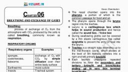
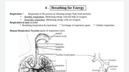
CHAPTER 17- BREATHING AND EXCHANGE, EXCHAN E OF GASES, Q. Difference between breathing and respiration, BREATHING, RESPIRATION, Breathing is the process of inhaling oxygen from Cellular respiration is the process of breaking down, the environment and exhaling carbon dioxide of glucose to produce energy, which is then used by, back to it with the help of the lungs., cells to carry out the cellular function., It is a biophysical voluntary process, , It is a biochemical, ical involuntary process, , It takes place in the lungs or gills., , It takes place in cells., , No energy is produced in this process., , Energy is produced in the form of ATP, , Breathing is an extracellular process., , Respiration is an intracellular process., , There are no enzymes used in this process., , Many enzymes play a major role in respiration., , Q. Difference between aerobic and anaerobic respiration, , Human Respiratory System: The respiratory system in humans is made up of the following parts:, External nostrils – For the intake of air., filter the air from dust and dirt and act as the, Nasal chamber – This is lined with hair and mucus to filter, body’s first line of defense against foreign pathogens
Page 2 :
, , , , , , , , Pharynx – It is a passage behind the nasal chamber and serves as the common passageway for both, air and food., Larynx – Also known as the soundbox as it houses the vocal chords, Epiglottis – It is a flap-like structure that covers the glottis and prevents the entry of food into the, windpipe., Trachea – It is a long tube passing through the mid-thoracic cavity., Bronchi – The trachea splits into two tubes called the bronchi, which enter each lung individually., Bronchioles – Each bronchus is further divided into finer channels known as bronchioles., Alveoli – The bronchioles terminate in balloon-like structures known as the alveoli. It facilitates the, gaseous exchange (oxygen and carbon dioxide) between atmosphere and lungs, Lungs – Lungs are the primary organs of respiration in humans and other vertebrates. They are, located on either side of the heart, in the thoracic cavity of the chest. Humans have a pair of lungs,, which are sac-like structures and covered by a double-layered membrane known as pleura (pleural, membrane. Lungs are present in a thoracic chamber. Thoracic chamber is formed of:, Vertebral column at dorsal side., Ventrally by sternum., Ribs at the lateral side., At the lower side, a dome-shaped diaphragm., , Q. What is the role or function of pleural fluid?, Pleural fluid keeps the pleura moist thus preventing the lungs from desiccation, It acts as a lubricant and reduces friction between the parietal and visceral pleura during breathing, It also acts as shock absorber, ANATOMY OF LUNGS, The lungs are the major organs of the respiratory system. They are roughly traiangular, paired and, non muscular structure located on either side of the heart, in the thoracic cavity of the chest.
Page 3 :
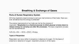
, , , , , Both the lungs are divided into sections, or lobes. The right lung has three lobes – superior, median, and inferior and is slightly larger than the left lung, which has two lobes- superior lobe and inferior, lobe, The lobes are divided from each other by two types of fissures: Oblique fissure and, Horizontal fissure, The lungs are covered by a protective membrane known as pleura and are separated from, the abdominal cavity by the muscular diaphragm., , Parts of Respiratory system - Respiratory system consists of two parts1. Conducting part/zone, The conducting portion provides a passage for the air. It brings the air from outside to the site, of respiration., The conducting part of the respiratory system serves to filter, warm and humidify air on its, way to the lungs., It includes the nose, nasopharynx, larynx, trachea, bronchi and bronchioles., 2. Exchange or respiratory part/zone, The respiratory portion helps in the exchange of gases and oxygenation of the blood., It includes alveolar ducts and alveoli, Q. What is the structure and function of alveoli?, Alveoli are tiny air sacs present in the lungs which appear as a bunch of grapes. They are roughly, spherical in shape having higher surface area for gas exchange, The alveoli have very thin epithelial layer (one cell thick) which is richly supplied with a network of, blood capillaries, Alveoli are highly vascularised structures i.e. each alveolus is surrounded by a rich capillary network, which increases the gas exchange with the blood, Their internal surface is covered with a layer of fluid, as dissolved gases are better able to diffuse, into the bloodstream, Function- The alveoli are the functional unit of lungs. It helps in the exchange of respiratory gases between, the air filled in alveoli and the surrounding pulmonary capillaries.
Page 4 :
Q. What is respiratory membrane or diffusion membrane?, Respiratory/diffusion membrane is a very thin membrane that separates the alveoli from the, pulmonary capillaries (0.5 - 0.6 mm thick). It is the primary site for gaseous exchange between the, alveoli and the blood capillary, The diffusion membrane is made up of three major layers One celled thin alveolar epithelium (squamous epithelium), Capillary endothelium (squamous epithelium) of alveolar capillaries and, Basement membrane in between them, , STEPS OF RESPIRATION- Respiration involves the following steps:, 1. Breathing or pulmonary ventilation by which atmospheric air is drawn in and CO2 rich alveolar air is, released out. (Gas exchange between atmosphere and lungs), 2. Diffusion of gases (O2 and CO2) across alveolar membrane. (Gas exchange between lungs and blood), 3. Transport of gases by the blood., 4. Diffusion of O2 and CO2 between blood and tissues. (Gas exchange between blood and tissues), 5. Utilization of O2 by the cells for catabolic reactions and resultant release of CO2 (Cellular respiration), Q. Describe the mechanism of breathing (pulmonary ventilation), The process of inspiration (breathing in) and expiration (breathing out) is known as breathing. Stuctures, involed in normal breathing- Ribs, external intercostal muscles and daiphragm (phrenic muscle), Principle of breathing- Boyle’s law, It states that the volume of gas is inversely proportional to pressure (when temperature is constant) i.e. P ∝, (1/V), The breathing mechanism involves two processes:, Inspiration- The process of intake of atmospheric air is known as inspiration. It is an active process., Expiration- This expulsion of air from the lungs is called expiration. It is a passive process., Mechanism of inspiration, a) The external intercostal muscle contracts, b) Rib cage moves upward and outward, c) Daiphragm contracts and flattens (antero- posterior axis), d) Volume of thoracic cavity increases (dorsoventral axis), e) Intrapulmonary pressure i.e. Pressure in alveoli decreases, f) Air moves in
Page 5 :
Mechanism of expiration, a) The external intercostal muscle relaxes, b) Rib cage moves downward and inward, c) Daiphragm relaxes (antero- posterior axis), d) Volume of thoracic cavity decreases, creases (dorsoventral axis), e) Intrapulmonary pressure i.e. Pressure in alveoli increases, f) Air moves out, Exchange or Diffusion of gases (O2 and CO2), Exchange of gases (O2 and CO2) takes place between alveoli and blood across the respiratory membrane, (external respiration) and between blood and tissues (internal respiration) by the process of diffusion., Exchange of O2 and CO2 depends upon the following factors:, a) Pressure/ concentration gradient: The atmospheric air is a mixture of gases. Each individual gas, contributes independently to its total pressure. The pressure of the individual gas contributed in the air is, called partial pressure.. The difference in the partial pres, pressure, sure determines the movement of gas from one, area to another. A gas always travels from higher to lower partial pressure., pO2 in alveoli is more (104 mm Hg) than that in the surrounding blood capillaries (40 mm Hg). So, O2 diffuses into capillary blood., pCO2 in deoxygenated blood within the capillaries is more (45 mm Hg) than that in alveoli (40 mm, Hg). So, CO2 diffuses to alveoli., The Partial pressures of O2 and CO2 (pO2 and pCO2) are given below., , b) Solubility of gases:, Solubility of CO2 is 20-25 times higher than that of O2. So, the amount of CO2 that can diffuse through the, diffusion membrane is higher than that of O2., c) Thickness of diffusion membrane::, The respiratory membrane is highly permeable to gases., gases The respiratory membrane or diffusion membrane, is less than 1 micrometer thick which enables easy exchange of respiratory gases, d) Surface area:, Presence of alveoli increases the surface area of lungs, lungs. It increases the gas exchange
Page 6 :
TRANSPORT OF RESPIRATORY GASES (O2 AND CO2), It is the transport of respiratory gases (O2 & CO2) from alveoli to the systemic tissues and vice versa., Oxygen transport, It is the transport of O2 from lungs to various tissues. It occurs in 2 ways:, a. In physical solution (blood plasma): About 3% of O2 is carried in a dissolved state through plasma., b. As oxyhaemoglobin:, , About 97% of O2 is transported by RBC in the form of oxyheamoglobin, O2 binds with haemoglobin to form oxyhaemoglobin. This is called oxygenation. Hb has 4 haem units. So,, each Hb molecule can carry 4 oxygen molecules. Binding of O2 depends upon pO2, pCO2, H+ ion, concentration (pH) and temperature., In the alveoli,, high pO2, low pCO2, lesser H+ ion concentration and lower temperature exist. These factors, are favourable for thee formation of oxyhaemoglobin, In tissues,, low pO2, high pCO2, high H+ ions and high temperature exist. So oxyhaemoglobin dissociates, to release O2, , CO2 transport, It is the transport of CO2 from tissues to lungs. In tissues, pCO2 is high and pO2 is low. In lungs, pCO2 is, low and pO2 is high., a) As carbonic acid (Dissolved form in plasma), plasma):: In tissues, 7% of CO2 is dissolved in plasma to form, carbonic acid and carried to lungs., b) As carbamino-haemoglobin:: In tissues, 23, 23% of CO2 binds to Hb to form carbamino, rbamino-haemoglobin. In, alveoli, CO2 dissociates from carbamino-haemoglobin., carbamino, c) As bicarbonates: Majority of CO2 about 70% is transported in the blood in the form of bicarbonate ion., ion, RBCs and plasma contain an enzyme, carbonic anhydrase. In this system, carbon dioxide diffuses into, the red blood cells. Carbonic anhydrase (CA) within the RBC quickly converts the carbon dioxide into, carbonic acid (H2CO3). Carbonic acid is an unstable intermediate molecule that immediately dissociates, into bicarbonate ions (HCO−3), −3) and hydrogen (H+) ions., These bicarbonate ions are then quickly transported out of the RBC via membrane proteins. At the, same time chloride ions are pumped into the RBC to maintain a constant electric charge. This exchange, of ions is known as chloride shift. Once inside the lungs, these bicarbonate ions are converted back into, carbon dioxide and then expelled out of the lungs via exhalation.
Page 7 :
What is Bohr effect?, Bohr Effect is the decrease of the oxygen binding capacity of haemoglobin with the increase in, concentration of carbon dioxide or decrease in pH, As the cells carry out their metabolic processes at a higher rate, they will produce more CO2., Carbon dioxide exits the cells of the tissue and enters into the red blood cells (RBC) found in the nearby, capillaries., Inside the RBC, CO2 and water combines together to form Carbonic, nic acid in presence of the enzyme, carbonic anhydrase, Carbonic acid is a weak acid. It readily dissociates into a hydrogen ion and bicarbonate ion., As the concentration of CO2 inside the blood increases, the pH decreases making the blood more acidic., This in turn decreases the heamoglobin’s affinity for oxygen and shifts the oxygen-hemoglobin, oxygen, dissociation curve to the right. This phenomenon is known as Bohr’s effect, eff, What is Haldane effect?, Haldane effect is the decrease of the carbon dioxide binding capacity of haemoglobin with the rise in, the concentration of oxygen., This effect promotes the release of carbon dioxide from the tissues to the blood and stimulates the, release of carbon dioxide from the blood to the lungs., OXYGEN HEAMOGLOBIN DISSOCIATION CURVE
Page 8 :
, , , , , , The oxygen heamoglobin dissociation curve is a graphical representation of the percentage of saturation, of haemoglobin at various partial pressures of oxygen. Mainly it tells us about the affinity of oxygen to, the hemoglobin and how the hemoglobin carries oxygen., In the lungs, the partial pressure of oxygen is high. Hence, haemoglobin binds to oxygen and forms, oxyhaemoglobin. Tissues have a low oxygen concentration, therefore the partial pressure of oxygen is, low. Therefore, at the tissues, oxyhaemoglobin releases oxygen to form haemoglobin., The oxygen dissociation curve is sigmoid shaped or S-shaped because of the co-operative binding of, oxygen to haemoglobin. As the first oxygen molecule binds to haemoglobin, it increases the affinity for, the second molecule of oxygen to bind. Subsequently, haemoglobin attracts more oxygen., , FACTORS AFFECTING OXYHEMOGLOBIN DISSOCIATION CURVE, There are several important factors that affect the affinity of hemoglobin to oxygen as therefore affect the, oxygen-hemoglobin dissociation curve. These factors include the (1) pH (2) temperature (3) carbon dioxide, (4) 2, 3-BPG and (5) carbon monoxide., 1. Effect of pH, Exercising tissue contains cells that produce more CO2. When CO2 leaves the tissue and enters the RBC of, the capillaries, it readily converts into bicarbonate and hydrogen ions, thereby decreasing the pH., A decrease in pH (increase in H-ions) shifts the Oxy-Hb curve to the right., 2. Effect of Carbon-dioxide, Metabolically active cells of our body produce more CO2; some of which can directly bind to the Hb and, decrease its affinity for Hemoglobin. Thus high PCO2 in the blood means more O2 will be delivered to the, tissue. Thus increase in CO2 concentration in the blood shifts the Oxy-Hb curve to the right., 3. Effect of 2,3 BPG, 2,3 BPG is an intermediate of glycolysis. Excess of 2,3 BPG can escape into the blood plasma where it binds, with Hb and decreases its affinity for O2. Therefore 2,3 BPG can cause the Hb to unload more O2 to the, tissues. Thus an increase in the concentration of 2,3 BPG in the blood shifts the Oxy-Hb curve to the right., 4. Effect of temperature, An increase in temperature decreases the affinity decreases the affinity of Hb with Oxygen and makes Hb, release more O2 in the tissue side. Thus an increase in temperature shifts the Oxy-Hb curve to the right., Q 3. Diffusion of gases occurs in the alveolar region only and not in the other parts of the respiratory, system. Why?, For efficient exchange of gases, the respiratory surface must have certain characteristics such as, It must be thin, moist and permeable to respiratory gases, It must be very large, It must be highly vascular, Only alveolar region has these characteristics. Thus, diffusion of gases occurs in this region only., Q4. What are the major transport mechanisms for CO2? Explain., The major transport mechanisms for CO2 are in the form of sodium bicarbonate (HCO 3-). About 70% of, carbon dioxide is transported as sodium bicarbonate. See explanation above., Q 5. What will be the pO2 and pCO2 in the atmospheric air compared to those in the alveolar air?, (i) pO2 lesser, pCO2 higher, (ii) pO2 higher, pCO2 lesser, (iii) pO2 higher, pCO2 higher, (iv) pO2 lesser, pCO2 lesser, As the partial pressure of oxygen in atmospheric air is higher than that of oxygen in alveolar air. So, in, atmospheric air, PO2 is about 159 mm Hg and in alveolar air, it is about 104 mm Hg. The partial pressure of
Page 9 :
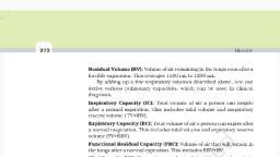
carbon dioxide in atmospheric air is lesser than that of carbon dioxide in alveolar air. In atmospheric air,, PCO2 is about 0.3 mmHg and in alveolar air, it is a, Q 6. Explain the process of inspiration under normal conditions., Inspiration is the process during which atmospheric air is drawn inside the body. When the intrapulmonary, pressure (pressure in the lungs) is lesser than the atmospheric pressure, inspiration takes place. The muscles, of the diaphragm, external intercostal muscles and abdominal muscles are referred to as the inspiratory, muscles which bring about the process of inspiration. See explanation above (add the diagram fro book), 7.How is respiration regulated?, Breathing is an automatic and rhythmic act produced by networks of neurons in the hindbrain (the pons and, medulla)., Q 8. What is the effect of pCO2 on oxygen transport?, The pCO2 plays an important role in the transportation of oxygen. At the alveolus, the low pCO2, and high pO2 favors the formation of oxyhaemoglobin., At the tissues, the high pCO2 and low pO2 favor the dissociation of oxygen from oxyhaemoglobin., So, the affinity of haemoglobin for oxygen is enhanced by the decrease of pCO2 in blood. Therefore,, oxygen is transported in the blood as oxyhaemoglobin and oxygen dissociate from it at the tissue, where pO2 is low., Q 9. What happens to the respiratory process in a man going up a hill?, When a man goes uphill, he gets less oxygen with each breath and as we know that when altitude increases,, the oxygen level in the atmosphere decreases. Because of this, the amount of oxygen in the blood starts to, decline. The respiratory rate increases in response to the decrease in oxygen content of the blood., Simultaneously, the rate of heartbeat increases to increase the supply of oxygen to the blood., Q10. What is the site of gaseous exchange in an insect?, In insects, gaseous exchange takes place through a network of tubes collectively known as the tracheal, system. The small openings on the sides of an insect’s body are called spiracles and oxygen-rich air enters, through the spiracles. These spiracles are connected to the network of tubes. From the spiracles, oxygen, enters the tracheae and from here, oxygen diffuses into the cells of the body. The movement of carbon, dioxide follows the reverse path and the CO 2 from the cells of the body first enters the tracheae and then, leaves the body through the spiracles, , Q11. Define oxygen dissociation curve. Can you suggest any reason for its sigmoidal pattern?, Explanation given above, 3. Distinguish between, (a) IRV and ERV, IRV (Inspiratory reserve volume), , ERV (Expiratory reserve volume), , It is the volume of air that a person can additionally It is the volume of air that a person can expire, inspire through a compelled inspiration, through an expelled expiration, For a healthy individual, the IRV is approximately For a healthy individual, the, 2500ml – 3000ml, approximately 1000ml to 1100ml, , ERV, , is
Page 10 :
(b) Inspiratory capacity and expiratory capacity, Inspiratory capacity (IC), , Expiratory capacity (EC), , Inspiratory capacity is the volume of air that can Expiratory capacity is the volume of air that, be inhaled post a normal expiration, can be exhaled post a normal inspiration, It is given by the sum of tidal volume and the It is given by the tidal volume and the, inspiratory reserve volume, expiratory reserve volume, i.e, IC = TV + IRV, i.e., EC = TV + ERV, (c) Vital capacity and Total lung capacity, Vital capacity (VC), After a maximum inspiration, it is the maximum, volume of air that can be exhaled. It includes IC, and ERV., The vital capacity in the lungs of humans is about, 4000ml, , Total lung cap acity (TLC), After maximum inspiration, it is the volume, of air in the lungs. It includes ERV, IC and, residual volume, The total lung capacity in the lungs of, humans is nearly 5000ml to 6000ml, , Q14. What is Tidal volume? Find out the Tidal volume (approximate value) for a healthy human in an, hour., During a normal respiration, the volume of air expired or inspired is referred to as tidal volume (TV). The, tidal volume is approximately 500ml for a healthy individual. A healthy individual can breathe around 12-16, times a minute and expire or inspire nearly 6000-8000ml of air per minute or, We can calculate the hourly tidal volume for a healthy human., If, Tidal volume = 6000 to 8000 mL/minute, So, the Tidal volume in an hour will be: = 6000 to 8000 mL × (60 min) = 360000 ml-480000 ml per hour., Hence, the hourly tidal volume for a healthy human is approximately 360000 ml-480000 ml.
 Learn better on this topic