Page 1 :
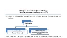
BODY FLUIDS AND CIRCULATION, Circulation is the transport of nutrients, oxygen, CO2 and excretory products to the, concerned tissues or organs. For circulation, simple organisms (sponges, coelenterates, etc.) use water from their surroundings. Complex organisms use body fluids (blood &, lymph) for circulation., CIRCULATORY PATHWAYS, Circulatory system is 2 types- Open and Closed., a), , Open circulatory system: Here, the blood pumped by the heart passes through large, vessels into open spaces or cavities called sinuses. E.g. Arthropods and molluscs., , b), , Closed circulatory system: Here, the blood pumped by the heart is always circulated, through, ugh blood vessels. This is more advantageous as the flow of fluid can be more, precisely regulated. E.g. Annelids and chordates., , All vertebrates have, ave a muscular chambered heart., Fishes: 2-chambered, chambered heart (an atrium + a ventricle)., Amphibians: 3-chambered heart, eart (2 atria + a ventricle)., Reptiles (except crocodiles): 3-chambered, chambered heart (2 atria + a ventricle). Ventricle is, incompletely partitioned., Crocodiles, birds & mammals: 4-chambered heart.
Page 2 :
Types of circulation, a) Single circulation: In fishes. In this, heart receives impure blood only (venous heart)., Deoxygenated blood → to heart → to gills → oxygenated blood → to body parts →, deoxygenated blood → to heart., b) Incomplete double circulation: In amphibians & reptiles. In this, left atrium gets, oxygenated blood from gills/ lungs/skin and right atrium gets deoxygenated blood from, other body parts. However, they get mixed up in the single, single ventricle. It pumps out mixed, blood., c) Double circulation: In birds & mammals. Right atrium gets deoxygenated blood and, passes to right ventricle and left atrium gets oxygenated blood and passes to left, ventricle. The ventricles pump it out separately without any mixing up., , COMPOSITION OF BLOOD, Formed of plasma (55%) & formed, ed elements (45%)., 1) PLASMA – It is a straw-colored, colored, slightly alkaline (pH 7.4) viscous fluid., Constituents of plasma, , , Water (90-92%): It is a good solvent., , , , Plasma proteins (6-8 %): Itt iincludes Fibrinogen: For blood coagulation., Globulins: Gamma globulins act, a as antibodies (for defense of the body)., Albumins: For osmotic balance. Regulate blood pressure., , , , Glucose, amino acids, lipids & cholesterol., , , , Inorganic constituents: Na+, Ca2+, Mg2+, Cl, Cl-, HCO3- etc., , , , Gases like CO2, O2, N2 etc., , Plasma without clotting factors is known as Serum.
Page 3 :
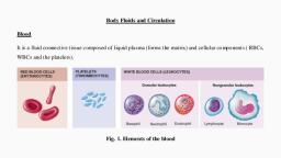
2) FORMED ELEMENTS (RBC, WBC & PLATELETS), , Red Blood Cells (RBC) or Erythrocytes:, , , Biconcave non-nucleated cells., ls. No mitochondria, Golgi complex etc., , , , Red colour is due to Haemoglobin (iron containing protein)., , , , Normal Hb level is 12-16, 16 g/ 100 ml., , , , RBC Count: 5 - 5.5 millions/ mm3 of blood., , , , Formed in: Red Bone marrow., , , , Average lifespan: 120 days. Worn, Worn-out, out RBCs are destroyed in spleen (graveyard of, RBCs)., , , , Function: CO2 and O2 transports., , White Blood Cells (WBC) or Leucocytes:, , , Colourless nucleated cells., , , , Count: 6000-8000 /mm3., , , , Formed in: Bone marrow but mature in lymph glands, spleen., , , , Average lifespan: Generally short lived (7, (7- 15 days)., , , , Function: Part of immune system., , Types of WBC: Granulocytes & Agranulocytes, Granulocytes- contains cytoplasmic granules in them, They are of 3 types:, a) Neutrophils (Heterophils): 60--65%. Soldier of the body. Causes phagocytosis., hagocytosis., b) Eosinophils (Acidophils): 2-3%., 3%. Resist parasitic infections as well as causes, c, allergic, reactions., c) Basophils, , (Cyanophils): 0.5-1%., 1%. Secrete histamine,, serotonin, heparin etc. Causes, Cause, , inflammatory reactions, 2. Agranulocytes- doesn’t contain cytoplasmic granules in them. They are of 2 types:
Page 4 :
a) Lymphocytes (20-25%): Smallest WBC with largest nucleus., Includes B- lymphocytes & T- lymphocytes., Function: Cause immune responses. Secrete antibodies., b) Monocytes (6-8%): Largest WBC., Function: Phagocytosis., Platelets (Thrombocytes):, , , Colourless non-nucleated cell fragments., , , , Count: 1.5 - 3.5 lakhs /mm3 of blood., , , , Formed in: Bone marrow from megakaryocytes., , , , Average lifespan: 7 days., , , , Function: Blood clotting., , BLOOD COAGULATION, It is the mechanism for haemostasis (prevention of blood loss through injuries). At the site, of injury, following events occur:, Clumped platelets & tissues release thromboplastin, ↓, It forms thrombokinase (Prothrombinase) enzyme, ↓, Thrombokinase hydrolyses prothrombin to thrombin enzyme in presence of Ca2+, ↓, Thrombin converts soluble fibrinogen to insoluble fibrin, ↓, Fibrin threads trap dead & damaged blood cells to form clot (coagulum)., , ABO BLOOD GROUPS, Karl Landsteiner (1900) recognized four types of blood groups in human beings, commonly, known as ABO blood groups. The grouping is based on the presence or absence of two, surface antigens on the RBCs namely A and B., The ABO grouping system is subdivided into 4 types based on the presence or absence of, antigens A and B on the red cell surface as shown below., a) BLOOD GROUP A - Red blood cells having antigen A and antibody B, b) BLOOD GROUP B - Red blood cells having antigen B and antibody A, c) BLOOD GROUP AB - Red blood cells having both antigen A and B on their surface, but no antibodies, d) BLOOD GROUP O - Red blood cells having no antigen on their surface but both A, and B antibodies, Antigen A reacts with anti-A. Antigen B reacts with anti-B.
Page 5 :
If bloods with interactive antigens & antibodies are mixed together, it causes clumping, (agglutination) of RBCs., Group ‘O’ blood can be donated to persons with any other blood group and hence ‘O’ group, individuals are called ‘universal donors’., Persons with ‘AB’ group can accept blood from persons with AB as well as the other, groups of blood, and such persons are called ‘universal recipients’., Blood group, , Antigens, , Antibodies, , Can donate blood to, , Can receive, blood from (Donor’s, group), , A, , A, , Anti-B, , A & AB, , A, O, , B, , B, , Anti-A, , B & AB, , B, O, , AB, , A, B, , Nil, , AB only, , A, B, AB & O, , O, , Nil, , Anti-A & Anti-B, , A, B, AB & O, , O only, , Rh Blood grouping, The Rh antigen similar to one present in Rhesus monkeys is also observed on the surface, of RBCs of majority of humans, hence the antigen is known as Rh antigen., The individuals having Rh antigen on their membrane of RBC are called Rh positive, (Rh+ve) and those in whom this antigen is absent are called Rh negative (Rh-ve)., Nearly 80% of humans are Rh+ve., Anti-Rh antibodies are not naturally found. So Rh-ve person can receive Rh+ve blood only, once but it causes the development of anti-Rh antibodies in his blood. So, a second, transfusion of Rh+ve blood causes agglutination of RBC. Therefore, Rh-group should be, matched before transfusion., , Erythroblastosis foetalis, A special case of Rh incompatibility has been observed between the Rh-ve blood of a, pregnant mother with Rh+ve blood of the foetus , which leads to a disease known, as erythroblastosis foetalis., Rh antigens of the foetus do not get exposed to the Rh-ve blood of the mother in the, first pregnancy as the two bloods are well separated by the placenta, During the delivery of the first child, maternal blood may get exposed to small amounts, of the Rh+ve blood from the foetus and the mother starts preparing antibodies against, Rh in her blood., In case of subsequent pregnancies, the Rh antibodies from the mother (Rh-ve) can leak, into the blood of the foetus (Rh+ve) and destroy the foetal RBCs, which cause severe, anaemia and jaundice to the baby leading to a condition known erythroblastosis foetalis., Erythroblastosis foetalis can be avoided by administering anti-Rh antibodies to the, mother immediately after the delivery of the first child.
Page 6 :
Structure of the Human Heart, The human heart is a four-chambered, chambered muscular organ,, shaped and sized roughly like a man's, closed fist.. The heart is reddish brown in colour and somewhat conical in form. It is located, almost in the middle of the thoracic cavity between the two lungs. Its broad base faces, fac, upward and its narrow apex is directed downward, slightly to the left and rests on the, diaphragm.
Page 7 :
Protective covering of the heart, The heart is enclosed in a tough 2 layered sac called pericardium - the outer parietal, pericardium and the inner visceral pericardium. The space between the two layers is called, the pericardial cavity which is filled with pericardial fluid. The pericardium protects the, heart from mechanical injury., Function of pericardial fluid :, Keeps the heart moist, Allows free movement of heart, Prevents friction between the heart wall and the surrounding tissue, It protects the heart from any kind of external jerk or shock., Layers of the Heart Wall, The heart wall is made up of three layers of tissue., The outer layer of the heart wall is the epicardium, made up of simple squamous, epithelium, The, , middle, , layer, , is, , the myocardium., , This, , is, , composed, , of "cardiac, , muscle, , fibers" which help the heart to contract., The inner layer is the endocardium, made up of simple squamous epithelium, , The internal cavity of the heart is divided into four chambers - two relatively small upper, chambers called atria and two larger lower chambers called ventricles. The walls of the, ventricles are relatively thicker than atrial walls., , The two atria are thin-walled chambers that receive blood from the veins. The two, ventricles are thick-walled chambers that forcefully pump blood out of the heart. The, right atrium receives deoxygenated blood from systemic veins; the left atrium receives, oxygenated blood from the pulmonary veins., , The two atria are separated from each other by a thin, muscular wall called the interatrial septum and the right and left ventricles are by a thick-walled, inter-ventricular, septum. The inter-atrial septum and inter-ventricular septum prevent mixing of, deoxygenated blood in the right side of the heart with oxygenated blood in the left side, of the heart., , The atria and ventricle of the same side are separated by a thick fibrous tissue called, the atrio-ventricular septum., , The opening between the right atrium and the right ventricle is guarded by a valve called, as the tricuspid valve, whereas a bicuspid valve guards the opening between the left, atrium and the left ventricle., , The largest artery is the aorta which arises from the left ventricle supplies blood to all, the body parts except lungs. Pulmonary artery that arises from the right ventricle, carries deoxygenated blood to lungs.
Page 8 :
provi, with, The openings of pulmonary artery and the systemic aorta respectively are provided, the semilunar valves. The valves allow the unidirectional flow of blood of flow, Blood flow of the heart, , , , The right atrium receives deoxygenated blood from the body’s largest veins — superior, vena cava and inferior vena cava — and pumps it through, ugh the tricuspid valve to the right, ventricle., , , , The right ventricle pumps the blood through the pulmonary valve to the lungs, where it, becomes oxygenated., , , , The left atrium receives oxygenated blood from the lungs and pumps it through the, bicuspid valve to the, he left ventricle., , , , The left ventricle pumps oxygen-rich, oxygen rich blood through the aortic valve to the aorta and the, rest of the body.
Page 9 :
The cardiac conduction system includes nodal tissues, bundles & fibres., Nodal tissues are specialized cardiac muscule present in heart wall. They are 2 types:, a) Sino-atrial node (SAN) in the right upper corner of the right atrium near the opening, of superior vena cava., b) Atrio-ventricular, ventricular node (AVN) in the lower left corner of the right atrium close to, the atrio-ventricular, ventricular septum., From the AVN, a bundle of fibrous atrio-ventricular, ventricular bundle (AV bundle) or Bundle of, His passes through atrio-ventricular, ventricular septa and divides into right & left bra, branches. Each, branch passes through the ventricular walls of its side., In the ventricular wall, AV bundle breaks up into minute fibres called Purkinje fibres, fibres., Nodal tissues generate action potential withoutt any external stimuli, i.e. it, is autoexcitable. SAN initiates and maintains contraction of heart by generating action, potential (70-75/min). So, it is called the pacemaker.
Page 10 :
CARDIAC CYCLE, The cardiac cycle describes all the activities of the heart through one complete heartbeat—, heartbeat, that is, through one contraction, raction and relaxation of both the atria and ventricles. A, contraction event (of either the atria or ventricles) is referred to as systole, and a, relaxation event is referred to as diastole, Average heart beat per minute= 75 beats, beats., Then, cardiac cycle= 60secs/75, ecs/75 beats= 0.8 sec/beat., Each cardiac cycle has 3 major events:, a) Joint diastole:, Joint diastole refers to phase in cardiac cycle of heart when both atria and ventricles are, relaxed. During joint diastole blood is poured into left and right atria. Due to this filling of, blood during joint diastole, an atrioventricular valve (Bicuspid and Tricuspid valves) open and, allows blood to flow from atria to ventricles. During joint diastole there is 70% filling of, ventricles., b) Atrial (Auricular) systole:, SAN generates an action potential. As a result, both the atria contract. It is called atrial, systole. This increases the flow of blood into the ventricles by about 30%., c) Ventricular systole:, The action potential is conducted to ventricular side by AVN & AV bundle from, f, where bundle, of His transmits it through the ventricular muscle. As a result, ventricles contract. It is, called ventricular systole. During this, the atria undergo diastole., sou, (lub) is, Heart sounds: During each cardiac cycle, 2 sounds are produced. The first sound, due to the closure of tricuspid and bicuspid valves. The second sound (dub) is due to the, closure of the semilunar valves. One heartbeat = a lub + a dub., , Stroke volume: It is the volume of blood pumped out by each ventricle during a cardiac, cycle. It is about 70 ml., , Cardiac output: It is the volume of blood pumped out by each ventricle per minute,, i.e. stroke volume x heart rate (70 x 72). It is about 5000 ml (5 litres)., , Cardiac output of an athlete is very high.
Page 11 :
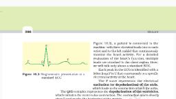
REGULATION OF CARDIAC ACTIVITY, Normal activities of heart are auto, auto-regulated, regulated by nodal tissues. So, it is called myogenic, heart. The special neural centre located in medulla oblongata of brain can moderate cardiac, function through autonomic nervous system (ANS).. Therefore, ANS helps in controlling, heart regulation., Sympathetic nerves of ANS increase the, e rate of heartbeat, the strength of, ventricular contraction and cardiac output., Parasympathetic nerves of ANS decrease the heartbeat, conduction of action, potential and the cardiac output., , , Adrenal medullary hormone adrenaline and nor-adrenaline increase the cardiac output, while acetylcholine decreases the cardiac output, , Electrocardiograph, It is an instrument used to obtain electrocardiogram., Electrocardiogram is the graphical representation of the electrical activity of the heart, during a cardiac cycle. To get an ECG, a patient is connected to the machine with 3 electrical, leads (one to each wrist and to left ankle) that monitor heart activity. For a detailed, evaluation of heart’s function, multiple leads are attached to the chest region., An ECG consists of the following waves:, , , P-wave: Represents the excitation (depolarization) of atria during atrial, atria systole., , , , QRS-complex: Represents depolarization of ventricles during Ventricular systole., , , , T-wave: Represents the repolarization of ventricles or ventricular diastole, , Deviation in the ECG indicates the abnormality or disease. So, ECG has great clinical, significance.
Page 12 :
DOUBLE CIRCULATION, It is the circulation in which blood flows through the heart twice. It includes pulmonary, circulation and systemic circulation., Pulmonary circulation: Circulation b/w lungs and heart., Deoxygenated blood from right ventr, ventricle → to pulmonary artery → to lungs → oxygenated, blood → to pulmonary veins → left atrium., Systemic circulation: Circulation b/w heart and various body parts., Oxygenated blood from left ventricle → to aorta → arteries → arterioles → capillaries →, tissues → deoxygenated, oxygenated blood from tissues → venules → veins → vena cava → to right, atrium., Systemic circulation provides nutrients, O2 and other substances to the tissues and takes, CO2 and other harmful substances away for elimination., Blood vessels are 3 types: Arteries,, ries, Veins & Capillaries., , BLOOD VESSELS, 1) Arteries, They carry blood from heart to other tissues. They contain oxygenated blood (except, pulmonary artery). Their smaller branches are called arterioles. Arteries are 3, 3-layeredinner tunica intima (squamous endothelium), middle tunica media (smooth muscles & elastic, fibres) and outer tunica externa (fibrous connective tissue)., 2) Veins, They carry blood towards heart. They contain deoxygenated blood (except pulmonary vein)., Their smaller branches are called venules. Veins are also 3-layered, layered but tunica media is, comparatively thin., 3) Capillaries, In tissues, arterioles divide into thin walled and single layered vessels. They are, called capillaries. They unite into venules.
Page 13 :
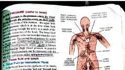
BLOOD PRESSURE, Blood pressure is the force, rce or pressure that the blood exerts on the wall of the blood, vessels. It is of two types:, , , he blood pressure in the arteries during ventricular, Systolic blood pressure - the, systole, ole is called systolic pressure. In a healthy resting adult man, it is about 120mm, Hg., , , , Diastolic blood pressure - the blood pressure in the arteries during ventricular, diastole is called diastolic pressure. In a healthy resting adult man, it is about 80mm, Hg, , , , The normal blood pressure for an adult human is 120/80 mm Hg., , Pulse pressure: The difference between the systolic and diastolic pressure is called pulse, pressure which is 40mm Hg.
Page 14 :
LYMPHATIC SYSTEM, It includes Lymph, Lymph vessels & Lymph nodes (glands)., As the blood passes through the capillaries in tissues, some water and soluble substances are, filtered out from plasma to the intercellular spaces, to form tissue (interstitial) fluid. It, has same mineral distribution as that in plasma., Some tissue fluid enters lymphatic system and the tissue fluid in them is called lymph. It, drains back to major veins. Lymph is a colourless fluid containing lymphocytes., Functions of lymph, , , It is the middleman between blood & tissues. Tissue fluid helps to exchange nutrients,, gases, etc. b/w blood and cells., , , , It carries plasma proteins synthesized in liver to the blood., , , , Transports digested fats (through lacteals in the intestinal villi), fat soluble vitamins,, hormones etc., , , , Filtration of bacteria and foreign particles., , , , Lymph nodes produce WBC (lymphocytes) & antibodies.