Page 1 :
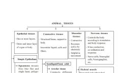
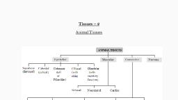
.c, om, , Tissue: A organized group of cells similar in structure, function and origin., • In a tissue cells may be dissimilar in structure and function but they are always similar in origin. Word, , w, , sp, ot, , p., b, , •, , The cells are of two types, somatic cells and germ cells., The word somatic is derived from the Greek word 'soma' means 'body'., All body cells of an organism except sperm and ova are somatic cells., The sperm and ova are germ cells., Various tissues combine together in an orderly manner to form large functional unit called organs., These organs combine together and form organ-system., , lo, g, , •, •, •, •, , io, , lo, , gy, ch, am, , Histology: The study of the structure and arrangement of tissue is called histology., There are four types of tissues present in animals namely – 1) Epithelial Tissue,, 2) Connective Tissue, 3) Muscular Tissue, 4) Nervous Tissue., , .b, , Epithelial tissue (epi : upon, thelio : grows), , A tissue which grows upon another tissue is called Epithelium, , w, , Epithelial tissue forms a covering on inner and outer surface of body and organs., The characteristics of epithelial tissues are as follows, 1. The cells of this tissue are compactly arranged with little intercellular matrix., 2. Cells rest on non-cellular basement membrane., 3. Cells are polygonal, cuboidal or columnar in shape., 4. A Single nucleus is present at the center or at the base., 5. This tissue is avascular. It has good capacity of regeneration., FunctionMajor function is protection and it also helps in absorption, transport, filtration and secretion., Epithelial tissue is classified into two types: 1. Simple epithelium, 2. Compound epithelium., , w, , Y.H.D., , XI-BIOLOGY 2020-2021 ( 9975761329 ), , animal tissue was coined by – Bichat, N. Grew coined the term for Plant Anatomy., Study of tissue – Histology, Histology word was given by – Mayer, Father of Histology – Bichat, Study of tissue is also called Microscopic anatomy., Founder of microscopic anatomy – Marcello Malpighi
Page 2 :
Simple epithelium- is made up of single layer of cells., Compound epithelium -is made up of two or more layers of cells. Lowermost layer lies on basement, membrane., A. Simple epithelial tissue:, , .c, om, , Simple epithelial tissue is further classified into, 1. squamous epithelium, 2. Cuboidal epithelium, 3. Columnar epithelium, 4. Ciliated Epithelium, 5. Glandular Epithelium, 6. Sensory epithelial tissue, 7. Germinal epithelial tissue, , io, .b, , w, , Cuboidal epithelial tissue, 3. Columnar epithelium:, 1. Columnar epithelial cells are tall, pillar like. Inner ends of the cells are narrow while free ends are, broad and flat., 2. Free surface shows large number of microvilli., 3. Nucleus is oval and is present in the lower half of the cell., 4. It is found in inner lining of intestine, gall bladder, gastric glands, intestinal glands, etc., Function: Secretion, absorption, , w, , w, , sp, ot, lo, g, , lo, , gy, ch, am, , p., b, , Squamous epithelial tissue, 1. Cells of this tissue are flat, thin, polygonal with serrated margin., 2. Cells of this tissue fit together like tiles of footpath. Hence it is called pavement epithelium., 3. Prominent spherical or oval nucleus is present at the centre of the cell., 4. It is found in blood vessels, alveoli, coelom, etc., Function: Protection, absorption, transport, filtration, secretion, 2. Cuboidal epithelial tissue:, 1. The cells are cuboidal in shape., 2. Spherical nucleus is present at the centre of cell., 3. It is found in lining of pancreatic duct, salivary duct, proximal and distal convoluted tubules of, nephron., Function: Absorption, secretion, , Y.H.D., , XI-BIOLOGY 2020-2021 ( 9975761329 ), , 1. Squamous epithelial tissue:
Page 3 :
lo, , .c, om, , gy, ch, am, , p., b, , lo, g, , sp, ot, , 4. Ciliated epithelium:, 1. Cells of this tissue are cuboidal or columnar. Free ends of cells are broad while narrow ends rest on, a basement membrane., 2. Free ends show hair like cilia., 3. Nucleus is oval and placed at basal end of cell., Function:, a) To create a movement of materials in contact in a specific direction and thus able to prevent, entry of foreign particles in the trachea,, b) push the ovum through oviduct., c) It is found in inner lining of buccal cavity of frog, nasal cavity, trachea, oviduct of vertebrates,, etc., , io, , .b, , w, , w, , Ciliated epithelial tissue, , Glandular epithelium:, 1. The cells may be columnar, cuboidal or pyramidal in shape., 2. Nucleus is large and situated towards the base., 3. Secretory granules are present in the cytoplasm., 4. The glands may be unicellular (goblet cells of intestine) or multicellular (salivary gland), depending on the number of cells., 5. Depending on mode of secretion, multicellular glands can be classified as duct bearing glands, (exocrine glands) and ductless glands (endocrine glands)., 6. Exocrine glands pour their secretions at a specific sites e.g. Salivary gland, sweat glands etc., 7. Endocrine glands release their secretions directly into blood stream. e.g. thyroid gland, pituitary, gland, etc., Function:, Secrete the mucus that trap the dust particles, lubricates the inner surface of respiratory and, digestive tracts, secretion of enzymes and hormones., , w, , Y.H.D., , XI-BIOLOGY 2020-2021 ( 9975761329 ), , Columnar epithelial tissue
Page 4 :
.c, om, , sp, ot, , lo, g, p., b, gy, ch, am, , Sensory epithelial tissue, , io, , lo, , 7. Germinal epithelial tissue:, 1. Cells of this epithelium divide meiotically to produce haploid gametes., Ex.: Lining of seminiferous tubules, inner lining of ovary., , w, , w, , w, , .b, , Y.H.D., , XI-BIOLOGY 2020-2021 ( 9975761329 ), , Glandular epithelial tissue, 6. Sensory epithelial tissue:, 1. It is composed of modified form of columnar cells and elongated neurosensory cells., 2. Sensory hairs are present at the free end of the cell., Function: It perceive external as well as internal stimuli. These are found in nose (Olfactory) Ear, (Auditory hair cells) Eye (photoreceptors)., , Germinal epithelial tissue, , B. Compound epithelial tissue:, 1. Compound epithelium consists of many layers of Cornified Squamous layer cells., 2. Only the lowermost layer of this tissue is based on the basement membrane.
Page 5 :
sp, ot, gy, ch, am, , p., b, , Protection, Ex.: Epidermis of skin, oesophagus cornea, vagina, rectum., b. Transitional epithelium:, , lo, g, , Function:, , lo, , 1. Structure of transitional epithelium is same like stratified epithelium., 2. The cells can undergo a change in their shape and structure depending on degree of stretch., , Function:, , .b, , io, , Distension of organ, Ex.: Urinary bladder, Cell junctions:, The epithelial cells are connected to each other laterally as well as to the basement membrane by, junctional complexes called cell junctions., , w, , Y.H.D., , XI-BIOLOGY 2020-2021 ( 9975761329 ), , .c, om, , Types of compound epithelium include:, a. Stratified epithelium:, 1. Nucleus is present in stratum germinativum (basal layer)., 2. Cells at free surface become flat and lack nucleus called stratum corneum., , The different types of cell junctions are as follows:, , w, , w, , Gap Junctions (GJs):, This intercellular connection allows passage of ions and small molecules between cells as well as, exchange of chemical messages between cells., Adherens Junctions (AJs):, It is involved in various signaling pathways and transcriptional regulations., Desmosomes (Ds):, These provide mechanical strength to epithelial tissue, cardiac muscles and meninges.
Page 6 :
sp, ot, , lo, g, , p., b, , gy, ch, am, , lo, io, , w, , .b, , Areolar tissue, Areolar tissue is a loose connective tissue found under the skin, between muscles, bones, around, organs, blood vessels and peritoneum. It is composed of fibers and cells., 1. Matrix of this tissue contains two types of fibers namely- white fibers and yellow fibers., a. White fibres are made up of collagen. They give tensile strength to the tissue., b. Yellow fibres are made up of elastin and are elastic in nature., 2.The tissue contains four different types of cells; a. Fibroblast the large flat cells having branching processes. They produce fibers as well, as polysaccharides that form the ground substance or matrix of the tissue., b. Mast cells are oval cells that secrete heparin and histamine., c. Macrophages are amoeboid, phagocytic cells., d. adipocytes, also called Fat cells have eccentric nucleus. These cells store fat., , w, , w, , Y.H.D., , XI-BIOLOGY 2020-2021 ( 9975761329 ), , .c, om, , Hemidesmosomes (HDs):, Allow the cells to strongly adhere to the underlying basement membrane. These maintain, tissue homeostasis by signaling., Tight junctions (TJs):, These junctions maintain cell polarity, prevent lateral diffusion of proteins and ions., Connective tissue:, It is most widely spread tissue in the body. It binds, supports and provides strength to other body tissues, and organs., Characters1. It consists of a variety of cells and fibers. These are embedded in the abundant intercellular, substance called matrix. Connective tissue protects the vital organs of the body., 2. It is highly vascular except cartilage., 3. Connective tissue is classified on the basis of matrix in to three typesa. connective tissue proper,, b. supporting connective tissue, c. fluid connective tissue., a. Connective tissue proper is further classified as loose connective tissue (ex. areolar connective, tissue and adipose tissue) and dense connective tissue (ex. ligament and tendon)., b. Supporting connective tissue also called skeletal tissue includes cartilage and bone. Fluid, connective tissue includes blood and lymph., FunctionConnective tissue protects the vital organs of the body. It acts as packing material and also helps in healing, process., A. Connective Tissue Proper, Loose connective tissue: Matrix of loose connective tissue is semisolid, jelly like, viscous matter made up, of gelatin., 1. Areolar tissue (Areola: air pockets):
Page 7 :
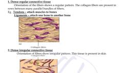
sp, ot, , lo, g, , p., b, gy, ch, am, , Adipose tissue, , lo, , B. Dense Connective Tissue, , w, , .b, , io, , 1. fibers and fibroblasts are compactly arranged in the dense connective tissue., 2. There are two types of dense connective tissue namely –, a. dense regular and dense irregular tissue., b. In dense regular connective tissue, collagen fibers are arranged in parallel manner., c. Two major examples of this tissue are tendons and ligaments., Tendons –, a) Tendons are a type of dense regular connective tissue, b) connect skeletal muscles to bones., c) To give tensile strength to the tissue,, d) Tendons contain bundles of white fibers., E.g. Achilles tendon, Hamstring tendon., , w, , w, , Y.H.D., , XI-BIOLOGY 2020-2021 ( 9975761329 ), , .c, om, , Function-This tissue acts as packing material, helps in healing process and connects different organs or, layers of tissues. It is found under the skin, between muscles, bones, around organs, blood vessels, and peritoneum., 2. Adipose tissue (adipo: fat):, Location- It is found in associated with alveolar connective tissue. Adipose tissue is present bellow skin,, around kidneys and between internal organs., Structure1. It contains large number of adipocytes., 2. The cells are rounded or polygonal., 3. Due to presence of fats stored in the form of droplets in adipocytes, the nucleus is shifted towards, the periphery., 4. Matrix is less and fibers and blood vessels are few in number., 5. The adipose tissue is of two types:, a. White adipose tissue:, i., It is opaque due to the presence of large number of adipocytes., ii., It is commonly present in adults., b. brown adipose tissue:, It is reddish brown in colour due to the presence of large number of blood vessels., Functions:, Adipose tissue is a good insulator, acts as a shock absorber and a good source of energy because it, stores fat. The tissue is found in the sole and palm region as well as around organs like kidneys., , Achilles Tendon: Achilles Tendon connects the calf muscles to heel bone. Pain at the back of ankle or, lower calf may signal a problem with an Achilles Tendon. Athletes who participate in track and field may, face Achilles tendon injury. Most tendon injuries occur near joints such as the shoulder, elbow, knee and, ankle.
Page 8 :
w, , .b, , io, , lo, , gy, ch, am, , p., b, , lo, g, , sp, ot, , Ligament, Ligaments are a type of dense regular connective tissue that are made up of elastic or yellow fibers, arranged in regular pattern. These fibers make ligaments elastic., Location- They are present at joints., Function- They prevent dislocation of bones., In dense irregular connective tissue fibers and fibroblast are not arranged in orderly manner., This tissue is found in dermis of skin., C. Supporting Connective Tissue, Supporting Connective Tissue is characterized by presence of hard matrix., It is classified into two types cartilage and bone., Cartilage: This is a pliable yet tough tissue., Structure1. It forms endoskeleton of cartilaginous fishes like shark. It is widely distributed in vertebrate, animals., 2. In cartilage, abundant matrix is delimited by a sheath of collagenous fibers called perichondrium., 3. Matrix is called chondrin. Just below the perichondrium, immature cartilage forming cells called, chondroblast are present., 4. Chondroblasts mature and get converted into chondrocytes., 5. Chondrocytes are seen scattered in the matrix. They are enclosed in lacunae., 6. Each lacuna contains 2-8 chondrocytes., 7. Based upon the type of matrix, there are four types of cartilage as explained below., A. Hyaline cartilage (Hyaline: Glass like): ., It is found at the ends of long bones, epiglottis, trachea, ribs, larynx and hyoid., Hyaline cartilage is elastic and compressible in nature., a) In this type of cartilage perichondrium is present., b) Matrix is bluish white and gel like., c) Very fine collagen fibers and chondrocytes are present., , w, , w, , Y.H.D., , XI-BIOLOGY 2020-2021 ( 9975761329 ), , .c, om, , Ligament-, , Hyaline Cartilage, FunctionIt acts as a good shock absorber as well as provide flexibility., It reduces friction
Page 9 :
w, , sp, ot, lo, g, , Elastic cartilage, , w, , .b, , io, , lo, , gy, ch, am, , p., b, , C. Fibrocartilage:, Location- Intervertebral discs are made up of fibrocartilage. It is also found at pubic symphysis., 1. The fibrocartilage is the most rigid cartilage., 2. Perichondrium is absent in fibrocartilage., 3. Matrix contains bundles of collagen fibers and few chondrocytes, scattered in fibers., Function- It maintains position of vertebrae., , w, , Y.H.D., , XI-BIOLOGY 2020-2021 ( 9975761329 ), , .c, om, , B. Elastic cartilage:, Location- It is found in ear lobe, tip of nose, etc., 1. In elastic cartilage perichondrium is present., 2. Matrix contain elastic fibers. Chondrocytes are few in number., Function- It gives support and maintains shape of the body part., , White fibrous cartilage, , D. Calcified cartilage:, This type of cartilage becomes rigid due to deposition of salts in the matrix. This reduces flexibility of, joints in old age, e.g. Head of long bones.
Page 10 :
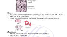
Bone:, Histological structure of bone, , sp, ot, , lo, g, , p., b, gy, ch, am, lo, io, .b, w, , w, , w, , Y.H.D., , XI-BIOLOGY 2020-2021 ( 9975761329 ), , .c, om, , 1. Bone is characterised by hard matrix called Ossein. Ossein is made up of mineral salt hydroxyapatite (Ca10(PO4)6(OH)2)., 2. Outer tough membrane called periosteum encloses the matrix., 3. Blood vessels and nerves pierce through periosteum., 4. Matrix is arranged in the form of concentric layers called lamellae., 5. Each lamella contains fluid filled cavities called lacunae. Fine canals that radiate from each lacuna, are called canaliculi., 6. Canaliculi of adjacent lamellae connect with each other as they traverse through the matrix., In the lacunae osteoblasts., 7. active bone cells and osteocytes, the inactive bone cells and osteoclasts are present., 8. Mammalian bone shows peculiar haversian system., 9. Haversian canal encloses an artery, vein and nerves., 10. According to presence of matrix there are two types of bones present in human body., In spongy bones, haversian system is absent. Reticular, matrix is arranged in the form of trabeculae., It contains red bone marrow., In compact bones, matrix shows haversian system without any space between lamellae., , Bone detailed structure
Page 11 :
D. Fluid Connective tissue (Vascular), Blood and lymph are fluid connective tissue present in the body of an animal., , Muscular tissue:, Characteristics of muscular tissue-, , w, , w, , .b, , io, , lo, , gy, ch, am, , p., b, , lo, g, , sp, ot, , .c, om, , The cells of this tissue are elongated and are called muscle fibers., Each muscle fibers are covered by a membrane sarcolemma., Cytoplasm of muscle cell is called sarcoplasm., Large number of contractile fibrils called myofibrils are present in sarcoplasm., One or many nuclei are present in muscle cell., Myofibrils are made up of proteins, actin and myosin., Muscle fibers contract and decrease in length on stimulation. Hence, muscular tissue is known as, contractile tissue., 8. This tissue is vascular tissue and is innervated by nerves too., 9. Muscle cells contain large number of mitochondria., A. Types of Muscular Tissue, 1. Skeletal muscles: skeletal muscles are also known as voluntary muscles, LocationThese muscles are found attached to bones., Structure1. Skeletal muscles consist of large number of fasciculi which are wrapped by connective tissue, sheath called epimysium or fascia. Each individual fasciculus is covered by perimysium., 2. Each fasciculus in turn consists of many muscle fibers called myofibers., , w, , Y.H.D., , XI-BIOLOGY 2020-2021 ( 9975761329 ), , 1., 2., 3., 4., 5., 6., 7., , Muscular tissue, 3. Each muscle fiber is a syncytial fiber that contains several nuclei., 4. The cell membrane called sarcolemma delimits the cytoplasm called sarcoplasm., 5. Sarcoplasm contains large number of parallelly arranged myofibrils hence nuclei get shifted to, periphery.
Page 12 :
sp, ot, , lo, g, , p., b, , gy, ch, am, , lo, io, .b, w, , w, , w, , Y.H.D., , XI-BIOLOGY 2020-2021 ( 9975761329 ), , .c, om, , 6. Each myofibril is made up of repeated functional units called sarcomeres., 7. Each sarcomere has a dark band called anisotropic or 'A' band in the center. In the center of 'A', band is light area called 'H' zone or 'Hensen's Zone'. In the center of 'H' zone there is 'M' line. 'A' band, are made up of myosin as well as actin., On either side of 'A' band are light bands called isotropic or 'I' bands that contain only actin., Myosin are thick and dark coloured while actin filaments are thin and light coloured. Adjacent light, bands are separated by 'Z' line (Z - Zwischenscheibe line). Dark and light bands on neighboring, myofibrils correspond with each other hence the muscle gets striated appearance., Skeletal muscles show quick and strong voluntary contractions., Function-They bring about voluntary movements of the body., Red and white muscles:, On the basis of amount of a red pigment, skeletal muscles are of two types – Red and white., Red muscles contain very high amount of myoglobin. Myoglobin is an iron containing red coloured, pigment only in muscles. It consists of one haeme and one polypeptide, globulin chain. It can carry one, molecule of oxygen., Due to presence of myoglobin, the muscles can obtain their oxygen from two sources, myoglobin and, haemoglobin., white muscles contain very low amount of this pigment., 2. Smooth or Non-striated muscles:, Location- These are found in the walls of visceral organs and blood vessels. Hence, they are also called as, visceral muscles. They may be arranged lengthwise (longitudinal muscles) or around circumference, (circular muscles) of any organ, 1. These muscles are present in the form of sheets or layers., 2. Each muscle cell is spindle shaped or fusiform., 3. The fibers are unbranched having single nucleus at the center., 4. Sarcoplasm contains myofibrils. Myofibrils are made up of contractile proteins actin and myosin., 5. Smooth muscles contain less myosin and more actin as compared to skeletal muscles., 6. Striations are absent. These muscles undergo slow and sustained involuntary contractions., 7. They are innervated by autonomous nervous system., , Smooth muscle
Page 13 :
sp, ot, lo, g, p., b, gy, ch, am, , Cardiac muscles, , The cardiac muscles are striated involuntary muscles. Some mammalian cardiac muscles are, modified are capable of generating impulse on their own. Hence mammalian heart is a myogenic heart. In, some animals, cardiac muscles need neural stimulus to initiate the contraction. Such a heart is called, neurogenic heart., , w, , .b, , io, , lo, , B. Nervous Tissue:, Characteristics of Nervous tissue1. Nervous tissue is composed of nerve cells or neurons and neuroglia., 2. Neuron is the structural and functional unit of nervous system., 3. Neuroglial cell are non-nervous supporting cells that fill in the interneuronal space. The neuroglial, cells are capable of regeneration and division but neurons are not capable of regeneration because, of lack of centriole. Intercellular matrix is absent in the neural tissue., 4. Neuron is an impulse generating and impulse conducting unit. They bring about quick, communication within the body., 5. Neurons change action potential of their membrane on receiving any external stimulus. This, property of neuron is called excitability., 6. They also carry a wave of electric impulse from dendron to axon, the processes of neuron. This is, called conductivity., Structure of Neuron7. A neuron is made up of-1. cyton or cell body, 2. cytoplasmic process or extensions., 1. cytona) It contains granular cytoplasm called neuroplasm and centrally placed nucleus., , w, , w, , Y.H.D., , XI-BIOLOGY 2020-2021 ( 9975761329 ), , .c, om, , .3. Cardiac Muscles:, LocationCardiac muscles form myocardium of the heart wall., Structure1. Muscles of this tissue show characters of both striated and non-striated fibers., 2. Sarcolemma is not distinct., 3. uni-nucleate muscle fibers appear to be multi-nucleate., 4. Adjacent muscle fibers join together to give branched appearance, 5. Points of adhesion of muscle fibers are formed by transverse thickenings of sarcolemma called, intercalated discs. These junctions at places allow cardiac muscles to contract as a unit. i.e. It, helps in quick transfer of stimulus.
Page 14 :
sp, ot, , lo, g, , w, , .b, , io, , lo, , gy, ch, am, , p., b, , Afferent Neuron:, It carries impulses from sense organ to central nervous system (CNS). Hence it is also called, sensory neuron. It is found in dorsal root of spinal cord., Efferent Neuron:, It carries impulses from CNS to effector organ. Hence it is also called motor neuron. It is found in, ventral root of spinal cord., Interneuron or association neuron:, These are located between sensory and motor neurons., These perform processing, integration of sensory impulses and activate appropriate motor neuron, to generate motor impulse., Depending on the presence or absence of myelin sheath, neurons are classified into two types. i.e., myelinated and non-myelinated nerve fiber., Myelinated or medullated nerve fibers have a insulating fatty layer called myelin sheath around, the axon. This makes the fiber appear white in colour. This sheath is secreted by Schwann cells. The, sheath is not continuous. It is interrupted at nodes of Ranvier. Neurilemma surrounds the axon., The impulse is conducted at a faster rate in such nerve fibers because it jumps from one node to the next., Such transmission of impulse is called saltatory conduction. Myelin sheath prevents the loss of the, impulse during conduction. Cranial nerves of vertebrates are myelinated. Schwann cell of a nonmedullated nerve fiber does not secrete myelin sheath. These fibers are grey in colour due to absence of, fatty layer. Conduction of impulse in a non-myelinated fiber is slower as compared to myelinated nerve, fiber. Nerves of autonomous nervous system are non-myelinated. Functional contact between axonal ends, and dendrites of adjacent neurons is called a synapse., 1. Unipolar/Monopolar Neuron: It has a single process originating from cyton. Both axon and dendron, arise from cyton at one point. They conduct impulses to central nervous system. Ex. Neurons of dorsal, root ganglion of spinal nerve., 2. Bipolar Neuron: It has two processes. A single dendron and an axon are given off from opposite poles, of the cyton. They bring about transmission of special senses like sight, smell, taste, hearing etc. Ex., Neurons of retina of eye, olfactory epithelium., 3. Multipolar Neuron : Cyton is star shaped and gives out more than two processes. There is only one, axon and remaining are dendrons. Axon initiates from a funnel shaped area called axon-hillock., , w, , w, , Y.H.D., , XI-BIOLOGY 2020-2021 ( 9975761329 ), , .c, om, , b) Neuroplasm contains mitochondria, Golgi apparatus, RER and granules called Nissl’s, granules., c) They are made up of RNA., 2. There are two types of cytoplasmic processes namely dendron and axon., Dendrons –, a) these are short, branched, processes., b) The fine branches of dendron are called dendrites., c) They carry impulse towards cyton., Axona) It is single, elongated, cylindrical process., b) Axon is bounded by axolemma., c) The protoplasm of the axon is axoplasm. It contains large number of mitochondria and, neurofibrils., d) Axon is enclosed in a fatty sheath called myelin sheath. Outer covering of myelin sheath is, neurilemma. Myelin sheath and neurilemma are parts of another cell called Schwann's cell., e) Schwann cell shows nucleus at periphery., f) The myelin sheath is absent at intervals along the axon and the place is called Node of Ranvier., g) The terminal arborization of an axon is called telodendron., Based on their functions, Neurons are classified into three types
Page 15 :
w, , w, gy, ch, am, , lo, , sp, ot, , lo, g, , p., b, , XI-BIOLOGY 2020-2021 ( 9975761329 ), , io, , .b, , w, , Y.H.D., , Multipolar Neuron, , .c, om