Page 1 :
Contents, , C H A P T E R, , 1, Introduction, , I, , Nature’s variety is boundless., , t seems to be an axiom of nature that where there is, diversity, there is also similarity. Indeed, nature’s variety, is boundless. When walking through the woods, across a, field, along a stream, through a zoo or wild life sanctuary, one, is impressed with the diversity of life. Even looking through a, microscope can be an elating experience. The universe of the, cell too is complex and diverse. Like the world around us, the, world of the cell is one of the forms specialized for a particular, type of existence. And as is in the larger universe of the plant and, animal kingdoms, where one can perceive basic life sustaining, processes common to all organisms, in the cellular world many, of the same processes and structures can be found in almost all, cells. This generalization often leads to one of the most fundamental and obvious statement that the cell is the microscopic, structural and functional unit of the living organisms. Thus,, there are many cell types among fungi, protozoans and higher, plants and animals. They differ in size, form and function,, degree of specialization and average generation time. Yet at the, ultrastructural level there is sameness about cells that is almost, tedious. The same basic structures—nuclei, cytoplasmic matrix, or cytsol, plastids, mitochondria, endoplasmic reticulum, Golgi, apparatus, plasma membrane, etc.,—all appear with predictable regularity. Such a sameness can also be observed at the, molecular level—all cell parts are made of highly organized, groups of few types of molecules, i.e., proteins, lipids, carbohydrates, nucleic acids, etc., , DEFINITION OF CELL BIOLOGY, The biological science which deals with the study of, structure, function, molecular organization, growth, reproduc-
Page 2 :
Contents, , CELL BIOLOGY, , 4, , tion and genetics of the cells, is called cytology (Gr., kytos = hollow vessel or cell; logous = to, discourse) or cell biology. Much of the cell biology is devoted to the study of structures and functions, of specialized cells. The results of these studies are used to formulate the generalization applied to, almost all cells as well as to provide the basic understanding of how a particular cell type carries out, its specific functions. The cell biologist, without losing sight of the cell as a morphologic and functional, unit within the organism, has to study biological phenomena at all levels of organization and to use all, the methods, techniques and concepts of other sciences (Table 1-1)., , Cytology versus Cell Biology, The cell biology has been studied by the following three avenues: classical cytology dealt with, only light microscopically visible structure of the cell; cell physiology studied biochemistry, biophysics, and functions of the cell; and cell biology interpreted the cell in terms of molecules (macromolecules such as nucleic acids and proteins). In recent years distinction between classical cytology, cell, physiology and cell biology has become blurred and outmoded and now two terms—cytology and cell, biology are used as the synonyms (Novikoff and Holtzmann, 1970)., , HISTORY OF CELL BIOLOGY, Ancient Greek philosophers such as Aristotle ( 384 —322 B.C.), and Paracelsus concluded that “all animals and plants, however, complicated, are constituted of a few elements which are repeated in each of, them.” They were referring to the macroscopic structures of an organism, such as roots, leaves and flowers common to different plants, or segments, and organs that are repeated in the animal kingdom. Many centuries later,, owing to the invention of magnifying lenses, the world of microscopic, dimensions was discovered. Da Vinci (1485) recommended the uses of, lenses in viewing small objects. In 1558, Swiss biologist, Conrad Gesner, (1516—1565) published results of his studies on the structure of a group, of protists called foraminifera. His sketches of these protozoa included, so many details that they could only have been made if he had used some, form of magnifying lenses. Perhaps this is earliest recorded use of a, magnifying instrument in a biological study., , Table 1-1., , 1., 2., , 3., 4., 5., , Aristotle ( 384 —322 B.C.), , Various levels of biological organization and instrumental resolving power (Source:, De Robertis and De Robertis, Jr., 1987)., , Dimension, , Biological, field, , Structures, , Method of study, , 0.1 mm or 100 µm, or larger, 100 µm to 10 µm, , Anatomy, , Organs, , Eyes and simple lenses, , Histology, , Tissues, , Cell biology, , Cell, bacteria, , Submicroscopic, morphology, Ultrastructure,, molecular and, atomic structure., , Cell components,, viruses, Arrangement, of atoms, , 10 µm to 0.2 µm, (200 nm), 200 nm to 1 nm, Smaller than, 1 nm (10A0), , }, , Various types of light, microscopes, X-ray, microscopy, , Polarization microscopy,, electron microscopy, X-ray diffraction
Page 3 :
Contents, , INTRODUCTION, , 5, , Further growth and development of cell biology are intimately associated with the development, of optical lenses and to the combination of these lenses in the construction of the compound, microscopes (Gr., mikros = samll; skopein = to see). Thus, the invention, of the microscope and its gradual improvement went hand-in-hand with, the development of cell biology., , 1. Growth of Cell Biology during 16th and 18th Centuries, The first useful compound microscope was invented in 1590 by, Francis Janssen and Zacharias Janssen. Their microscope had two, lenses and total magnifying power between 10X and 30X. Such types of, microscopes were called “flea glasses”, since they were primarily used, to examine small whole organisms such as fleas and other insects. In, 1610, an Italian Galileo Galilei (1564 —1642) invented a simple, microscope having only one magnifying lens. This microscope was used, to study the arrangement of the facets in the compound eye of insects., The Italian microanatomist Marcello Malpighi ( 1628—1694), was among the first to use a microscope to examine and describe thin, slices of animal tissues from such organs as the brain, liver, kidney,, spleen, lungs and tongue. He also studied plant tissues and suggested that, they were composed of structural units that he called “utricles”. An Fig. 1.1. Hooke’s compound, English microscopist Robert Hooke (1635—1703) is credited with, microscope., coining the term cell (L., Cella = hollow space) in 1665. He examined a, thin slice cut from a piece of dried cork under the compound microscopes (Fig. 1.1) which were built, by him. In 1665, Hooke published a collection of essays under the title Micrographia. One essay, described cork as a honey comb of chambers or “cells”. The chambers or cells are now recognized to, be empty spaces left behind after the living portions of the cell had disintegrated. Hooke thought of the, cells, he observed as something, similar to veins and arteries of, animals—they were filled with, “juices” in living plants. But his, crude microscopes did not permit the observation of any intracellular structure., Dutch microscopist,, Anton van Leeuwenhoek, (1632—1723) had succeeded, in greatly improving the art of, Anton van Leeuwenhoek, Fig. 1.2. Leeuwenhoek’s microscope. polishing lenses of short focal, (1632—1723), length. He used his lenses in, building numerous microscopes, some with magnifications approaching 300X (Fig. 1.2). Leeuwenhoek, was the first to observe living free-living cells; he described in 1675, microscopic organisms in, rainwater collected from tubes inserted into the soil during rainfall. His sketches included numerous, bacteria (bacilli, cocci, spirilla and other Monera), protozoa, rotifers, and Hydra. Leeuwenhoek was, also first to describe the sperm cells of humans, dogs, rabbits, frogs, fish and insects and to observe the, movement of blood cells of mammals, birds, amphibians and fish, noting that those of fish and, amphibians were oval in shape and contained a central body (the nucleus); while those of humans and, other mammals were round. He also observed the striated muscles. Leeuwenhoek’s observations were, recorded in a series of reports that he sent during 1675—1683 to the Royal Society of London., An English plant microanatomist Nehemiah Grew (1641—1721) published accounts of the
Page 4 :
Contents, , 6, , CELL BIOLOGY, , microscopic examination of sections through the flowers, roots and stems of plants and clearly, indicated that he recognized the cellular nature of plant tissues., , 2. Growth of Cell Biology during 19th Century, Nineteenth century witnessed various cell biological inventions and formulations of various, landmark theories such as cell theory and protoplasm theory. In 1807, Mirbel stated that all plant, tissues were composed of cells. French biologist, Rene Dutrochet (1776—1827) correctly concluded, in 1824, that all animal and plant tissues were “aggregates of globular cells.” In 1831, an English, botanist Robert Brown ( 1773—1858) discovered and named the nucleus in the cells (e.g., epidermis,, stigmas and pollen grains) of the plant Tradescantia. He established that the nucleus was the, fundamental and constant component of the cells., , Cell Theory, In 1838, a German botanist Mathias Jacob Schleiden (1804—1881) put forth the idea that cells, were the units of structure in the plants. In 1839, his coworker, a German zoologist , Theodor Schwann, (1810—1882) applied Schleiden’s thesis to the animals. Both of them, thus, postulated that the cell is, the basic unit of structure and function in all life. This simple, basic and formal biological generalization, is known as cell theory or cell doctrine. In fact, both Schleiden and Schwann are incorrectly credited, for the formulation of the cell theory; they merely made the generalizations which were based on the, works of their predecesors such as Oken (1805), Mirbel (1807), Lamarck (1809), Dutrochet (1824),, Turpin (1826), etc., (see Sheeler and Bianchi, 1987). However, Schleiden was the first to describe, the nucleoli and to appreciate the fact that each cell leads a double life—one independent, pertaining, to its own development, and another as integral part of a multicellular plant. Schwann studied both, plant and animal tissues and his work with the connective tissues such as bone and cartilage led him, to modify the evolving cell theory to include the idea that living things are composed of both cells and, the products or secretions of the cells. Schwann also introduced the, term metabolism to describe the activities of the cells., In the coming years, the cell theory was to be extended and, refined further. K. Nageli (1817—1891) showed in 1846 that plant, cells arise from the division of pre-existing cells. In 1855, a German, pathologist Rudolf Virchow (1821—1902) confirmed the Nageli’s, principle of the cellular basis of life’s continuity. He stated in Latin, that the cells arise only from the pre-existing cells (viz., his actual, aphorism was “omnis cellula e cellula” —every cell from a cell)., Virchow, thus, established the significance of cell division in the, reproduction of organisms. In 1858, Virchow published his classical textbook Cellular Pathology and in it he correctly asserted that, Louis Pasteur (1822—1895), as functional units of life, the cells were the primary sites of disease, and cancer. Later , in 1865, Louis Pasteur (1822—1895) in France, gave experimental evidence to support Virchow’s extension of the cell theory., The modern version of cell theory states that (1) All living organisms (animals, plants and, microbes) are made up of one or more cells and cell products. (2) All metabolic reactions in unicellular, and multicellular organisms take place in cells. (3) Cells originate only from other cells, i.e., no cell, can originate spontaneously or de novo, but comes into being only by division and duplication of, already existing cells. (4) The smallest clearly defined unit of life is the cell., The cell theory had its wide biological applications. With the progress of biochemistry, it was, shown that there were fundamental similarities in the chemical composition and metabolic activities, of all cells. Kolliker applied the cell theory to embryology—after it was demonstrated that the, organisms developed from the fusion of two cells—the spermatozoon and the ovum. However, in the, recent years, large number of sub-cellular structures such as ribosomes, lysosomes, mitochondria,, chloroplasts, etc., have been discovered and studied in detail. Consequently, it may appear that cell is
Page 5 :
Contents, , INTRODUCTION, , 7, , no longer a basic unit of life, because life may exist without cells also. Even then, the cell theory remains, a useful concept., Exception to cell theory. Cell theory does not have universal application, i.e., there are certain, living organisms which do not have true cells. All kinds of true cells share the following three basic, characteristics: 1. A set of genes which constitute the blueprints for regulating cellular activities and, making new cells. 2. A limiting plasma membrane that permits controlled exchange of matter and, energy with the external world. 3.A metabolic machinery for sustaining life activities such as growth,, reproduction and repair of parts. Viruses do not easily fit in these parameters of a true cell. Thus, they, lack a plasma membrane and a metabolic machinery for energy production and for the synthesis of, proteins. However, like any other cellular organism, viruses have (1) a definite genetically determined, macromolecular organization; (2) a genetic or hereditary material in the form of either DNA or RNA;, (3) a capacity of auto-reproduction; and (4) a capacity of mutation in their genetic substance. In, consequence, viruses can only reproduce inside the host cells which may belong to animals, plants or, bacteria. They use their own genetic programme for reproduction but rely on the raw materials (i.e.,, amino acids, nucleotides) and biosynthetic machinery of the host cells ( i.e., ribosomes, tRNA,, enzymes) for their multiplication. Thus, a virus may be defined as an infectious, subcellular and, ultramicroscopic particle representing an obligate cellular parasite and a potential pathogen whose, reproduction (replication) in the host cell and transmission by infection cause characteristic reaction, in the host cells. Outside the host cells, viruses are just like non-living inert particles and like the salt, or sugar, they can be purified, crystallized and placed into jars on a shelf for years. Due to this fact,, viruses have been variously described such as “naked genes that had somehow acquired the ability to, move from one cell to another (Alberts et al., 1989), or as “cellular forms that have degenerated, through parasitism”, or as “primitive organisms that have not reached a cellular state.”, , Tobacco mosaic virus, , A, Paramyxovirus, (Mumps virus), , T-even phage, , Rhizopus, Paramecium, , Fig. 1.3., , Vaucheria, , B, , Organisms forming exceptions to the cell theory : A—Three types of viruses;, B—Three cases of cellular organization.
Page 6 :
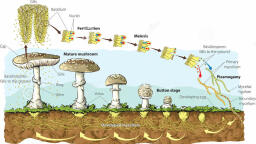
Contents, , 8, , CELL BIOLOGY, , There are certain other organisms such as the protozoan Paramecium, the fungus Rhizopus and, the alga Vaucheria (Fig. 1.3B) which do not fit into the purview of the cell theory. All of these organisms, have bodies containing undivided mass of protoplasm which lacks cell-like organization and has more, than one nucleus. They tend to raise the question that whether cell is a basic unit of structure in them., , Protoplasm Theory, Up to middle of the 19th century, greater emphasis was given to the cell wall and less to the, cellular content. But soon cell biologists started to recognize the importance of “juicy” or “slimy”, contents of the cells. In 1835, Felix Dujardin termed the jelly-like material within protozoans as, sarcode. In 1835, H.von Mohl (1805—1875) described cell division. In 1839, the Czech biologist, J.E. Purkinje (1787—1869) coined the term protoplasm to describe the contents of cells (animal, embryos). Von Mohl, in 1846, applied the name protoplasm to the contents of embryonic cells of the, plants. Max Schultze, in 1861, established similarity between sarcode and protoplasm of animal and, plant cells and, thus, offering a theory which later on was improved and called protoplasm theory by, O.Hertwig (1849—1922) in 1892., Protoplasm theory holds that all living matter, out of which animals and plants are formed, is the, protoplasm. The cell is an accumulation of living substance or protoplasm which is limited in space by, an outer membrane and possesses a nucleus. The protoplasm which is filled in the nucleus is called, nucleoplasm and that exists between the nucleus and the plasma membrane is called cytoplasm., The last quarter of 19th century is usually considered as “classical period of cell biology”. Since, various significant cell biological discoveries have been made during this period. Certain landmark cell, biological discoveries of second half of the 19th century have been tabulated in a chronological order, in the Table 1-2., , 3. Growth of Cell Biology in 20th Century, 20th century has witnessed great advancement in cell biological knowledge due to the following, two main reasons: (1) the increased resolving power of instrumental analysis due to the introduction, , Table 1-2., , Chronological tabulation of certain important investigations of 19th century in cell, biology., , Year, , Name of contributor, , Cell biological contribution, , 1855, 1857 –, 1881, 1857, , C.Nageli and C. Cramer, H.M.Edwards, , 1865, , G.Mendel, , 1866, 1870, , Haeckel, W.His, , 1871, , F.Miescher, , 1873, , A.Schneider, , Coined the term cell membrane., Explained division of labour, in body cells., Discovered mitochondria (“sarcosomes”), in muscle and in 1888 he isloated them., Developed the fundamental principles of, heredity., Named plastids., Developed the microtome for cutting serial, sections of tissue for cell study., Isolated nuclei and nucleoprotein from pus, cells, spermatozoa and from haemolyzed erythrocytes of birds., Described chromosomes (nuclear filaments), for the first time., Described the spindle and astral rays and, showed in 1879 that only one sperm enters, the egg in fertilization., , A.Kolliker, , H.Fol
Page 7 :
Contents, , INTRODUCTION, Year, , Name of contributor, , Cell biological contribution, , 1875, , E.Strasburger, , 1876, , E. van Beneden, O.Hertwig, , 1878, 1879, , Schleicher, W.Flemming, , 1881, , Reinke and Rodewald, E.G. Balbiani, , Described mitosis in plant cells and in 1882, introduced the terms cytoplasm and nucleoplasm. In 1884, he described fertilization in, angiosperms., First observed the centriole., Studied reproduction in sea urchin and concluded that fertilization involves the union of, sperm and egg pronuclei., Coined the term karyokinesis., Introduced the term chromatin and described, the longitudinal splitting of chromosomes, during nuclear division of animal cells. In 1882,, he coined the term mitosis., Performed chemical analysis of protoplasm., Discovered the larval salivary gland chromosomes (i.e.,giant or polytene chromosomes), in Chironomus., Described many animal tissues with a detail, that has not been surpassed by any other, light microscopist. In the next two decades, he,, Cajal and other histologists developed staining methods and laid the foundations of, microscopic anatomy., Discovered chromomeres in the chromosomes., Showed that in Ascaris the number of chromosomes in the gametes is half that of in the body, cell., Proposed that chromosomes contain the units of, heredity., Introduced the term chloroplast., Described details of chloroplast structure., Observed and named phagocytosis., Discovered cytochromes., Described the structure of centrioles and coined, the term centrosome. In 1892, he described, spermatogenesis and oogenesis., Introduced the term chromosome., Stained mitochondria with a specific stain, (1886), recognised their role in cellular respiration and considered them as autonomous organelles. In 1894, he coined the term bioblasts, for mitochondria., Published his monograph—Die Zelle und das, Gewebe (The cell and the tissue) in which he, attempted to achieve a general synthesis of, biological phenomena based on characteristics of the cell, its structure and function. He,, thus, created cytology as a separate branch, of biology., , Retzius, , 1882, 1883, , W.Pfitzner, E. van Benden, , W. Roux, , 1886, 1888, , Schimper, Meyer, E. Metchnikoff, C.A. MacMunn, T.Boveri, , 1890, , W.Waldeyer, R.Altmann, , 1892, , O.Hertwig, , 9
Page 8 :
Contents, , 10, , CELL BIOLOGY, 1897, , C.Benda, , 1898, , Camillo Golgi, , Coined the term mitochondrion and studied it, in spermatozoa and other cells., Described and coined the term Golgi complex, for the reticular structure found in the cytoplasm of nerve cells of owls and cats. He used, the silver staining method in studies., , of electron microscopy and X-ray diffraction techniques, and (2) the convergence of cytology with, other fields of biological research, especially genetics (cytogenetics), physiology (cell physiology) and, biochemistry (cytochemistry). Consequently new histochemical, cytochemical and immunocytochemical (using antibodies to localise antigens) techniques have been developed to detect various, molecular components of the cell. Likewise, various cellular components have been separated by, ultracentrifugation; different biochemical events of the cell could be known in detail by autoradiography; and methods of tissue culturing have made possible the study of living cells. Phase contrast, microscopy and interference microscopy have been used to study the living cells. The ultrastructure, of a cellular membrane could be observed by the techniques of freeze-fracturing and freeze-etching., Moreover, micromanipulators, micromanometric methods (e.g., by Cartesian diver balance of Zeuthen, weight of a single amoeba can be determined), chromatography, electrophoresis, spectrophotometry,, etc., have provided new opportunities to cell biologists to investigate minute details of cell and its, components. Due to the employment of various improved ultratechniques in the study of the cells, the, validity of the cell theory and protoplasm theory has become vague. Therefore, presently, both of these, theories have been replaced by another new theory called organismal theory., , Organismal Theory, The organismal theory holds that the body of all multicellular organisms is a continuous mass of, protoplasm which remains divided incompletely into small centres, the cells, for the various biological, activities. Thus, a multicellular organism is a highly differentiated protoplasmic individual, differing, with a unicellular Protozoa only in size and degree of differentiation of the protoplasm. The, differentiation involves separation of the protoplasm into subordinate semi-independent compartments, the so-called cells. Even the embryological development of a multicellular individual includes, only growth and progressive internal differentiation of a small single protoplasmic individual (egg)., Organismal theory too fails to ascertain the position of viruses., Certain landmark cell biological discoveries and Nobel Prize winning investigations of 20th, century have been tabulated in a chronological way in the Table 1-3., , Table 1-3., , Chronological tabulation of certain important cell biological investigations of, 20th century., , Year, , Name of contributor, , Cell biological contribution, , 1900, , C.Garnier, J.Loeb, E.Strasburger, T.H. Montgomery, , Introduced the term ergastoplasm., Discovered artificial parthenogenesis., Introduced the term plasmodesmata., Showed that homologous chromosomes undergo pairing or synapsis during the reduction division., Got Nobel Prize for his pioneering studies of the, proteins., Discovered the enzymes and got Nobel Prize for it., Demonstrated the presence of mitochondria in, plant cells., Coined the term meiosis for the reduction and, cell division., , 1901, , 1902, , E.Fischer, , 1903, 1904, , E.Buchner, F. Meves, , 1905, , J.B.Farmer, J.E.Moore
Page 9 :
Contents, , INTRODUCTION, , 11, , Year, , Name of contributor, , Cell biological contribution, , 1906, , M.Tswett, C.Golgi and, S.R. Cajal, R.G. Harrison, , Invented column chromatography., Got Nobel Prize for their contributions, regarding the structure of nerve cells., Developed the technique of tissue culture;, cultivated amphibian spinal cord in a lymph, clot., Got Nobel Prize for their contributions on, phagocytosis of bacteria during infection;, staining procedures for bacteria, and studies, on immunity., Investigated the chemistry of the nucleus and, got Nobel Prize for this contribution., Got Nobel Prize for his studies on chlorophyll, and other plant pigments., Got Nobel Prize for their studies on the metabolism of muscle tissue and for relationship between muscle metabolism and lactic acid., Developed the first autoradiographic method, to localize radioactive polonium in biological, specimens., Got Nobel Prize for his studies on properties of, colloids,especially proteins and for the development of analytical ultracentrifugation., Got Nobel Prize for the discovery of human, blood groups and for studies of cellular agglutinins or antigens., Designed and built the first interference microscope., Got Nobel Prize for his studies on the nature and, mode of action of respiratory enzymes and for, the studies of oxidation and reduction in, metabolism., Built the first transmission electron, microscope., Coined the term plasmalemma., Discovered pinocytosis., Invented phase contrast microscope and got, Nobel Prize for this invention in 1953., Got Nobel Prize for the discoveries concerning, the role of chromosomes in the transmission of, heredity., Introduced the technique of electrophoresis, for separating proteins in solution., Proposed protein-lipid-protein structure, (sandwich model) of plasma membrane., Demonstrated the feasibility of the electron, microscope., Isolated tobacco mosaic virus (TMV) in crystalline form., Got Nobel Prize for his studies on biological, oxidation and the involvement of vitamine C., , 1907, , 1908, , E.Metchnikoff, and P.Ehrlich, , 1910, , A.Kossel, , 1915, , R.Wilstatter, , 1922, , A.V.Hill and, O.Meyerhof, , 1924, , Lacassagne, and coworkers, , 1926, , T.Svedberg, , 1930, , K.Landsteiner, , Lebedeff, 1931, , O.Warburg, , 1932, , E.Ruska and, M.Knoll, J.Q. Plowe, W.H.Lewis, F.Zernike, , 1933, , T.H. Morgan, , A.Tiselius, 1935, , J.Danielli, and H.Davson, M.Knoll, M.W.Stanley, , 1937, , A. von SzentGyorgyi
Page 10 :
Contents, , 12, , CELL BIOLOGY, Year, , Name of contributor, , Cell biological contribution, , H.A.Krebs, , Discovered the citric acid cycle or tricarboxylic, acid cycle and got Nobel Prize for this work in, 1953., , 1938, , Behrens, , 1939, 1941, , F.A.Lipman, Coons, , 1942, , Martin and, Synge, A.Claude, , Employed differential centrifugation to separate nuclei and cytoplasm from liver cells., Proposed a central metabolic role for ATP., Used antibodies coupled to fluorescent dyes, to detect cellular antigens., Developed partition chromatography, leading, to paper chromatography two years later., Isolated cell components such as ribosomes,, mitochondria and nucleus in relatively pure, form by differential centrifugation., Introduced the metal shadowing technique., , 1943, , 1944, , 1945, , Williams and, Wyckoff, C.F. Robinow, K.R.Porter, F.A. Lipman, , 1947, 1948, , C.F.Cori and, G.T.Cori, A.Tiselius, , 1948, , C.de Duve, , 1952, , 1953, 1954, , Grigg and Hodge, G.E. Palade, A. Morten and, R. Synge, Manton et al., Robinson and, Brown, J.Rhodin, L.Pauling, , Fawcett and, Porter, , Demonstrated the nucleus (= nucleoid) in the, bacteria., Discovered and named the endoplasmic, reticulum., Discovered coenzyme A (a key compound in, cell metabolism) and got Nobel Prize in 1953 for, his studies on this coenzyme., Got Nobel Prize for their studies of the, metabolism of glycogen., Got Nobel Prize for his studies on the chemistry, of proteins and for development of electrophoresis., Isolated lysosomes and identified their enzymatic properties. In 1955, he coined the term, lysosome., Studied fine structure of the flagellum of sperm., Described the ultrastructure of mitochondria., Got Nobel Prize for the development of, chromatographic procedures for the separation of, biological substances., Studied fine structure of cilia of higher plants., Reported ribosomes in the plant cells (i.e.,, bean root)., Described and named microbody in mouse, kidney tissue., Got Nobel Prize for his studies on the nature of, chemical bonds, especially the peptide bond of, proteins., Confirmed the 9 + 2 fibrillar arrangement of, cilia and flagella.
Page 11 :
Contents, , INTRODUCTION, , 13, , Year, , Name of contributor, , Cell biological contribution, , 1955, , G.E. Palade, , Observed ribosomes in animal cells and in 1956, he detected RNA in the isolated ribosomes., Completed the analysis of the amino acid sequence of bovine insulin; the first protein to, be sequenced; got Nobel Prize in 1958 for this, contribution., Gave the first correct human chromosome, count (46 chromosomes in diploid condition)., Used negative staining technique in visualizing viruses, bacteria and protein filaments., Forwarded the concept of unit membrane., Discovered quantosomes in the chloroplast., Independently verified the existence of DNA, fibrils in mitochondria and chloroplasts., Got Nobel Prize for his work on the assimilation, of CO2 by plants, photosynthesis, the “Calvin, cycle.”, Got Nobel Prize on their studies of the, structure of globular proteins, especially myoglobin and haemoglobin., Got Nobel Prize for their work on the role of, sodium and potassium ions in the conduction, of nerve impulses along the nerve cell membrane., Got Nobel Prize for their studies on the, metabolism of cholesterol and fatty acids., Obtained a complete carrot plant from a single, carrot root cell by tissue culture technique., Coined the term peroxisome for catalase enzyme, containing microbody., Produced first heterokaryons of mammalian, cells by the virus-induced fusion of human and, mouse cells., Coined the term glyoxisome for the glyoxylate, cycle containing microbody of plant cells., Got Nobel Prize for studies on the mechanism, of action of hormones and role of cyclic AMP., Proposed the fluid mosaic model of cell, membrane., Got Nobel Prize for isolation and characterization of sub-cellular organelles and other particles., Got Nobel Prize for the studies of bioenergetics., Perfected video-enhanced contrast light, microscopy., Got Nobel Prize for his studies on the structure, of complicated biological molecules (e.g., proteins and nucleic acid in TMV and histone core, of nucleosome) by using electron microscopy, and X-ray crystallography., , F.Sanger, , 1956, 1959, , 1960, , 1961, , J.H.Tjio and, A.Levan, Brenner and, Horne, G.D. Robertson, Park and Pon, H.Ris and M., Nass, M.Calvin, , 1962, , M.F. Perutz, and J.C.Kendrew, , 1963, , J.Eccles, A., Hodkin and, A. Huxley, K. Bloch and, F.Lynem, Kato and, Takeuchi, De Duve, , 1964, , 1965, , Harris and, Watkins, 1967, 1971, 1972, , R.W.Breidenbach, and H.Beevers, E.A. Sutherland, , 1978, 1981, , S.J. Singer and, G.L. Nicolson, A.Claude, C.de, Duve and G. Palade, P.Mitchell, Allen and Inoue, , 1982, , Aaron Klug, , 1974
Page 12 :
Contents, , CELL BIOLOGY, , 14, , Year, , Name of contributor, , Cell biological contribution, , 1984, , Schwartz and, Cantor, C.Milstein, J., F. Kohler and J.K. Jerne, S.Tonegawa, , Developed pulsed field gel electrophoresis, for the separation of very large DNA molecules., Got Nobel Prize for his studies on molecular, immunology., Got Nobel Prize for primary discoveries in the, field of antibodies ( immunobiology)., , 1987, , UNIT OF MEASUREMENT OF CELL, The viruses and cells of most bacteria, blue green algae, animals and plants are minute in size and, are measured by the fractions of standard units. The standard units are metres, grams, litres and, seconds. The value of different units of measurements has been tabulated in, Table 1- 4., , Table 1-4., , Units of measurement used in cell biology (Avers, 1978)., A. Length, , Metre, (m), , Millimetre, (mm), , 1, 1,000 (1 × 10 3), 0.001, 1, 0.000001, 0.001, 1 × 10 -9, 1 × 10⁻⁶, 1 × 10⁻⁷, 1 × 10⁻¹⁰, , Micrometre, µm), or Micron (µ, , Namometre or, µ), Millimicron (nm or mµ), , Angstrom, (Α 0), , 1,000,000 (1 × 10 6), 1,000, 1, 0.001, 1 × 10⁻⁴, , 1,000,000,000 (1 × 10⁹), 1,000,000, 1,000, 1, 0.1, , 1 × 10 10, 1 × 10⁷, 1 × 10⁴, 10, 1, , Microgram, µg), (µ, , Nanogram, (ng), , Picogram, ( pg)), , 1,000,000, 1,000, 1, 0.001, 1 × 10⁻⁶, , 1 × 10⁹, 1 × 10⁶, 1 × 10³, 1, 0.001, , B. Weight, Gram, (g), 1, 0.001, 1 × 10⁻⁶, 1 × 10⁻⁹, 1 × 10-12, , Milligram, (mg), 1,000, 1, 0.001, 1 × 10⁻⁶, 1 × 10⁻⁹, , 1 × 10 12, 1 × 10⁹, 1 × 10⁶, 1 × 10³, 1, , CELL BIOLOGY AND OTHER BIOLOGICAL SCIENCES, The cell biology has helped the biologists to understand various complicated life activities such, as metabolism, growth, differentiation, heredity and evolution at the cellular and molecular levels. Due, to its wide application in various branches of biological science, many new hybrid biological sciences,, have sprung up. Some of them are as follows:, 1. Cytotaxonomy (Cytology and Taxonomy). Each plant and animal species has a definite, number of chromosomes in its cells and the chromosomes of the individuals of a species resemble, closely with one another in shape and size. These characteristics of the chromosomes help a taxonomist, in determining the taxonomical position of a species. Further, cell biology furnishes strong support to, the manner of origin of certain taxonomic units. Therefore, the cytotaxonomy can be defined as a, cytological science which provides cytological support to the taxonomic position of any species., 2. Cytogenetics (Cytology and Genetics). Cytogenetics is that branch of cell biology which is, concerned with the cytological and molecular bases of heredity, variation, mutation, phylogeny,
Page 13 :
Contents, , INTRODUCTION, , 15, , morphogenesis and evolution of organisms. The Weismann’s germ plasm theory, Mendel’s laws of, inheritance and the concept of gene could be well understood only after the application of cytological, concept to the genetics., 3. Cell Physiology (Cytology and Physiology). The cell physiology is the study of life activities,, viz., nutrition, metabolism, excitability, growth, reproduction or cell division and differentiation of the, cell. The cell physiology has helped in understanding various complicated physiological activities at, cellular level., 4. Cytochemistry (Cytology and Biochemistry). The cytochemistry is that branch of cytology, which deals with the chemical and physico-chemical analysis of living matter. For example, the, cytochemical analysis has revealed the presence of carbohydrates, lipids, proteins, nucleic acids and, other organic and inorganic chemical compounds in the cells., 5. Ultrastructure and Molecular Biology. These are the most modern branches of, biology in which the merging of cytology with, biochemistry, physico-chemistry and especially, macromolecular and colloidal chemistry become increasingly complex. Knowledge of the, submicroscopic organization or ultrastructure, of the cell is of fundamental importance because practically all the functional and physicochemical transformations take place with the, molecular architecture of the cell and at a, molecular level. The recent discoveries in, molecular biology such as the discovery of, molecular model of DNA by Waston and, Waston and Crick., Crick in 1953, molecular interpretation of protein synthetic mechanism, genetic code, etc., have an extraordinary impact on modern cell biology and, biology., 6. Cytopathology (Cytology and Pathology). The application of molecular biology to pathological science has helped in understanding various human diseases at molecular level. Because most, diseases are caused due to disorder of genetic codes in DNA molecule which alter the synthetic process, of enzymes and ultimately disturb metabolic activities of the cell., 7. Cytoecology (Cytology and Ecology). The cytoecology is the science in which one studies the, effects of ecological changes on the chromosome number of the cell. The cytological studies on plants, and animals have revealed that the ecological habitat and geographical distribution have the correlation, with chromosome numbers., , REVISION QUESTIONS, 1., 2., 3., 4., , Who had discovered the cell ? Explain, how is the growth of cell biology linked with the, improvement in instrumental analysis ?, What is cell theory ? Describe the cell theory and explain the exceptions of cell theory., What is meant by ‘classical period of cell biology’ ? Write about certain landmark discoveries of, this period., Write short notes on the following:, (i) Protoplasm theory;, (iii) Branches of cell biology;, (ii) Organismal theory;, (iv) Scope of cell biology.