Notes of PUC 2nd Year, Biology 2nd pu pract. exp. 5 to 9.pdf - Study Material
Page 1 :
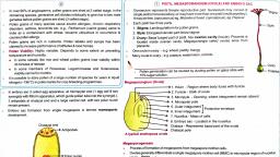
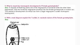
II PUC Biology lab manual, [EMBRYOSAC], EXERCISE - 12 : FEMALE GAMETOPHYTE DEVELOPMENT IN PLANTS, Aim: To identify development stages of female gametophyte in ovary of ar angios perm flower., Principle: Female gametophyte (embryo sacs) present inside the ovule. Female gametophyte develops from, the megaspore mother cell. It contains 8 nuclei and 7 cells., Tdiploid, Requirements: Permanent slide or photographs showing femalegametophyte anda compound microscepe., OBSERVATION, (1) Development of female gametophyte:, a), The, megaspore mother cell undergoes meiosis resulting in 4 haploid megaspores ., b) Out of which 3 disintergrates and the remaining one divides thrice to produce 8 haploid nuclei., c) Of these 8 nuclei, 6 get surrounded by cell wall to form cells. They are 3 antipodals at chalazal end 2, synergids with an egg cell at the micropylar end., by Mitos i3, d) The remaining 2 nuclei remain in the center as polar nuclei. Thus before fertilization the embroy sac, has 8 nuclei and 7 cells., e) – since he embryosac is developed from single,, tunctional haploid megaspore ("), 't is called, monosporie "development., 2) v.S. of ovule (megasprangium), a. Ovule develops inside the ovary., b. It is attached to the placenta by stalk funicle.", c. It consists of two integuments and nucellus., d. Nucellus cells contain food material., e. Female gametophyte develops inside the nucellus., f. Theembryosae forms the megaspore mother cell., g. A typical anatropous "ovule has 8-nucleated aud 7-celled condition,, h. 3 cells at Michopolar end - one egg cell amd two bynergids, 3cells at chalazal, end called antipodals, and ané central cell w tw. poláe nuclei,, 32, od put, 1st page
Page 3 :
II PUC Biology lab manual, EXERCISE- 13: GETOGENESIS SPERMATOG ENESIS, Aim: To study the different stages of gametogenesis in mammalian testis and every slide,, Principle: In all male and female organisms gamete formation takes place in their gonads, i.e.,testis and, ovary respectively. The process of gamete formation, called gametogenesis involves meiotic cell division., The gametogenic development in testis is called spermatogenesis and in ovary it is oogenesis. They exhibit, marked differences and can be examined in transverse section (T.S.) of the gonads., Requirement: Permanent slides of T 8. of testis and ovary, compound microseope,, 'lens-cleaning paper and cleaning ffuid, XProcedure : 1. Clean thế slide and microscope's éye and objęctive lenses with the help of lens cleaning, paper using any cleaning fluid., 2. Place the slide on the stage of the microşcope and obsérve first under lower magnification and then in, higher magnífication. Obşerve various stages of gamete development., 3. Récord your observations in the notebook and draw labelled diagrams.', AY, + Each testis is surrounded by a thick, fibrous, vascularised connective tissue capsule called tunica, albuginea., 2 Each testis is made up of about 250 compartments called testicular labules., 3. Each lobule contains 2-3 coiled seminiferous tubules -which are, lined w two typesof germ cets,, Spermatogonia undergo spermatogenesis to produce sperms.-, Supportive and Nutritive Sertoli er sustentacular cells. are present. pse SSc, Spermatdi also Seel, Stroma of testis contains intersitial cells called Leydig cells. they secrete testosterone (male sex, hormone(ea ndrogens)., (A, T.S of Mammalian Testis: Testes represent a pair of male gonads., namaly Spermatogonia & šertoli cells., y different stages dike., y, (in between Ie seumini felous, tubules), * Next page- Spermatogenesis Flaw.chart + points.
 Learn better on this topic