Notes of Bsc I Zoology, Zoology Cell Biology.pdf - Study Material
Page 1 :
BSC, BSCZO102, , B. Sc. I YEAR, CELL & MOLECULAR BIOLOGY, , DEPARTMENT OF ZOOLOGY, SCHOOL OF SCIENCES, UTTARAKHAND OPEN UNIVERSITY
Page 2 :
BSCZO-102, , Cell and Molecular Biology, , DEPARTMENT OF ZOOLOGY, SCHOOL OF SCIENCES, UTTARAKHAND OPEN UNIVERSITY, Phone No. 05946-261122, 261123, Toll free No. 18001804025, Fax No. 05946-264232, E. mail
[email protected], htpp://uou.ac.in
Page 4 :
Contents, Course 1: Cell and Molecular Biology, Course code: BSCZO102, , Credit: 3, , Unit, Block and Unit title, number, , Block 1 Cell Biology or Cytology, 1, 2, , 3, 4, 5, 6, 7, 8, , Cell Type: History and origin. Prokaryotic and Eukaryotic cell. Difference, between Prokaryotic and Eukaryotic cell., Plasma Membrane: History, Ultra structure, and chemical composition of plasma, membrane (Lamellar-models, micellar models and fluid mosaic model). Functions, of plasma membrane ., Mitochondria: History and structure of mitochondria, biogenesis and functions of, mitochondria (Respiratory chain complex and Electron transport mechanism)., Endoplasmic Recticulum, Ribosome, Golgi Bodies: History, structure, functions, and importance., Lysosomes, Centrioles, Microtubules: History, structure, functions and, Importance., Nucleus: History, structure, functions and importance., Chromosomes: History, types and functions of chromosomes. Giant, chromosomes, Polytene chromosome and Lampbrush chromosome., Cell Division: Mitosis (cell cycle stages, cytokinesis) Meiosis (reproductive cycle, stages, synoptonemal complex, recombination nodules). Comparison between, meiosis and mitosis., , BLOCK 2 Molecular Biology:, 9, , 10, , 11, , 12, , Page, Number, 1-128, 1-16, 17-31, , 32-44, 45-65, 66-79, 80-91, 92-104, 105-128, , 129-204, 129-152, , Structure and Type of DNA: Structure, functions and type of DNA, Watson, And Crick’s structural model of DNA, chemical composition of DNA, replication, of DNA and recombinant DNA., Structure of RNA: Structure of RNA (primary, secondary and tertiary structure) and, 153-172, types of RNA (transfer RNA, messenger RNA, ribosomal RNA). Biosynthesis of mRNA, t-RNA. Function and importance of RNA., Protein Synthesis and Regulation: Protein Synthesis, mechanism (initiation,, elongation and termination) of protein synthesis. Gene regulation (Operon hypothesis:, regulator gene, promoter gene, operator gene, structural gene, repressor gene, corepressor gene and inducer gene), regulation at transcription, regulation by gene, arrangement and reversible phosphorylation, types of control mechanisms, regulation of, gene activity in eukaryotes., Genetic Code: Properties of genetic code, codons and anti codon, The Wobble, Hypothesis, Mutation and the triplet code., , 173-194, , 195-204
Page 5 :
ZO-102 Cell & Molecular Biology, , Uttarakhand Open University, , UNIT: 1 CELL TYPE, Contents, 1.1 Objectives, 1.2 Introduction, 1.3 History and Origin, 1.4 Basic Components of Prokaryotic and Eukaryotic Cells, 1.4.1 Prokaryotic Cells, 1.4.2 Eukaryotic Cells, 1.4.3 Differences between Prokaryotic Cells and Eukaryotic Cells, 1.5 Summary, 1.6 Glossary, 1.7 Self Assessment Questions and Possible Answers, 1.7.1 Multiple Choice Questions, 1.7.2 Very Short Questions, 1.8 References and Suggested Readings, 1.9 Terminal and Model Questions, , Page 1
Page 6 :
ZO-102 Cell & Molecular Biology, , Uttarakhand Open University, , 1.1 Objectives, Study of this unit will let the students to:, •, •, •, •, •, , Define Prokaryotic cell;, Explain the structure of prokaryotic cell;, Write about Eukaryotic cell;, Elucidate the structure of Eukaryotic cell;, Differentiate between prokaryotic and eukaryotic cell., , 1.2 Introduction, A structure containing a mass of cytoplasm surrounded by semi-permeable membrane, called plasma membrane is called a cell. It encloses cytoplasm, many cell organelles, along with nucleus or nuclear material. On the basis of organization of membranes,, variety and structure of cytoplasmic organelles and complexity of nuclear region, the, cells are classified into two types: Prokaryotic cell and Eukaryotic cell. These terms, were suggested by Hans Ris in 1960s., , 1.3 History and Origin, A cell was defined as “unit of biological activity delimited by a semi permeable, membrane and capable of self-reproduction in a medium free of other living systems”, by Loewy and Siekevitz (1963)., The study of cell has been made possible with the help of light microscope. Robert, Hooke (1665) with the help of light microscope discovered that a section of cork is, made up of small cavities surrounded by firm walls. He used the term “cell” for the, first time to describe his investigations on the “texture of a piece of cork”. Later on A., Van Leeuwenhoek (1632-1723) observed various unicellular organisms and cells, like bacteria, protozoan’s, red blood cells and sperm etc. He observed nucleus in, some erythrocytes and all this was made possible with the improved microscopes. In, 1809, Mirble M. stated that all plant tissues are composed of cells. In the same year,, importance of cells in living organisms was described by J.B. Lamarck. Robert, Brown in 1831 observed nucleus in certain plant cells. Mimosa cells were boiled in, nitric acid by Dutrochet (1837) to separate the cells to conclude that all organic, tissues are composed of globular cells, united by simple adhesive forces. “All living, organism are composed of cells” was stated by Schwann, T. (1839) after examining a, variety of animals and plant tissues., , Page 2
Page 7 :
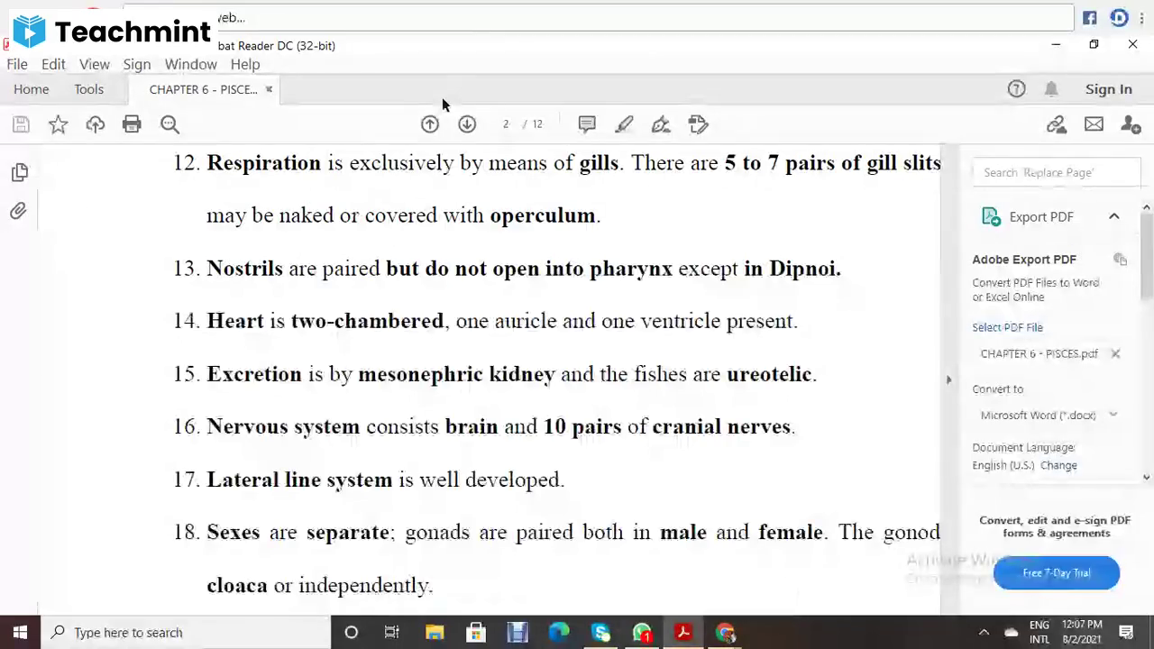
ZO-102 Cell & Molecular Biology, , Uttarakhand Open University, , Fig. 1.1: A Bacterial Cell, , 1.4: BASIC COMPONENTS, EUKARYOTIC CELL, , OF, , PROKARYOTIC, , AND, , 1.4.1 Prokaryotic Cells, Prokaryotic cells are the most primitive cells and have simple structural organization., It has a single membrane system. They include bacteria, viruses, blue-green algae,, mycoplasmas, rickettsias, spirochetes etc. Cyanobacteria or blue green algae are the, largest and most complex prokaryote, in which photosynthesis of higher plants type, have evolved. Prokaryotes are included in the kingdom Monera and the super, kingdom Prokaryota. The Prokaryotes have the following characters:, 1., The size of prokaryotic cells ranges between 1 to 10 µm. They occur in a, variety of forms., 2., , Prokaryotic cell consists of three main components:, , (I), Outer covering: It is composed of inner cell or plasma membrane, middle cell, wall and outer slimy capsule., a. Cell membrane: Cell membrane made up of lipids and proteins, is thin and, flexible and controls the movement of molecules across the cell. Respiratory enzymes, are carried by it for energy releasing reactions. Mesosomes, the in-folds of plasma, Page 3
Page 8 :
ZO-102 Cell & Molecular Biology, , Uttarakhand Open University, , membrane bears respiratory enzymes and these are considered analogous to, mitochondria of eukaryotic cells. Similarly, the pigments and enzymes molecules that, absorb and convert the light into chemical energy in photosynthetic cells are also, associated with the plasma membrane’s in-folds called photosynthetic lamella., These lamellae are analogous to the chloroplast of eukaryotic cells. Plasma membrane, plays role in replication and division of nuclear material. Since the in-folds remain, continuous with the cell membrane, they are not considered as separate, compartments. Thus, prokaryotic cell is non-compartmentalized., b. Cell wall : It is a rigid or semi-rigid non-living structure that surrounds the cell, membrane and its thickness ranges between 1.5 to 100 µm. Chemically it is composed, of peptidoglycans. . Some bacteria such as mycoplasmas lack cell wall., c. Slimy capsule: A gelatinous coat outside the cell wall is the slimy capsule. It is, composed of largely of polysaccharides and sometimes it may have polypeptides and, other compounds also. It protects the cell against desiccation, virus attacks,, phagocytosis and antibiotics, (II) Cytoplasm: Prokaryotic cytoplasm contains proteins, lipids, glycogen and, inorganic ions along with enzymes for biosynthetic reactions and ribosomes, tRNA, and mRNA for protein synthesis. Prokaryotic cytoplasm has some special features as, follows:, a. It lacks cell organelles like endoplasmic reticulum, mitochondria, Golgi apparatus,, Centrosomes, vacuoles, Lysosomes, microfilaments, intermediate filaments and, microtubules., b. The only cytoplasmic organelle found in prokaryotic cells is the ribosomes. They, are smaller than eukaryotic ribosomes i.e., 70S and lie free in the cytoplasm. They, form poly-ribosomes at the time of protein synthesis. They are the sites of protein, synthesis., c. Like eukaryotic cells, the cytoplasm of prokaryotic cell does not show streaming, movement or cyclosis., d. Gas vacuoles are also formed in some prokaryotic cells., e. The cell does not show phagocytosis, pinocytosis and exocytose, substances enter, and leave the cell through the cell membrane., f. They may contain deposits of polysaccharides or inorganic phosphates., (III) Nucleoid: Nuclear envelope is absent in prokaryotic cell and the genetic material, lies directly into the cytoplasm. Such nuclear material is known as nucleoid., Nucleoid consists of greatly coiled single pro-chromosome. It shows the following, special features:, , Page 4
Page 9 :
ZO-102 Cell & Molecular Biology, , Uttarakhand Open University, , a. A short and simple pro-chromosome is present which is attached at least at one, point on cell membrane., b. Mostly there is single copy of chromosome, the prokaryotic cell is haploid., c. The DNA is naked as it is not associated with basic histone proteins. It is double, stranded, helical and circular., d. The amount of DNA is lesser than eukaryotic cell and it codes fewer proteins., Replication of DNA is continuous throughout the cell cycle. Transcription and, translation occurs in cytoplasm and processing of mRNA is not required., e. The processes like meiosis, gamete formation or fertilization are absent., Conjugation is seen in some bacteria., f. Mitotic apparatus absent., g. There is no nucleolus., h. Cell membrane folds or mesosomes help to segregate the replicated products of, chromosomes into daughter cells., 3. Plasmids: In some prokaryotic cells, in addition to nucleoid, a small circular, double stranded DNA molecule is present. It is called plasmid. Plasmids have 1000 to, 30,000 base pairs and they generally encode proteins required by the organism to, resist antibiotic and other toxic material., 4. Flagellum: It is a whip like locomotory structure found in many bacteria. It is, 150Å thick and 10 to 15µm long. As the flagellum does not have any surrounding, membrane, it grows at the tip., It has two main parts: Filament and basal body., (i), , Filament- Filament extends out of cell into the medium and it is composed of, many intertwined spiral chains of the subunits of a protein called flagellin., Flagellin differs from actins or tubulin., , (ii), Basal Body- The basal body attaches the flagellum to the cell and generates, the force to rotate it. It is composed of many components and numerous proteins. It, has two parts: shaft and hook., 5. Pili: These are short, rod like non-motile processes or fimbriae present on many, bacteria. These are formed of pilin protein. They are usually less than 10 nm thick., They help in attachment of bacteria to surfaces or food or to one another. Tubular sex, Pili are present in some bacteria., Prokaryotic cells have all the biochemical mechanisms required to synthesize, complex organic materials from simple organic precursors necessary for life. Thus,, Page 5
Page 10 :
ZO-102 Cell & Molecular Biology, , Uttarakhand Open University, , inspite of being simple in structure prokaryotes are more versatile in their synthetic, activities than eukaryotes., , 1.4.2 Eukaryotic Cells, The internal organization of eukaryotic cell is more developed than prokaryotic cells, from which they are believed to have been evolved. They are evolved to have double, membrane system. Primary membranes are the one that surrounds the cell, celled cell, or plasma membrane and the secondary membrane surround the nucleus and other, cellular organelles. Eukaryotic cells occur in protists, fungi, plants and animals., Eukaryotic cells have the following characteristics:, 1., Number- In multicellular organisms the numbers of cells are correlated with, the body size. The human blood contains about 30 quadrillion (3 × 10¹⁵) corpuscles, and a 60 kg human being has about 60 × 1015 cells. All multicellular organisms begin, their life with a single cell “Zygote” and then become multicellular by its mitotic, division during development., 2., Shape- A cell may be spherical, cuboidal, oval, disc-like, polygonal,, columnar, spindle like or irregular. Thus, cells acquire a variety of shapes not only in, various organisms but also in different tissues of the same organism. The shape of cell, is correlated with its functions like the shape of muscles and nerve cells are well, adapted to their functions. Many factors such as cell functions, age of cell, presence or, absence of cell wall, viscosity of cytoplasm etc. are responsible for various shapes of, cells., 3., Size- Most of the eukaryotic cells is microscopic and their size ranges between, 10 to 100µm. Sporozoits of malaria parasite (Plasmodium vivax) is among the, smallest cells having the size equal to 2µm long. While the Ostrich egg measures 175, × 120mm. Nerve cells are the longest having the size of its fiber to be of few meters, long. Human cells generally range from 20 to 30µm., 4., Components of a cell- Three main components of the eukaryotic cells are cell, membrane, cytoplasm and nucleus. The cytoplasm and the nucleus further have, several components. Various cell components are discussed below:, (i), Cell membrane- Cell membrane, plasma membrane or plasmalemma is a thin, elastic living covering that surrounds the cell keeping the cell contents in place,, provides shape to the cell and controls the transfer of materials across it. It is, composed of lipid-protein complex. It lacks respiratory enzymes. In many protists and, animal cells it allows endocytosis and exocytosis., In certain protists, many fungi and all plant cells, the cell membrane is covered by a, thick, rigid non-living cell wall that protects and supports the cell. In prokaryotes the, cell wall surrounding the plasma membrane has a different structure in comparison to, eukaryotes., Page 6
Page 11 :
ZO-102 Cell & Molecular Biology, , Uttarakhand Open University, , (ii), Cytoplasm- The cytoplasm or the cytosome is a semi-fluid, homogeneous,, translucent ground substance known as cytoplasmic matrix or cytosol which is present, between the cell membrane and the nucleus. In the protozoan cell the outer firm layer, of cytoplasm is called ectoplasm and the inner layer around the central fluid mass is, called the endoplasm. The cytosol shows “cyclosis” or the streaming movement. The, eukaryotic cytoplasm has the following features:a. Organelles: The organized structures having the specific functions and capacity of, growth and multiplication in some cases are known as organelles. Mitochondria,, centrosomes, Golgi bodies, plastids and vacuoles are the organelles that can be, observed under light microscope, while endoplasmic reticulum, ribosome,, microfilaments, microtubules, intermediate filaments and micro bodies can only be, seen under electron microscope. These organelles are often described as protoplasmic, structures. The cells having cilia or flagella have their basal bodies at the bases are in, the cytoplasm while rest of its part extends out of cytoplasm. These organelles are, described as follows:, I. Mitochondria: The rod like or globule shaped structures scattered in the cytoplasm, are found singly or in groups. They are bounded by double membrane of, lipoproteins. The inner membrane gives out finger like structure known as cristae, which partially subdivide the inner chamber of mitochondrion. On the inner surface of, cristae are present mushroom like structures, oxysomes that are related to, phosphorylation. The space between the membranes and its lumen is filled with, mitochondrial matrix. Both the membranes and the matrix contain many oxidative, enzymes and coenzymes. Since mitochondria contain DNA molecules and ribosomes,, they synthesize certain proteins. They produce the energy and reserve it in the form of, adenosine triphosphate (ATP). Due to the presence of its own DNA and ability of, protein synthesis along with its duplication, the mitochondria are called semi, autonomous organelle. The DNA of mitochondria resembles that of bacterial cell;, hence it is also called as endo-symbiotic organelle., II. Centrosomes: (9+0) there is a clear zone around centrioles, near the nucleus, that, includes a specialized portion of cytoplasm, called centrospheres. Its matrix is called, kinoplasm that bears two rounded bodies the “centrioles”. Each centriole consists of, nine fibrillar units and each of them is found to contain three microtubules arranged, in a circle. Both the centrioles are arranged at right angle to each other. Centrioles, form the spindles of microtubules at the time of cell division. Centrioles are absent in, plant cell and the spindle is formed without their help., III., Golgi bodies: These are the stack of flattened parallel-arranged sacs and, vesicles found in association of endoplasmic reticulum. They are composed of many, lamellae, tubules, vesicles and vacuoles. Their membranes are supposed to be, originated from ER and are composed of lipoproteins. In plant cells the Golgi, complex is called dictyosome that secretes required materials for the formation of cell, Page 7
Page 12 :
ZO-102 Cell & Molecular Biology, , Uttarakhand Open University, , wall at the time of cell division. It helps in the formation of acrosome of sperms,, release of hormones, enzymes and other synthetic materials., IV., Plastids: These organelles are found in plant cells and are absent in animal, cells. They may be colored like chloroplast or chromoplasts or colorless like, leucoplast. Since the leucoplast store and metabolise the starch and lipids, they are, called amyloplast and lipoplast respectively. Chloroplast contains the green pigment, the chlorophyll that helps in photosynthesis and protein storage. Chloroplast has a, double outer membrane, the stroma, that bears many soluble enzymes, and a, complex system of membrane bound compartments called thalakoids constituting, granna. Like mitochondria, chloroplast also has their own DNA, ribosomes and, complete protein synthetic machinery. Hence these are also called endo-symbiotic and, semi-autonomous organelle., V. Metaplasm: The particles like vacuoles, granules and other cytoplasmic bodies, such as ribonucleoprotein molecules are represented by it., VI., Cilia, basal bodies and flagella: Cilia are the minute structures covering the, surface in some cells. Both cilia and flagella originate from the basal bodies or, blepharoplast lying in cytoplasm. They consist of nine outer fibrils with the two, larger fibrils in the centre. Each fibril consists of two microtubules, or has 9+2, arrangement. Cilia and Flagella are the structure born by certain cells. They are, composed of microtubules made of the protein tubulin. They have 9 + 2 plan of, microtubule. Both grow at the base. They act as locomotory organelles, moves by, their beats or undulations for they get the energy by breakdown of ATP molecule., VII. Microtubules: The ultra fine tubules of protein (tubulin) traversing the, cytoplasm of plant and animal cells providing the structural framework to the cell,, determine the cell shape and general organization of the cytoplasm are known as, microtubules. Tubules are made up of 13 individual filaments. Microtubules help in, transport of water and ions, cytoplasmic streaming (cyclosis) and the formation of, spindles during cell division., VIII. Basal granules: The spherical bodies found at the base of cilia and flagella, are called the basal bodies. Each of them is composed of nine fibrils and each fibril, consists of the three microtubules, out of which two enter the cilia or flagella., IX., Ribosome’s: Ribosome is the minute spherical structures that originate in, nucleolus and are found attached with the membrane of endoplasmic reticulum and in, the cytoplasm. They are mainly composed of ribonucleic acids (RNA) and protein., They are mainly responsible for protein synthesis., b., Inclusions: These are the non-living or deutoplasmic structures which are, incapable of growth and multiplication. Common cell inclusions are stored organic, materials such as starch grains, glycogen granules, aleuron grains, fat droplets,, pigment granules and inorganic crystals.Cytoplasm is stores raw materials needed for, Page 8
Page 13 :
ZO-102 Cell & Molecular Biology, , Uttarakhand Open University, , the metabolism in both the cytoplasm and the nucleus. Many metabolic processes like, biosynthesis of fatty acids, nucleotides, proteins and oxidation take place in, cytoplasm. It distributes the nutrients, metabolites and enzymes in a cell and brings, about exchange of materials between the organelles as well as with the environment, or extracellular fluid also., c., Nucleus: In a eukaryotic cell the genetic material is enclosed by a distinct, nuclear envelope that forms a prominent spherical organelle the “Nucleus”. The, nuclear envelope bears pores for the exchange of materials between the cytoplasm, and the nucleoplasm., , Fig. 1.2: An animal cell as shown by electron microscope, , Page 9
Page 14 :
ZO-102 Cell & Molecular Biology, , Uttarakhand Open University, , 1.4.3 Differences between Prokaryotic Cells and Eukaryotic Cells, The internal organization of eukaryotic cell is more developed than prokaryotic cells, from which they are believed to have been evolved., S. No., , Prokaryotic Cells, , S. No., , Eukaryotic Cells, , 1., , A prokaryotic cell is, surrounded by a single, membrane layer., , 1., , A eukaryotic cell is, surrounded, by, a, double, membrane, layer., , 2., , In most cases the cell wall, surrounds, the, plasma, membrane and it is, composed, of, carbohydrates,, lipids, proteins and certain amino, acids., , 2., , Cell wall is present in, protists, most fungi, and plants and is, composed of chitin in, most fungi and or, cellulose in others., , 3., , Respiratory enzymes are, present on cell membranes., , 3., , Absent on the cell, membrane, , 4., , Thalakoids occurs free in, cytoplasm., , 4., , They occur within the, chloroplast., , 5., , Cytoplasm lacks organelles, like, centrosomes,, endoplasmic, reticulum,, mitochondria,, Golgi, apparatus, microfilaments,, intermediate, filaments,, microtubules and micro, bodies. While ribosomes, are present, , 5., , All the cell organelles, are present in the cell, along with ribosomes., , 6., , Gas vacuoles may occur, while sap vacuoles are, absent., , 6., , Sap, vacuoles, are, commonly present., , 7., , 70S ribosomes are present, that lie free in cytoplasm or, attached to mRNA., , 7., , 80S ribosome’s are, present, either free or, bound to ER and, nuclear envelope or, mRNA., , Page 10
Page 15 :
ZO-102 Cell & Molecular Biology, S. No., , Uttarakhand Open University, , Prokaryotic Cells, , S. No., , Eukaryotic Cells, , 8., , Endocytosis and exocytose, do not occur., , 8., , These processes take, place in many protists, and in animals., , 9., , Process of meiosis or, gamete formation or true, fertilization does not occur., , 9., , In these cells the, process of meiosis,, gamete formation and, true fertilization occur, in most cases of sexual, reproduction., , 10., , Cells are haploid., , 10., , Cells are diploid, while, haploid cells also, occur., , 11., , Nuclear envelope is absent, and nuclear material lie in, cytoplasm and is called, nucleoid., Nucleoid, contains, a, single, chromosome., , 11., , Nuclear, envelope, surrounds the nuclear, material. The structure, is called nucleus. It, contains two to many, chromosomes., , 12., , Nucleolus absent., , 12., , One or more nucleoli, are present within the, nucleus., , 13., , Circular DNA is present, without, associated, proteins., , 13., , Nuclear DNA is linear, and is associated with, proteins, while extra, nuclear, DNA, is, present, without, proteins., , 14., , Flagella if present are, simple, consist of a single, fibril and are formed of a, protein flagellin., , 14., , Flagella, if present are, complex, have 9+2, pattern of microtubules, formed of a protein, tubulin., , 15., , Plasmids and pili occur in, many prokaryotic cells., , 15., , These structures are, absent., , 16., , Most, , 16., , Most, , prokaryotes, , are, , of, , them, , are, , Page 11
Page 16 :
ZO-102 Cell & Molecular Biology, S. No., , Prokaryotic Cells, asexual organisms., , Uttarakhand Open University, S. No., , Eukaryotic Cells, sexual organisms., , 1.5 SUMMARY, Robert Hook (1665) for the first time described the texture of a piece of cork as, “cell”. Similar structures were observed by many scientists while studying many, living organisms. It was Schwann T. (1839) who stated that all living organisms are, composed of cells after examining a variety of plant and animal tissues. Basically two, types of cells are there, “Prokaryotic” and “Eukaryotic”. Prokaryotic cells are the, primitive cells that include bacteria, blue-green algae, viruses and photosynthetic cells, cyanobacteria etc. Their size varies from 1 to 10 um and they consist of mainly three, components: the outer covering that includes all cell membrane, cell wall and a slimy, capsule. Another component is cytoplasm which lacks cell organelles except, ribosomes. The processes like phagocytosis and endocytosis are absent. The third, component is nucleoid that lacks nuclear membrane. Additional small circular DNA, the plasmid may also be present. Flagella and pili like structure are also seen in some, prokaryotic cells. Eukaryotic cells are more developed and are surrounded by double, membranes. Shape and size of these cells and their number in multicellular organisms, varies. It is also composed of three main components. Cell membrane or plasma, membrane is a thin elastic living covering. The cytoplasm is a semi fluid,, homogenous, translucent consisting of many cell organelles, inclusions, cilia, flagella,, basal bodies and microtubules., , 1.6 GLOSSARY, Cytoplasm: Gel like substance enclosed within the cell membrane excluding nucleus., Plasma membrane: It is the biological membrane that separates the interior of the, cell from the outside environment., Prokaryote: The cell that lacks a distinct nucleus and other specialized membrane bound, organelles., Eukaryote: an organism whose cell contains a membrane bound distinct nucleus, along with other specialized organelles enclosed in membranes., Mesosome: The in-folding of plasma membrane in some bacterial cells that carry, respiratory enzymes., Poly-ribosome: It is a group of ribosomes associated with a single messenger RNA, during the translation process., Page 12
Page 17 :
ZO-102 Cell & Molecular Biology, , Uttarakhand Open University, , Phagocytosis: The process by which a cell engulfs a solid particle to form an internal, vesicle known as phagosome is called phagocytosis, also called eating of cell., Pinocytosis: The process of intake of liquid into a cell by the budding of small, vesicles from the cell membrane is called pinocytosis, also called drinking of cell., Exocytosis: In the process of exocytosis materials are exported outside the cell by, using energy from ATP molecules., Conjugation: When the genetic material is transferred from one bacterial cell to other, either by direct contact or by a bridge like connection between two cells is called, conjugation., , 1.7:- Self Assessment Questions and Possible Answers, , 1.7.1 Multiple Choice Questions:, 1., , 2., , 3., , 4., , 5., , There is no organized nucleus in:, (a), , Bacterial cell (b), , Green algae cell, , (c), , Animal cell, , Plant cell, , (d), , The prokaryotic cells are characterized by:, (a), , A distinct nuclear membrane (b), , (c), , Distinct chromosome (d), , Absence of chromatin material, , Absence of nuclear membrane, , In a prokaryotic cell, DNA is:, (a), , Enclosed by nuclear envelop, , (b), , Lacking, , (c), , Not a genetic material, , (d), , without a membrane, , Cell wall is found around the:, (a), , Prokaryotic cells, , (b), , (c), , Plant cells, , All the above, , (d), , Algal cells, , Chemical energy of food stuffs is converted into biologically useful forms by:, (a), , Ribosomes, , (b), , (c), , Mitochondria (d), , Golgi complex, Plastids, Page 13
Page 18 :
ZO-102 Cell & Molecular Biology, , 6., by:, , 7., , 8., , 9., , 10., , Uttarakhand Open University, , Sun radiant energy is converted into chemical energy of organic compound, , (a), , Mitochondria (b), , Chloroplast, , (c), , Ribosomes, , Centrosomes, , (d), , Which structure is present only in animal cell?, (a), , Cell membrane, , (b), , (c), , Centrioles, , Ribosomes, , (d), , Lysosomes, , Single envelope system is characteristic of:, (a), , Prokaryotic cell, , (c), , None (d), , (b), , Eukaryotic cell, , Both, , Prokaryote and eukaryotes have the common:, (a), , Mitotic apparatus, , (b), , Histone, , (c), , Genetic code, , (d) Mitochondria, , Unicellular microscopic organisms were first studied by:, (a), , Robert Hooke (b), , (c), , Pasteur (d), , Priestley, , Leeuwenhoek, , ANSWERS:1. (a), , 5.(c), , 9. (c), , 2. (d), , 6.(b), , 10.(d), , 3. (d), , 7.(c), , 4. (d), , 8.(a), , 1.7.2 Very Short Questions:, 1., , What are prokaryotes? Give an example., , 2., , What are eukaryotes? Give few examples., , 3., , Cell is an open dynamic system. Is it correct?, , 4., , Prokaryotic cells are haploid. Is it so?, Page 14
Page 19 :
ZO-102 Cell & Molecular Biology, , Uttarakhand Open University, , 5., , What are cyanobacteria?, , 6., , Give three essential characteristics of cell?, , 7., , Where is nucleolus found?, , 8., , What are the power houses of the cell?, , 9., , Name the protein factories of prokaryotic and eukaryotic cells?, , 10., , What is the control centre of a cell?, , Answer:1. Organisms without an organized nucleus e.g., Bacteria, 2. Organisms with an organized nucleus. Plants, yeast;, 3. Yes, 4. Yes, 5. Blue green algae, 6. Cell membrane, cytoplasm, nuclear material, 7. Nucleus, 8. Mitochondria, 9. Ribosome, 10. Nucleus, , 1.8 References and Suggested Readings:Brown, R. (1831). Observations on the organs and mode of fecundation in Orchideae and, Asclepiadeae. Trans. Linn. Soc. London, 16: 685-746., Dutrochet, H. (1837). Memoires pour servir á l’ histoire anatomique et physiologique des, végétaux et des animaux. Bailliere, Paris., Hooke, R. (1665). Micrographia: or some physiological descriptions of minute bodies made, by magnifying glasses with observations and inquiries thereupon. Royal Society,, London, UK., Page 15
Page 21 :
ZO-102 Cell & Molecular Biology, , Uttarakhand Open University, , UNIT 2: PLASMA MEMBRANE, Contents, 2.1 Objectives, 2.2 Introduction, 2.3 History, 2.4 Plasma Membrane, 2.4.1 Ultra Structure of Plasma Membrane, 2.4.1.1 Symmetrical Molecular Structure of Plasma Membrane, 2.4.1.2 Asymmetrical Molecular Structure of Plasma Membrane, 2.4.2 Chemical Composition of the Plasma Membrane, 2.4.2.1 Lipids, 2.4.2.2 Proteins, 2.4.2.3 Enzymes, 2.4.2.4 Carbohydrates, 2.4.2.5 Salts, 2.4.3 Lamella-model of plasma membrane (Danielli-Davson model), 2.4.4 Miceller model of plasma membrane, 2.4.5 Fluid Mosaic Model of plasma membrane, 2.5 Functions of Plasma Membrane, 2.6 Summary, 2.7 Glossary, 2.8 Self assessment question and possible answers, 2.8.1 Multiple Choice Questions, 2.8.2 Very Short Questions, 2.9 References and Suggested Reading, 2.10 Terminal and model questions, Page 17
Page 22 :
ZO-102 Cell & Molecular Biology, , Uttarakhand Open University, , 2.1 Objectives:After reading this unit the readers should be able to:, Define plasma membrane, Describe the ultra structure of plasma membrane, Explain the chemical composition of plasma membrane, Outline the various theories of plasma membrane, Discuss the functions of plasma membrane, , 2.2 Introduction:Every cell, prokaryotic or eukaryotic, is surrounded by a thin layer of outermost, boundary called the plasma membrane or cell membrane or plasma - lemma. The plasma, membrane is a discrete structure and is remarkably complex in its molecular organization. It, maintains the difference of the internal environment of the cell from its external environment, by controlling the entrance and exit of the molecules and ions. It checks the loss of, metabolically useful substances and encourages the release of toxic metabolic byproducts of, the cell. Thus, it functions as semi-permeable or selectively permeable membrane. It is, about 70-100Å in thickness. In plant cells plasma lemma is further covered by cellulosic cell, wall. It is an important cell organelle composed of lipids and proteins. It possesses devices, for attachment to other cells for cell-to-cell communications, ion pumps for controlling, internal milieu of the cell, receptors for hormones and mechanisms for the production of, secondary messengers that activates the cell's physiological response., , 2.3 History:It had been shown by Karl W. Nageli (1817-1891) that the cell membrane is semipermeable and is responsible for the osmotic and other related phenomena exhibited by living, cells. Before 1855, he used the term zellen membrane in his early papers. The term plasma, membrane was used in 1855 by him to describe the membrane as a firm protective film that is, formed by out flowing cytoplasm of an injured cell when protein rich cell sap came in contact, with water., , Page 18
Page 23 :
ZO-102, 102 Cell & Molecular Biology, , Uttarakhand Open University, , 2.4 Plasma Membrane, embrane:2.4.1 Ultra Structure of Plasma Membrane, , 2.4.1.1 Symmetrical Molecular Structure of Plasma Membrane:Membrane, Plasma membrane is a tripartite structure and is made up of three layers, having total, thickness of 75Å. Two di-electronic, electronic layers are there, each of 25Å thickness, enclosing a, middle dielectronic layer which is also 25Å thick. The middle layer is a tri-molecular, tri, layer of, lipids having its non-polar, ar hydrophobic groups facing inwards, whereas polar hydrophilic, groups facing outwards. The hydrophilic polar groups are covered by a protein layer which is, 20 to 25Å thick. The protein chains lie at right angles to the lipids., , Fig. 2.1: Symmetrical pattern of molecules in plasma membrane (Source: Singh and Tomar, 2008), , 2.4.1.2 Asymmetrical Molecular Structure of Plasma Membrane :It is also a tripartite structure having a thick inner dielectronic component of 35-40, 35, Å,, a narrow outer dielectronic component of 25Å thickness, and a central dielectronic layer, (bimolecular layer of lipids) which is 30Å wide; thus total thickness comes to 90-95Å., 90, , Fig. 2.2: Asymmetrical pattern of plasma membrane, , Page 19
Page 24 :
ZO-102, 102 Cell & Molecular Biology, , Uttarakhand Open University, , In different types of cells the thickness of plasma membrane varies. For example, in red, blood corpuscles of rabbit, the plasma membrane is about 215 Å thick whereas, in intestinal, epithelial cells it is 105 Å in thickness. Very small pores measuring about 10Å in diameter, (smaller than, an pores of nuclear membrane) have been discovered in the membranes., , 2.4.2 Chemical Composition of the Plasma Membrane:Membrane:, Plasma membrane is primarily composed of protein and lipid, although carbohydrate, is often present in association with protein (as glycoprotein) or lipid (as glycolipid)., However, the relative proportions of protein and lipid vary considerably in membranes from, different sources., , 2.4.2.1:-, , Lipids, , The plasma membrane contains about 20 to 79% lipids mainly of three types like, phospholipids,, holipids, cholesterol and glycolipids. The phospholipids which make up between 55%, and 75% of the total lipid content, consists chiefly of lecithin and cephalin. The remainder, consists of sphingolipids (with an amino group) and glycolipid conjugates with, carbohydrates., arbohydrates. Phospholipids derived from glycerol are called phosphoglycerides., A phosphoglycerides is made up of two fatty acid chains, a glycerol backbone and a, phosphorylated alcohol. The outer layer of phospholipids consists mainly of lecithin and, sphingomyeline,, ingomyeline, while the inner layer is composed mainly of phosphatidyl ethanolamine and, phosphatidyl serine (both are phosphoglycerides). The glycolipids (sugar containing lipids), are mainly in the outer half of the bilayer., , Fig. 2.3: A phospholipids cholesterol, cholesterol complex of cell membrane, , Cholesterol is present in eukaryotes but not in prokaryotes. Plasma membrane of cells such as, erythrocyte, liver cells and myelinated nerve cells are rich in cholesterol., Page 20
Page 25 :
ZO-102, 102 Cell & Molecular Biology, , Uttarakhand Open University, , Membrane lipids are amphipathic molecules. They contain, contain both a hydrophobic and, hydrophilic moiety. Hydrophilic unit is also called the polar head groups, is represented by a, circle and their hydrocarbon tails are depicted by straight or wavy lines. Polar head groups, have affinity for water, whereas their hydrocarbons tails avoid water. This can be, accomplished by forming a micelle, in which polar head groups are on the surface and, hydrocarbon tails are directed inside., , Fig.2. 4: A phospholipids molecule, , Another arrangement of lipid molecule in a membrane is a bimolecular sheet, which is, also called a lipid bilayer. Phospholipids and glycolipids are key membrane constituents of, bimolecular sheets. Hydrophobic interactions are the major driving force for the formation of, lipid bilayer. The lipid bilayer, ayer of the membrane is interrupted only by the proteins that, traverse it. This bilayer consists primarily of:, (a), , Neutral Phospholipids and Cholesterol:, Cholesterol: These include phosphatidylenoline, lecithin, cerebroside, and sphingomyeline and phosphatidyl ethanolamine., mine. They are without, any electric charge at neutral pH and are closely packed in the bilayer along with, cholesterol., , (b), , Acidic Phospholipids:: These constitute about 5% to 20% fractions of the total, phospholipids of plasma membrane. They are negatively charged and are associated, with proteins by way of lipid-protein, lipid protein interactions. Common examples are, phosphatidyl inositol, phosphatidylserine, sulpholipids, phosphatidyl glycerol and, Cardiolipin., , In plasma membrane, lipid fractions form permeability barrier and structural framework., , 2.4.2.2 Proteins:Proteins are the main component of plasma membrane. Myelin sheath (membrane, surrounding some nerve axons) is composed of about 80% lipids and 20% protein and, Page 21
Page 26 :
ZO-102 Cell & Molecular Biology, , Uttarakhand Open University, , presence of lipid makes myelin an excellent insulator. Eukaryotes membrane which serves, primarily as permeability barriers possesses about 50% proteins and 50% lipid. Plasma, membrane that are actively involved in energy transfer, such as inner membrane of, mitochondria, chloroplasts and membranes of aerobic prokaryotes have large amounts of, proteins i.e. about 75%. They not only provide mechanical support but also act as carriers or, channels, serving for transport. In addition numerous enzymes, antigens and various kinds of, receptor molecules are present in plasma membranes. Membrane proteins are classified as, integral (intrinsic) or peripheral (extrinsic) according to the degree of their association, with the membrane (Singer, 1971)., (a), , Peripheral Proteins: They are also called extrinsic proteins associated with membrane, surface. These can be separated by addition of salts, soluble in aqueous solutions and, usually free of lipids. They are bound to the surface by electrostatic and hydrogen, bond interactions. They form outer and inner layers of the lipid bilayer of plasma, membrane. Common examples are cytochrome-C found in mitochondria, acetyl, cholinesterase in electroplax membrane and spectrin found in erythrocytes., , (b), , Integral or Intrinsic Proteins: These proteins penetrate the lipid layer wholly or, partially and represent more than 70% of the two protein types. Their polar ends, protrude from the membrane surface while non-polar regions are embedded in the, interior of the membrane. Usually they are insoluble in water solutions and can be, separate them from the membrane by detergents or organic solvents. The major, integral proteins span the thickness of the membrane and have a small amount of, carbohydrates on the pole at the outer surface. This protein appears to be involved in, the diffusion of anions across the membrane. Integral proteins may be attached to the, oligosaccharides to form glycoprotein or to phospholipid to form lipoproteins or, proteolipids. Common intrinsic proteins are rhodopsin found in retinal rod cells and, cytochrome oxidase found in mitochondrial membranes., , Every protein in the cell membrane is distributed asymmetrically with respect to the lipid, bilayer., , 2.4.2.3 Enzymes:About 30 enzymes have been found in various membranes. Those most constantly, found are 5'-nucleotidase, Na+-K+ activated ATPase, alkaline phosphatase, adenylcyclase,, RNAse and acid phosphomonoestrase. Na+-K+ activated Mg+ ATPase plays an important role, in the ionic exchange and may also act as carrier protein or permease across the plasma, membrane. Some enzymes have a preferential localization. For example, alkaline, phosphatase and ATPase are more abundant in bile capillaries, while disaccharides are, present in microvilli of the intestine. Enzymes are asymmetrically distributed, for example in, the outer surface of erythrocytes there are acetylcholinestrase, nicotinamide adenine, dinucleotidase and Na+-K+ ATPase. In the inner surface there is NADH-diaphorase, G3PD,, adenylate cyclase, protein kinase and ATPase., , Page 22
Page 27 :
ZO-102, 102 Cell & Molecular Biology, , Uttarakhand Open University, , 2.4.2.4 Carbohydrates, The membranes of eukaryotic cells usually, u, contain 2%, % to 10% carbohydrates in the, form of glycolipids and glycoproteins. Hexose, hexosamine, fucose and sialic acid are the, commonest carbohydrates found in the membrane. Plasma membranes of neuronal surface, contain gangliosides (Lapertina, 1967), 1967) and are probably involved in the ion transfers. The, distribution of oligosaccharides is also highly asymmetrical., , 2.4.2.5 Salts and water:water, They are also present in cell membranes. Water in cell membranes forms parts of, membrane structure as it does in all cell constituents., , 2.4.3 Lamella-model, model of plasma membrane (Danielli-Davson, (Danielli Davson model), Danielli-Davson, Davson model (1934) suggested that the plasma membrane consists of two, layers of lipid molecules arranged radially with their hydrophobic hydrocarbon chains toward, each other and with their respective polar groups arranged outwardly and inwardly, throughout the entire double layer of lipid molecules. The polar ends of the lipid molecules, are associated with a monomolecular layer of polar globular protein molecule. The entire, structure thus consisted of double layer of lipid molecule sandwiched between two, continuous layers of protein. The lipid molecules are set at right angles to the surface and are, so arranged in two layers that their non-polar, non polar hydrophobic fatty acid tails face each other and, their polar hydrophilic phosphate heads face the protein layer. The proteins involved, involve were, thought to be globular. Moreover, lamellar theory assumed the cell membrane to be a stable, structure with little functional specificity and variability., , Fig. 2.5: A schematic diagram of Davson-Danielli, Davson Danielli model of membrane structure, , Page 23
Page 28 :
ZO-102, 102 Cell & Molecular Biology, , Uttarakhand Open University, , Fig. 2.6: A modification, ification of original Danielli-Davson, Danielli Davson model, showing pores lined by polar protein, molecules extending through the lipid bilayer, , 2.4.4 Miceller model of plasma Membrane:According to the view of Hiller and Hoffman (1953),, ), plasma membrane consists of a, mosaic of globular subunits or micelles. If fatty acid molecules are completely surrounded by, water, they may form aggregate called micelles in which the hydrophobic regions of fatty, acid molecules are oriented toward the interior of the micelle away from the aqueous phase, and their hydrophilic groups are at the surface in contact with the surrounding water. Micelles, may be in the form of small spheres of bimolecular layers. These micelles are closely packed, together having a central core of lipid molecules and hydrophilic shell of polar groups. Each, lipid micelle measures 40Å to 70Å in diameter. Protein component of the plasma membrane, forms a monolayer on either side of the lipid micelles and is represented by globular type., The spaces between the globular micelles are thought to represent water filled pores which, measures about 4Å in diameter. These pores are bounded partly by the polar groups of, micelles and partly by the polar groups of associated protein molecules., , Fig.2.7: Plasma membrane based on Miceller, M, theory (diagrammatic), atic), , Page 24
Page 29 :
ZO-102, 102 Cell & Molecular Biology, , Uttarakhand Open University, , 2.4.5 Fluid Mosaic Model of plasma membrane:It was proposed by Singer and Nicholson (1972)., (1972). The lipids are thought to be, arranged primarily in a bilayer in which proteins are embedded to varying degrees. Singer, classifies membrane proteins as peripheral or integral. The proteins varied in size and, dissolved to varying degrees in the lipid matrix, matrix are able to diffuse laterally in the plane of, membrane, and the entire structure is hence dynamic. In this model, lipid molecules may, exhibit intra molecular movement or may rotate about their axis or may display flip-flop, flip, movement including transfer from, fr, one side of bilayer to the other., , Fig. 2.8: Plasma membrane based upon Fluid-mosaic, Fluid mosaic model, , The lipids, glycoprotein and many of the intrinsic proteins of the membranes are, amphipathic molecules. These amphipathic molecules constitute liquid crystalline, crystall, aggregates, in which the polar groups are directed toward the water phase and the non-polar, non, groups are, situated inside the bilayer. The lipid bilayer forms the structural matrix which serves as the, permeability barrier of the membrane. In membranes with high lipid content, lipid bilayer is, extensive and interrupted only occasionally by protein molecules, whereas in membranes, with high protein content, the extent of lipid bilayer is reduced. Thus, fluid mosaic model, may describe the chemical composition of, of the molecular organization and ultra structure of, plasma membranes. This arrangement allows various enzymes and antigenic glycoprotein to, have their active sites exposed to the outer surface of the membrane. The fluidity of, membrane also implies that both, both the lipid and the protein have considerable freedom of, movement within the bilayer. The fluidity of the lipid depends on the degree of saturation of, the hydrocarbon chains and on the ambient temperature. A considerable proportion of the, lipids in the membrane, brane are unsaturated, so that melting point of the bilayer is below body, temperature., , Page 25
Page 30 :
ZO-102 Cell & Molecular Biology, , Uttarakhand Open University, , 2.5 Functions of Plasma Membrane:The plasma membrane serves many functions such as:, It maintains the individuality and form of the cell., It keeps the cell contents in place and distinct from the environmental materials., It protects the cell from injury., It regulates the flow of materials into and out of the cell to maintain the concentration, and kinds of molecules and ions in the cell. A cell remains alive as long as the cell, membrane is able to determine which materials should enter or leave the cell., It forms organelles within the cytoplasm., Its junctions keep the cells together., It’s infolds help in the intake of materials by endocytosis (pinocytosis and, phagocytosis)., It’s out folds (microvilli) increase the surface area for absorption of nutrients. The out, folds also form protective sheaths around cilia and flagella., Its receptor molecules permit flow of information into the cell., Its oligosaccharide molecule helps in recognizing self from non-self., By controlling flow of material and information into the cell, the plasma membrane, makes metabolism possible., It permits exit of secretions and wastes by exocytosis., It controls cellular interactions necessary for tissue formation and defense against, microbes., It helps certain cells in movement by forming pseudopodia as in Amoeba and, leucocytes., The bio-membranes around the organelles help the latter to:, (1), , Maintain their identity and functional individuality., , (2), , Receive and turn out required material., , 2.6 SUMMARY, The plasma membrane constitutes the outermost boundary of the cell and it is, remarkably complex in its molecular organization. It is composed of almost equal parts of, proteins and lipids. It allows only selected ions and macromolecules to enter or leave the cell,, thus it functions as a semi permeable membrane., Ultra structure of plasma membrane may be of symmetrical or asymmetrical molecular, structure in nature. Plasma membrane is a tripartite structure in both of the above types, the, difference lies in the thickness of the three layers. In symmetrical molecular structure all the, Page 26
Page 31 :
ZO-102 Cell & Molecular Biology, , Uttarakhand Open University, , three layers, the outer and inner adielectronic along with the middle di-electronic layer are of, 25Å thickness each having total thickness of 75Å. While in asymmetrical structure the inner, adielectronic component is of 35Å to 40Å thickness, the outer dielectronic component is of, 25Å thick and the central dielectronic layer is 30Å wide, thus total thickness becomes 9095Å., Plasma membrane is primarily composed of proteins and lipids, although, carbohydrate is often present in association with proteins (as glycoproteins) or lipids (as, glycolipids). However, the relative proportions of proteins and lipids vary considerably in, membranes. Enzymes are also found in plasma membranes which play an important role in, ionic exchange. Besides, salt and water are also present. The arrangement of lipids and, proteins molecules is explained through various theories., Lamella model of plasma membrane is consisted of a double layer of lipid molecules, arranged radially with their hydrophobic hydrocarbon chains towards each other and with, their respective polar groups arranged outwardly and inwardly. The double layer of lipids is, sandwiched between two continuous layers of proteins. According to miceller theory, plasma, membrane consists of a mosaic of globular subunits or micelles. These micelles are closely, packed together having the lipid molecules in the central core. Protein components form a, monolayer on the entire surface of the lipid micelles forming a globule. The widely accepted, theory is fluid mosaic models of membrane as it can be used to describe the structure of, different membranes. In this model the lipids are arranged in a bilayer in which proteins are, embedded as peripheral or integral. The proteins varied in size and dissolved to varying, degrees in the lipid matrix, diffuse laterally in the plane of membranes and the entire structure, is hence dynamic., Plasma membrane performs variety of functions as they impart shape to the cell and, protects the cell contents. It regulates the cellular semi permeability, resorption, excretion and, secretion. It contributes to the formation of various cell organelles within the cell. Its, junctions keep the cells together., , 2.7 GLOSSARY:Plasma membrane - A microscopic membrane made up of lipids and proteins which forms, the external boundary of the cytoplasm of a cell or encloses a vacuole, and regulates the, passage of molecules in and out of the cytoplasm., Permeability- The ability of a barrier to let any substance pass through it., Ions- An atom or molecule with a net electric charge due to the loss or gain of one or more, electrons., Semi permeable- Allowing certain substances especially small molecules or ions to pass, through it but not others, especially allowing the passage of a solvent but not of certain, solutes., Receptor- A receptor is a protein molecule in a cell or on the surface of a cell to which a, substance such as a hormone, a drug, or an antigen can bind, causing a change in the activity, of the cell., Page 27
Page 32 :
ZO-102 Cell & Molecular Biology, , Uttarakhand Open University, , Dielectric- Having the property of transmitting electric force without conduction., Hydrophobic- The substances that have an affinity for water due to the formation of, hydrogen bonds., Hydrophilic- Hydrophilic molecules typically have polar groups enabling them to readily, absorb or dissolve in water as well as in other polar solvents., Micelles- It is an aggregate of molecules in a colloidal solution, such as those formed by, detergents., Peripheral proteins/extrinsic proteins- Peripheral membrane proteins are proteins that, adhere only temporarily to the biological membrane with which they are associated. These, molecules attach to integral membrane proteins, or penetrate the peripheral regions of the, lipid bilayer., Integral proteins/intrinsic proteins- An integral membrane protein (IMP) is a type of, membrane protein that is permanently attached to the biological membrane. All trans, membrane proteins are IMPs, but not all IMPs are trans membrane proteins., Amphipathy- It is the property of a molecule having both polar (water-soluble) and non, polar (not water-soluble) affinities in its structure., Enzyme- The proteins which acts as catalysts within living cells and increases the rate of, biochemical reactions., , 2.8 SELF ASSESSMENT QUESTIONS AND POSSIBLE ANSWERS, , 2.8.1 Multiple Choice Questions:, 1., , 2., , 3., , According to Fluid mosaic model, the correct sequences of substances in, plasmalemma is:, (a), , L-P-P-L, , (b), , P-L-L-P, , (c), , P-P-L-L, , (d), , L-P-L-P, , Membrane occurs in:, (a), , Chromosomes, nuclei and mitochondria, , (b), , Cytoplasm, chloroplasts and mitochondria, , (c), , Cytoplasm, nuclei and starch grains, , (d), , Chromosomes, chloroplasts and starch grains, , Plasma membrane is:, (a), , Non-selective barrier, , (b), , Selective barrier, , (c), , Impermeable, , (d), , made of cellulose, , Page 28
Page 33 :
ZO-102 Cell & Molecular Biology, 4., , 5., , 6., , 7., , 8., , 9., , 10., , Uttarakhand Open University, , What limits Animal cells from outside?, (a), , Cell wall, , (b), , Basement membrane, , (c), , Shell membrane, , (d), , Plasma membrane, , Cell membrane consists of:, (a), , Protein double layer, , (b), , Phospholipid proteins, , (c), , Phosphoproteins, , (d), , Glycoproteins, , Non-membranous cell organelles are:, (a), , Ribosomes, , (b), , centrioles and ribosomes, , (c), , E.R., , (d), , Mitochondria, , Which of the following theories explain that plasma membrane is selectively, permeable:, (a), , Unit membrane theory, , (b), , Cascade theory, , (c), , Sandwich theory, , (d), , Fluid Mosaic theory, , The hydrophobic ends of phospholipid molecules are:, (a), , Polar, , (b), , Non-polar, , (c), , Neutral, , (d), , Bipolar, , The membrane protein that extend through both sides of lipid bilayer., (a), , Acidic protein, , (b), , Glycoprotein, , (c), , Intrinsic protein, , (d), , Glycolic acid, , Two plant cells are connected with the help of:, (a), , Cell wall, , (b), , Plasma membrane, , (c), , Plasmodesmata, , (d), , None of these, , M.C.Q:- ANSWERS, 1. (b) 2. (b) 3. (b) 4. (d) 5. (b) 6. (b) 7. (d) 8. (b) 9. (c) 10.(c), , 2.8.2 Very short questions:, 1., , What is the thickness of plasma membrane?, , 2., , Who proposed the fluid mosaic hypothesis for the molecular structure of cell, membrane?, , 3., , What is the structure of plasma membrane?, Page 29
Page 34 :
ZO-102 Cell & Molecular Biology, , Uttarakhand Open University, , 4., , What are the main lipid components of the plasma membrane?, , 5., , What are the two types of proteins of the plasma membrane on the basis of their, association with the membrane and their solubility?, , 6., , What are tunnel proteins?, , 7., , Why Na+-K+ ATPase enzyme is most important?, , 8., , Who proposed that plasma membrane contained a lipid bilayer and protein adhering, to both lipid aqueous interfaces?, , 9., , Who gave the unit membrane model of plasma membrane?, , 10., , Give the two alternative name of cell membrane., , Answers:1. 70 - 100Å., 2. Singer and Nicolson., 3. It is formed of bilayer of lipids into which protein complexes are embedded in, a kind of mosaic arrangement., 4. Phospholipids, cholesterol and galactolipids., 5. Integral or intrinsic proteins and peripheral or extrinsic proteins., 6. Large integral protein molecules that lie throughout the phospholipid matrix, and projects on both the surfaces., 7. It helps in ion transfer across the plasma membrane. This enzyme is, dependent on the presence of lipids and is inactivated when all lipids are, extracted., 8. Danielli and Davson in 1935., 9. Robertson, 1959., 10. Plasma membrane and plasmelemma., , 2.9 References:Ballowitz, E. (1990).Fibrillare Struktur and Contraktilitat.Pflugers Archiv, ges.Physiol., 46:433-464., Freud, S. (1982).Uber den Bau der Nervenfesern and Nervenzellen beim, Flusskrebs.Sitzungsb.d.kais.Akad.d.Wein.,math.naturw.Classe, 85, Abth.,3:9-46., Palay, S.L (1960). The fine structure of secretory neurons in the pre optic, nucleus of the goldfish (carassius auratus).Anat.Rec. 138: 417-443., Page 30
Page 35 :
ZO-102 Cell & Molecular Biology, , Uttarakhand Open University, , De Robertis, E. and Franchi, C.M (1953).The submicroscopic organization of, axon material isolated from myelin nerve fibers.J.Exp.Med. 98:269-275, , 2.10 Suggested Readings: Kimball's Biology pages, Cell Membranes., 1. Alberts B, Johnson A, Lewis J, et al. (2002). Molecular Biology of the, Cell (4th ed.). New York: Garland Science. ISBN 0-8153-3218-1., 2. Kleinzeller, A. 1999. Charles Ernest Overton’s concept of a cell membrane., In: Membrane permeability: 100 years since Ernest Overton (ed. Deamer, D.W., Kleinzeller A., Fambrough D.M.), pp. 1–18, Academic Press, San, Diego., 3. Sharp, L. W. (1921). Introduction to Cytology. New York: McGraw Hill, p., 42., 5. Singer SJ, Nicolson GL (Feb 1972). "The fluid mosaic model of the structure, of cell membranes". Science. 175 (4023): 720–31., , 2.10 TERMINAL AND MODEL QUESTIONS, 1., , Define plasma membrane., , 2., , Describe the structure and functions of plasma membrane., , 3., , Write notes on:, a., , Fluid Mosaic Theory, , b., , Miceller Model of Plasma Membrane, , c., , Lamella model of Plasma membrane, , d., , Define phagocytosis and pinocytosis., , 4., , Explain in detail the ultra structure of plasma membrane., , 5., , Differentiate between integral and peripheral proteins., , Page 31
Page 36 :
ZO-102 Cell & Molecular Biology, , Uttarakhand Open University, , UNIT 3: MITOCHONDRIA, Contents, 3.1 Objectives, 3.2 Introduction, 3.4 Structure of Mitochondria, 3.4.1 Morphology of Mitochondria, 3.4.2 Ultra structure of Mitochondria, 3.5 Biogenesis of Mitochondria, 3.6 Functions of Mitochondria, 3.7 Respiratory Chain Complex or Electron Transport System (ETS), 3.7.1 Complexes, 3.8 Electron Transport Mechanism, 3.9 Summary, 3.10 Glossary, 3.11 Self Assessment Questions and Possible Answers, 3.11.1 Multiple Choice Questions, 3.11.2 Very Short Questions, 3.12 References and suggested readings, 3.13 Terminal and Model Questions, , Page 32
Page 37 :
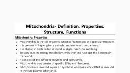
ZO-102 Cell & Molecular Biology, , Uttarakhand Open University, , 3.1 Objectives:After reading this unit the readers will be able to:, Define mitochondria, Illustrate the morphology and ultra structure of mitochondria, Describe the biogenesis of mitochondria, Explain the functions of mitochondria, Elucidate the respiratory chain complex and electron transport mechanism., , 3.2 Introduction:Mitochondria (Gr., mito, thread; chondrion, granule) are thread like or granular, structures of eukaryotic cells. These may assume rod-like shape called chondriosomes which, may enlarge or aggregate to form massive spheroidal bodies called chondriospheres. These, are not present in bacterial cells. Mitochondria are the 'power plants' which by oxidation, release the energy contained in the fuel molecules or nutrients and make other forms of, chemical energy. The main function of mitochondria is oxidative phosphorylation, which is, an exergonic reaction, meaning that it releases energy. In prokaryotes, oxidation of organic, material is carried out by plasma membrane enzymes., , 3.3 History :Kölliker (1880) was the first who observed the mitochondria in insects muscle cells., He called them as 'sarcosomes'. Flemming (1882) named the mitochondria as 'fila'. Altmann, in 1894 observed them and named them Altmann's granules or bioblasts. The term, 'mitochondria' was applied by Benda (1897-98). They were recognized as the sites of, respiration by Hogeboom and his coworkers in 1948. Lehninger and Kennedy (1948), reported that the mitochondria catalyze all the reactions of the citric acid cycle, fatty acid, oxidation and coupled phosphorylation., , 3.4 Structure of Mitochondria, 3.4.1 Morphology of Mitochondria:Morphologically mitochondria may be in the form of filaments or small granules., These may assume rod-like shape called chondriosomes which may enlarge or aggregate to, form massive spheroid bodies called chondriospheres., , Page 33
Page 38 :
ZO-102 Cell & Molecular Biology, , Granule, , Uttarakhand Open University, , Inner Membrane, Outer Chamber, , F1 Particle, DNA, Matrix Cristae, , Ribosome, , Outer Membrane, Fig. 3.1: Structure of a Mitochondrion, , 1. Position- Mitochondria lie freely in cytoplasm, possessing power of independent, movement and may take the form of filaments. In some cells they can move freely,, carrying ATP where needed, but in others they are located permanently near the, region of the cell where more energy is needed. E.g., in the rod and cone cells of, retina mitochondria are located in the inner segment, in cells of kidney tubules they, occur in the folds of basal regions near plasma membrane, in neurons they are located, in the transmitting region of impulse, in certain muscle cells (e.g. diaphragm),, mitochondria are grouped like rings or bracers around the I-band of myofibril. During, cell division they get concentrated around the spindle., 2. Number- The number of mitochondria varies a good deal from cell to cell and from, species to species. A few algae and some protozoan have only single mitochondria., Their number is related to the activity, age and type of the cell. Growing, dividing and, actively synthesizing cells contain more mitochondria than the other cells. In Amoeba, (Chaos chaos), there may be as many as 50,000 mitochondria. In rat liver cells, these, are few in number, about 1000 to 1600. Some Oocytes contain as many as 3, 00,000, mitochondria., 3., , Size- The average size of mitochondria is 0.5-1.0µ in diameter and about 2-8 µ in, length. In exocrine cells of mammalian pancreas they are about 10 µ long and in, oocytes of amphibian Rana pipiens are 20-40µ long. Yeast cells have the smallest, mitochondria., , 3.4.2 Ultra structure of Mitochondria:The electron microscope shows the mitochondrion as the vesicles bounded by an, envelope of two unit membranes and filled with a fluid matrix., Page 34
Page 39 :
ZO-102 Cell & Molecular Biology, 1., , Uttarakhand Open University, , Membranes- Both the inner and the outer mitochondrial membranes resemble the, plasma membrane in molecular structure. Each of them is 60-70Å, trilamellar and, composed of two layers of phospholipid molecules sandwiched between two layers of, protein molecules. However, the two membranes differ in the kinds of protein and, lipids they have and also in their properties. Both the outer and the inner membranes, contain specific pumps or channels, for the transport of molecules through them. The, membranes may be connected at adhesion sites through which proteins are transferred, from the outer to the inner membrane. The outer and the inner membrane are, separated from each other by a narrow space called the inter-membrane space or outer, chamber or peri-mitochondrial space. It is about 80Å wide. It contains a clear, homogeneous fluid., (i), , Outer Membrane- The outer membrane is smooth permeable to most small, molecules, having trans-membrane channels formed by the protein 'porin'. It, consists of about 50% lipid, including a large amount of cholesterol. It, contains some enzymes but is poor in protein., , (ii), , Inner Membrane- The inner membrane is selectively permeable and regulates, the movement of materials into and out of the mitochondrion. It is rich in, enzymes and carrier proteins permease. It has a very high protein/lipid ratio, (about 4:1 by weight). It lacks cholesterol. Cardiolipin is closely associated, with certain integral proteins and is apparently required for their activity., , 2., , Matrix- The space between the cristae called the inner chamber is filled with a gel, like material termed the mitochondrial matrix. It contains proteins, lipids, some, ribosomes, RNA, one or two DNA molecules and certain fibrils, crystals and dense, granules., , 3., , Cristae- The inner mitochondrial membrane bears plate like infoldings called the, cristae. They extend inwards to varying degrees, and may fuse with those from the, opposite side, dividing the mitochondrion into compartments. They are arranged in a, characteristic manner in different cells. Normally they run at right angles to the long, axis of the rod shaped mitochondria. In cells of the proximal parts of the kidney, tubules, the cristae are longitudinal folds parallel to the long axis of mitochondrion. In, many protozoans, in insect flight muscles cells and in adrenal endocrine cells the, cristae are tubular. Cristae are lamellar in hepatocytes. In heart muscle cells cristae are, zig-zag., They also vary in number. The active cells may have numerous cristae whereas the, inactive cells may have only a few. The cristae have in them a narrow intra-crista, space. It is continuous with the inter-membrane space. The cristae greatly increase the, inner surface of the mitochondrion to provide enough space for housing enzyme, assemblies. The cristae also allow for expansion or swelling of mitochondria under, different metabolic and environmental conditions., , Page 35
Page 40 :
ZO-102 Cell & Molecular Biology, , Cristae, , Uttarakhand Open University, , Matrix, 250 Å, 700 Å, 1400 Å, , Fig. 3.2: Cristae in a mitochondrion of an endothelial cell of human being, , 4., , Oxysomes- The inner mitochondrial membrane bears minute regularly spaced, particles known as the inner membrane subunits or elementary particles (EP) or, oxysomes. An oxysome consists of three parts- a rounded head piece or F1 subunit, joined by a short stalk to a base piece or F0 subunit located in the inner membrane., There may be 100,000 to 1000,000 oxysomes in a single mitochondrion., , Matrix in Inner Chamber With, Enzymes For Protein Synthesis, Lipid Synthesis and Krebs Cycle, , Matrix or M Face, of Inner Membrane, , Cytosol or C Face, of Inner Membrane, , Inter Membrane Space, (Outer Chamber), Crista, Inner Membrane, Subunit (ATPase), , Head, Piece, , 100Å, , Stalk, , Intra, Cristal, Space, , Outer Membrane, (60-70Å), , 33Å, Base, Piece, , 45Å, 15Å, , Inner Membrane (50-60Å), Electron Transport Chain, , Fig. 3.3: Detailed structure of a crista and an oxysome, , Page 36
Page 41 :
ZO-102 Cell & Molecular Biology, , Uttarakhand Open University, , 3.5 Biogenesis of Mitochondria:The formation of new mitochondria has been explained with the following hypothesis., 1., , De Novo Synthesis- According to this hypothesis mitochondria arises de novo from, precursors in the cytoplasm., , 2., , Origin from membrane- This hypothesis proposes that the mitochondria arises from, the invaginations of plasma membrane, endoplasmic reticulum, Golgi apparatus or, nuclear envelop. The membrane invaginates and extends into the cytoplasm as a, tubular structure. It gradually becomes curved and folded and forms a double walled, structure, the mitochondrion., , 3., , Develop from Micro bodies- It is held that they mitochondria are developed by the, accumulation of micro bodies in the cytoplasm. A micro body consists of a single, outer membrane and a dense matrix with a few cristae which eventually develops into, fully formed mitochondria., , 4., , Prokaryotic Origin- It is believed that mitochondria are originated from bacteria. It, is supported by many evidences., , 5., , (i), , First is the localization of enzymes of respiratory chain, which in case of, bacteria, are localized in plasma membrane which can be compared with the, inner membrane of the mitochondrion., , (ii), , In some bacteria, plasma membrane forms membranous projections (called, mesosomes) like cristae of mitochondria. These mesosomes possess, respiratory chain enzymes., , (iii), , The mitochondrial DNA is circular as it is in bacteria. Replication process of, mitochondria is similar to bacteria., , (iv), , Ribosomes in mitochondria are smaller and similar in size to that of bacterial, ribosomes., , (v), , Chloramphenicol inhibits the synthesis of protein in mitochondria as well as in, bacteria. Furthermore, in the process of protein synthesis, mitochondria, depend partially on mitochondrial matrix and DNA and partially on nucleus, and cytoplasm of the eukaryotic cells. It exhibits the symbiotic nature of, mitochondria. These evidences support the prokaryotic origin of mitochondria., , Replication- It is held that mitochondria are self-replicating organelles. New, mitochondria arise by some type of splitting process from pre-existing mitochondria., , The last hypothesis seems probable. Since the mitochondria have their own DNA and, ribosomes, they can replicate new mitochondria. However, there is a nuclear control over the, process as the mitochondria synthesize some of their proteins themselves and get others from, the cytoplasm of the cell formed under the direction of the nuclear DNA., Page 37
Page 42 :
ZO-102 Cell & Molecular Biology, , Uttarakhand Open University, , 3.6 Functions of Mitochondria:Mitochondria perform the following functions:1., , Cell respiration takes place in mitochondria and so they are known as the 'power, house' of the cell. They bring about stepwise oxidation of food stuffs or "low-grade", fuel of the cell and transfer the energy so released to the energy carrier ATP, the, "high-grade" fuel of the cell. ATP is used to bring about the energy-requiring, activities in the cells, namely, biosynthesis, active transport, transmission of nerve, impulse, muscle contraction, cell growth and division and bioluminescence., , 2., , Mitochondria provide intermediates for the synthesis of important biomolecules, such as chlorophyll, cytochromes, steroids etc., , 3., , Some amino acids are also formed in the mitochondria., , 4., , Mitochondria actively accumulate calcium ions as calcium phosphate precipitate., They regulate the calcium ions concentration in the cytoplasm by storing and, releasing Ca+. The calcium ions regulate numerous biochemical activities in the cell., , 3.7 Respiratory Chain Complex or Electron Transport System:Respiratory chain complex or electron transport system consists of a series of, complex proteins, which take part in the respiratory chain. There are five complexes formed, of lipoproteins and two mobile electron carriers — coenzyme Q (CoQ) or ubiquinone, (UQ) and cytochrome C., , 3.7.1 Complexes:Complexes are the sites where hydrogen ions released during Krebs’s cycle are, oxidized and their energy is trapped in ATP., 1., , 2., , Complex I (NADH-CoQ reductase). It consists of the following components., (a), , NADH dehydrogenase- It consists of flavoprotein with FMN as prosthetic, group. The protein is a single polypeptide chain with molecular weight 70,000., , (b), , Non-heme iron (NHI)- Protein with iron-sulphur centers (Fe-S).There are six, Fe-S centers, i.e., Fe-SN1a, Fe-SN1b, Fe-SN2, Fe-SN3, Fe-SN4 and Fe-SN5., It is the largest complex with molecular weight 8, 50,000 and includes a, flavoprotein containing FMN. This is the first step in the electron transport, chain. Electrons are taken into this complex by NAD+ which is located at the, matrix side of the membrane., , Complex II (Succinate-CoQ reductase). It has the following components., Page 38
Page 43 :
ZO-102 Cell & Molecular Biology, , Uttarakhand Open University, , (a), , Succinic dehydrogenase with the molecular weight 70,000 it has covalently, bound FAD as prosthetic group and two Fe-S centers, i.e., Fe-SS1 and Fe-SS2., , (b), , Fe-SS 3 protein of molecular weight 27, 000 and, , (c), , Cytochrome b with absorbance 557.5 nm, , Coenzyme Q (CoQ) or Ubiquinone (UQ) - It is mobile carrier between complex I, and III, and II and III. Complex II precedes the electron transport chain and is coupled, to succinate by way of FAD (flavinadenine dinucleotide)., 3., , Complex III (CoQH2-Cyt.C-reductase). This complex contains:, (a), , Cytochrome b of molecular weight 30,000, , (b), , Cytochrome e of molecular weight 50,000, , (c), , Cytochrome c1 having two polypeptides of molecular weight 29,000 and, 15,000., , (d), , NH1 protein with Fe-S centre and molecular weight 26,000, , (e), , Core proteins, , (f), , Antimycin-binding protein., , Cytochrome c- It is mobile carrier between complexes III and IV with molecular, weight 13,000., 4., , Complex IV (Cytochrome C-Oxidase). It contain cytochrome a (Cyt. a) not, inhibited by CO, cytochrome a3 (Cyt. a3) inhibited by CO and two atoms of copper, (Cu and Cu). The final oxidation of hydrogen occurs in it, resulting in water (H2O), formation., , 5., , Complex V (ATPase complex). It contains head piece, stalk and base piece. Head, piece (F1) consists of 5 subunits and inhibitor of molecular weight 3, 60,000., α — Subunits 2 or 3 with molecular weight 53,000, β — Subunits 2 or 3 with molecular weight 50,000, γ — Subunits 1 or 2 with molecular weight 33,000, δ — Subunits 1 or 2 with molecular weight 7,500, E — Subunits 1 or 2 with molecular weight 7,000, F1 inhibitor protein I with molecular weight 10,000, Stalk has F5 or oligomycin sensitivity conferring protein (OSCP) of molecular weight, 18,000 and F6 (Fe2) of molecular weight 8,000., Base piece (F0) is made of proteolipids — a hydrophobic protein complex forming, proton channel. There are four proteins of molecular weight 29,000, 22,000, 12,000, and 7,800., Page 39
Page 44 :
ZO-102 Cell & Molecular Biology, , Uttarakhand Open University, , All these complexes and the phosphorylation system are organized within the inner, mitochondrial membrane in an asymmetrical manner. The electron transport system is only, accessible to NADH and succinate from matrix side of the membrane, while cytochrome c is, reached from cytoplasm side of the membrane. This molecular organization is consistent with, the transfer of proton (H+) across the membrane from matrix side to cytoplasm side of the, membrane., The respiratory chain is coupled at three points with the system in which, phosphorylation of ADP to ATP takes place. The six protons that originated in the respiratory, chain are translocated across the inner mitochondrial membrane from matrix side to, cytoplasm side, and these six protons will give rise to three molecules of ATP through the use, of mitochondrial ATPase., , 3.8 Electron Transport Mechanism:In the electron transport chain electrons are transferred from a donor molecule to an, acceptor molecule, thus, it consists of a several electron receptors. Molecular oxygen is the, final hydrogen acceptor. The respiratory chain is located in the inner mitochondrial, membrane. In the respiratory chain, the electron transfer is done in stepwise fashion in which, the electron pairs are passed from one acceptor to another, thus, delivering energy more, gradually. Flow of electrons in mitochondria occurs as follows:, NADH, , FMN, , UBIQUINONE, , Cyt b, , Cyt c1, , FAD, , Cyt c, , SUCCINATE, , Cyt aa3, O2, , 3.9 Summary:The term Mitochondria was coined by Benda (1897-98). Mitochondria are the 'power, house' which by oxidation, release the energy contained in the fuel molecules or nutrients and, make other forms of chemical energy. Mitochondria are lacking in bacterial cells, where, oxidation of organic material is carried out in plasma membrane. They may move freely in, the cytoplasm in some cells or they are fixed permanently in others depending upon the, requirement of ATP energy in that particular part of the organ., Ultra structure of mitochondria reveals that it is a double membrane bounded, organelle. The outer membrane is smooth contoured and is freely permeable while, the inner, membrane is selectively permeable. It regulates the movement of materials into or out of the, mitochondria. The salient feature of the inner membrane is that it is thrown into a series of, infoldings in the cavity of mitochondrion. These infoldings are known as cristae. Between the, outer and the inner membrane is a space called peri-mitochondrial space or intracristal space., It contains a homogeneous fluid of low density. The cavity of mitochondria is filled with, dense fluid known as mitochondrial matrix. In the matrix are present proteins, lipids few, Page 40
Page 45 :
ZO-102 Cell & Molecular Biology, , Uttarakhand Open University, , ribosomes, one or two DNA molecules, RNA and certain other granules. A larger chunk of, the mitochondrial proteins represent enzymes., A number of functions are performed by mitochondria; these include oxidation,, dehydrogenation, oxidative phosphorylation and respiratory activity. A large number of, enzymes and numerous cofactors and metals essential to mitochondrial functions, work, together in an orderly fashion. Besides, oxygen the only fuel that a mitochondrial needs is, phosphate and adenosine diphosphate (ADP). The principal final products are ATP plus CO2, and H2O., The respiratory chain takes in succinic acid (succinate) and NADH from Krebs’s, cycle enzymes. These together with oxygen, respiratory chain produce many molecules of, ATP and finally CO2 and water. As the electrons carried by NADH and succinic acid travel, down the chain they give up their energy, which is used up for the conversion of ADP to, ATP. During respiratory chain a series of pigments, chemicals and enzymes are involved. In, the major pathway chief line of oxidation-reduction reactions of the cell is the removal of, hydrogen from substrate by dehydrogenases. Hydrogen is usually picked up by the coenzyme, part of dehydrogenase from substrate and carried to flavoprotein, which act as a hydrogen, carrier (i.e. FAD-flavin adenine dinucleotide). From FAD, each hydrogen is discharged as ion, in the cell fluid and electrons are passed on to the pigments — cytochromes which are a, b, c,, c1 and c3 types mainly. From cytochromes, electrons are given to the enzymes cytochrome, oxidase, which finally discharges electrons to oxygen. This oxygen unites with hydrogen ions, to form water., , 3.10 Glossary:Plasma membrane: The membrane forming the surface of cytoplasm and consisting of a, bimolecular phospholipids layer between an inner and outer layer of protein molecules., Neuron: The nerve cell with its outgrowths, structural unit of nervous system., Adenosine Triphosphate (ATP): a molecule containing high energy bonds that provides, energy for many biochemical cellular processes by undergoing enzymatic hydrolysis., Diaphragm: a muscular or ligamentous partition that separates the thorax from the abdomen, in mammals., I-band: In a sarcomere, I-band is the zone of thin filaments that is not superimposed by thick, filaments., Myofibril: Myofibrils are the rod like units of muscle cells. They are composed of repeating, sections of sarcomere, which appear under the as dark and light bands., Oocytes: Oocyte is a female gametocyte or an immature ovum involved in reproduction. It is, produced in the ovary during female gametogenesis and it undergoes meiotic division to form, an ovum., Vesicles: A vesicle is a small structure within a cell, consisting of fluid enclosed by a lipid, bilayer membrane., Page 41
Page 46 :
ZO-102 Cell & Molecular Biology, , Uttarakhand Open University, , Porin: The beta barrel proteins that acts as transport protein which cross a cellular membrane, and act as a pore through which molecules can diffuse. Porins are large enough to act as, channels that are specific to different types of molecules., Permease: The permease is membrane transport proteins that facilitate the diffusion of a, specific molecule in or out of the cell by passive transport., Cardiolipin: Cardiolipin is an important component of the inner mitochondrial membrane, where it constitutes about 20% of the total lipid composition. It is essential for the optimal, function of numerous enzymes that are involved in mitochondrial energy metabolism., Cristae: A cristae is a fold in the inner membrane of the mitochondrion. It provides a large, amount of surface area for the chemical reactions to occur on., Oxysomes: It is a structural unit of cellular cristae., De novo: De novo is a Latin expression meaning "from the beginning," "afresh," "anew,", "beginning again.", Microbody: A microbody is a type of organelle that is found in the cells of plants, protozoa, and animals and microbody include peroxisome, glyoxysome and glycosome., , 3.11 Self Assessment Questions and Possible Answers:3.11.1 Multiple Choice Questions:1., , 2., , 3., , Cell's power houses are its:, (a), Lysosomes, (c), Ribosomes, , (b), (d), , Mitochondria, Golgi apparatus, , Mitochondrion is bounded by:, (a), A single unit membrane, (c), No membranes, , (b), (d), , Two unit membranes, Plasma membranes, , New mitochondria arise:, (a), De novo, (c), From plasma membrane, , (b), (d), , By replication, from nuclear envelop, , 4., , The ATPase enzyme is located in the mitochondria in:, (a), Oxysomes, (b), Outer membrane, (c), Inner membranes, (d), Matrix, , 5., , The name mitochondria were given by:, (a), Altman, (c), Benda, , (b), (d), , Flemming, Kollikar, , ETS is located in:, (a), Outer mitochondrial membrane, (c), Inner mitochondrial membrane, , (b), (d), , Inter membrane space, mitochondrial matrix, , 6., , Page 42
Page 47 :
ZO-102 Cell & Molecular Biology, , Uttarakhand Open University, , 3.11.2 Very short questions:, 1., , Where are ETS enzymes located in mitochondria?, , 2., , Give the function of mitochondria., , 3., , What are cristae?, , 4., , What type of DNA do mitochondria have?, , 5., , Mention three parts of oxysome., , 6., , Who named mitochondria?, , 7., , What kind of enzymes is present in the mitochondria?, , 8., , Name the enzymes oxysomes represent., , 9., , Which is the most common energy carrier in cells?, , 10., , Give alternative names of oxysomes., , ANSWERS, , 3.11.1:1. (b), , 3. (b), , 5. (c), , 2. (b), , 4. (a, , 6. (c), , 3.11.2:- ANSWERS, , 1. Inner membrane, 2. ATP formation, 3. Infolds of inner mitochondrial membrane, 4. Circular, single molecule and double stranded, 5. Head piece, stalk and base piece (FO & F1), 6. Benda, 7. Respiratory enzymes, 8. ATPase (ATP Synthetase), 9. ATP, 10. Elementary particles, inner membrane subunits, F0-F1 Complex., , Page 43
Page 48 :
ZO-102 Cell & Molecular Biology, , Uttarakhand Open University, , 3.12 References and Suggested Readings, 1. Flemming, W. (1882). Zellsubstanz, Kern and Zellteilung, Leipzig (quoted from Cowdry)., 2. Hogeboom, G.H., Schneider, W.C. and Pallade, G.E. (1948). Cytochemical studies on, mammalian tissues. I. Isolation of intact mitochondria from rat liver; some, biochemical properties of mitochondria and submicroscopic particulate material. J., Biol. Chem., 172: 619-636., 3. Kölliker, R.A. (1880). Report on the Pennatulida dredged by H.M.S. Challenger during the, years 1873-1876. Challenger Reports Zool., 1(2): 1-41., 4. Lehninger, A.L. and Kennedy, E.P. (1948). The requirements of the fatty acid oxidase, complex of the rat liver. J. biol. Chem., 173(2): 753-771., , 3.13 Terminal and Model Questions, 1., , Give an account of history and structure of endoplasmic reticulum., , 2., , Show protein trafficking in a cell with the help of a labeled diagram., , 3., , Describe types and functions of ER., , 4., , Describe structure and functions of ribosomes., , 5., , Show the structure of 80S ribosome with the help of labeled diagram., , 6., , Give an account of history, structure and functions of Golgi bodies., , 7., , Describe the morphology of Golgi bodies., , Page 44
Page 49 :
ZO-102 Cell & Molecular Biology, , Uttarakhand Open University, , UNIT 4: ENDOPLASMIC RETICULUM, RIBOSOME,, GOLGI BODIES, Contents, 4.1 Objectives, 4.2 Introduction, 4.3 Endoplasmic Reticulum, 4.3.1 General History of Endoplasmic Reticulum, 4.3.2 Structure of Endoplasmic Reticulum, 4.3.2.1 Ultra structure of Endoplasmic Reticulum, 4.3.3 Functions of Endoplasmic Reticulum, 4.3.3.1 Functions of smooth endoplasmic reticulum, 4.3.3.2 Functions of rough endoplasmic reticulum, 4.3.4 Importance of Endoplasmic Reticulum, 4.4 Ribosomes, 4.4.1 General History of Ribosome, 4.4.2 Structure of Ribosome, 4.4.2.1 Ultra structure of ribosome, 4.4.3 Functions of Ribosome, 4.4.4 Importance of Ribosome, 4.5 Golgi Complex, 4.5.1 General History of Golgi Bodies, 4.5.2 Structure of Golgi Bodies, 4.5.3 Functions of Golgi Bodies, 4.5.4 Importance of Golgi Bodies, 4.6 Summary, 4.7 Glossary, 4.8 Self Assessment Questions and Possible Answers., 4.8.1 Multiple Choice Questions., 4.8.2 Very Short Questions., Page 45
Page 50 :
ZO-102 Cell & Molecular Biology, , Uttarakhand Open University, , 4.9 References and suggested readings., 4.10 Terminal and Model Questions., , Page 46
Page 51 :
ZO-102 Cell & Molecular Biology, , Uttarakhand Open University, , 4.1 Objectives:After reading this unit the readers will be able to:, Define endoplasmic reticulum (ER), Discuss the structure and functions of endoplasmic reticulum, Explain the importance of ER, Discuss the structure and functions of ribosome, Write the importance of ribosome, Explain the structure and functions of Golgi bodies, Tell the importance of Golgi bodies., , 4.2 Introduction:The matrix of cell contains various particles of different sizes called cytoplasmic, constituents or organelles. They include rounded, globular, filamentous or granular, mitochondria, network of endoplasmic reticulum, elongated secretary particles of Golgi, apparatus, ribosomes, plastids, centrosomes and lysosomes. Endoplasmic reticulum is a, complex, finely divided vacuolar or tubular system, extending from nucleus through, cytoplasm to the margins of the cells. This system is enclosed by double membrane., Ribosomes are small dense and granular ribonucleoprotein (i.e. RNA and proteins) particles, found attached to outer surface of endoplasmic reticulum and nucleus as well as freely, scattered in cytoplasm, mitochondrial matrix and chloroplast. Golgi bodies may consist of, many flattened sacs. In plant cells they are collectively called as ‘dictyosome’. They are, found scattered throughout the cytoplasm. Golgi complex occupies different positions in, different kinds of cells. In secretary and absorptive cells, it usually lies between the nucleus, and the cell surface where secretion and absorption occurs. In nerve cells it surrounds the, nucleus, and lies elsewhere in other cells., , Page 47
Page 52 :
ZO-102 Cell & Molecular Biology, , Uttarakhand Open University, , 4.3 Endoplasmic Reticulum, 4.3.1 General History of Endoplasmic Reticulum:Early cytologists held that some sort of supporting network or cytoskeleton was, present in the cells. It was given various names — Nissil substance, ergastoplasm,, basophilic bodies, etc. In 1945, Porter, Claude and Fullman with the help of electron, microscope noted a delicate membranous network in the cytoplasm. It was later called, endoplasmic reticulum (ER) by Keith Porter in 1953. The ER originally seemed to be, confined to the endoplasm of the cell, hence its name., , 4.3.2 Structure of Endoplasmic Reticulum:In eukaryotic cells endoplasmic reticulum is generally the largest membrane which, forms extensive system of intercommunicating membranous sacs or channels. It represents 30, to 60% of total membrane in a cell. The membrane of endoplasmic reticulum may or may not, have ribosomes attached to their outer membrane. Accordingly these are classified as rough, (RER) or smooth endoplasmic reticulum (SER). Rough endoplasmic reticulum is, characterized by the presence of ribosomes of about 150Å in diameter and rich in protein and, RNA. Smooth endoplasmic reticulum lacks ribosomes. It comprises three types of elements:, cisternae, tubules and vesicles (Fig. 4.1)., Cisternae- These are flattened, unbranched, sac like elements with about 40-50µm in, diameter. They lie in stacks (piles) parallel to but interconnected with one another. They are, separated from one another by cytosolic spaces. The small granular structures called the, ribosomes may or may not be present on the surface of cisternae., Tubules- These are irregular, branching elements, which form a network along with, other elements. They are about 50-100µm in diameter, and are often free of ribosomes., Vesicles- These are oval, vacuole like elements, about 25-500µm in diameter. They, often occur isolated in the cytoplasmic matrix. They are also free of ribosomes. A fluid called, the endoplasmic matrix is present in the lumen of ER. All the elements of ER freely, communicate with one another, , Page 48
Page 53 :
ZO-102 Cell & Molecular Biology, , Uttarakhand Open University, , Fig. 4.1: Various forms of ER., , 4.3.2.1 Ultra structure of Endoplasmic Reticulum:The membrane bounding the cisternae, tubules and vacuoles of the ER is similar to, the cell membrane. It is 50-60Å thick. The membranes of endoplasmic reticulum are, composed of two layers of phospholipids molecules sandwiched by two layers of protein, molecules like other membranes in the cell (Robertson, 1959). The ER membrane has a, relatively high protein/lipid ratio. It is continuous with the cell membrane, Golgi membranes, and outer membrane of the nuclear envelope. Certain cisternae open out by pores in the cell, membrane. In the lumen of endoplasmic reticulum, secretary granules were observed by, Palade (1956). The lumen acts as a passage for the secretary products. About 30-40 different, enzymes are associated with the ER for the various synthetic activities. These may be located, on the cytoplasmic surface or luminal surface or both. Membrane bound endoplasmic, reticulum spaces varies in shape and sizes in different cell types (Fig. 4.2)., On the basis of absence or presence of ribosomes, two kinds of ER are found in cells., 1., , Smooth Endoplasmic Reticulum: Ribosomes are absent on the walls of ER and so it, appears smooth and hence called smooth or agranular ER. It mainly occurs as, tubular forms. The tubules forms irregular lattices and measures about 500-1000Å in, diameter. Smooth ER is commonly found in the cells involved in the synthesis of, steroids or lipids i.e. non protein type of synthesis (Christensen and Fawcett, 1961), such as adrenal or sebaceous glands, gonadial interstitial cells. Certain cells with, carbohydrate metabolism (e.g. liver cells), impulse conduction (e.g. muscle cells),, with pigment production (e.g., retinal pigment cell) and electrolyte excretion (e.g.,, chloride cells of fish gills) are also have more of SER in them., , 2., , Rough Endoplasmic Reticulum (RER): It is characterized by the presence of, ribosomes on the surface of reticulum and so it is also known as granular ER. It is in, the form of flattened cisternae with the width of 400-500Å. RER occurs largely in the, cells that are actively involved in the synthesis of proteins such as enzymes (e.g., pancreatic cells, plasma cells and liver cells) or mucus (goblet cells). In exocrine cells, of pancreas, RER consists of reticular sheets and fenestrated cisternae in the basal, region of the cell. These cisternae measures about 5-10 micron in length and their, Page 49
Page 54 :
ZO-102 Cell & Molecular Biology, , Uttarakhand Open University, , groups are 400-1000Å in diameter. In apical region of the cells, granular reticulum, occurs in the form of vesicles. Granular and agranular ER are in continuity of their, membranes in the regions of contact., , Fig. 4.2: Various types of elements of endoplasmic reticulum., , 4.3.3 Functions of Endoplasmic Reticulum:ER serves many functions. These may be listed as follows., , 4.3.3.1 Functions of smooth endoplasmic reticulum:1., , Surface for Synthesis- The SER provides surface for the synthesis of fatty acids,, phospholipids, glycolipids, steroids and visual pigments., , 2., , Glycogen Metabolism- The SER carries enzymes for glycogen metabolism in liver, cells. Glycogen granules are attached in larger numbers to the outside of the SER’s, membranes in liver cells., , 3., , Detoxification- The SER has enzymes that are involved in the detoxification in the, liver, i.e., converts harmful materials such as carcinogens and pesticides, into, harmless ones for excretion by the cell., , 4., , Formation of organelles- The SER produces Golgi apparatus, lysosomes, micro, bodies and vacuoles., , 5., , Transport route- The proteins shift from RER through SER to Golgi apparatus for, further processing., Page 50
Page 55 :
ZO-102 Cell & Molecular Biology, , Uttarakhand Open University, , 6., , Skeletal Muscle Contraction- The sarcoplasmic reticulum in skeletal muscle cells, release Ca2+ ions to cause contraction and absorbs Ca2+ ions to bring about relaxation., , 7., , Fat Oxidation- The SER membranes carry out the initial reactions in the oxidation of, fats., , 4.3.3.2 Functions of rough endoplasmic reticulum, 1., , Surface for Ribosomes- The RER provides a large surface for the attachment of, ribosomes., , 2., , Surface for synthesis- The RER offers extensive surface on which protein synthesis, can be conveniently carried on by ribosomes. The newly formed proteins may enter, the ER membranes, becoming a part of the membrane structure or pass into the ER, lumen. The proteins becoming a part of ER membrane eventually move from the ER, via membranes of other cell organelles, namely Golgi apparatus, secretary vesicles to, become permanent plasma membrane proteins. The proteins entering ER lumen are, packed for export., , 3., , Packaging- The proteins in ER lumen are processed and get enclosed in spherical, membrane bound vesicles which get pinch off from the ER. These vesicles have, various fates. Some remain in the cytoplasm as storage vesicles while others migrate, to the plasma membrane and expel their contents by exocytosis. Some fuse with Golgi, apparatus for further processing of their proteins for storage or release from the cell., , 4., , Smooth ER Formation- The RER gives rise to the smooth ER by loss of ribosomes., , 5., , Formation of Nuclear Envelope- The RER forms nuclear envelope around daughter, cells in cell division., , 6., , Formation of Glycoproteins- The process of linking sugars to proteins to form, glycoproteins starts in the RER and is completed in Golgi apparatus., , Page 51
Page 56 :
ZO-102 Cell & Molecular Biology, , Uttarakhand Open University, , Fig. 4.3: Transport of proteins from Golgi apparatus. Proteins are sorted and transformed in Golgi, network and transported in vesicles to their final destination., , 4.3.4 Importance of Endoplasmic Reticulum:1., , Transport of Materials- The ER facilitates transport of materials from one part of, the cell to another thus forming the cell's circulatory system., , 2., , Formation of Desmotubule- Tubular extension, called desmotubule, extends through, plasmodesmata to make ER continuous in the two adjacent plant cells., , 3., , Support- The ER acts as an intracellular supporting framework, the cytoskeleton that, also maintains the form of the cell., , 4., , Localization of Organelles- It keeps the cell organelles properly stationed and, distributed in relation to one another., , 5., , Surface for Synthesis- The ER offers extensive surface for the synthesis of a variety, of materials., , 6., , Storage of Materials- The ER provides space for temporary storage of synthetic, products such as proteins and glycogen., , 7., , Exchange of materials- The ER helps in the exchange of materials between the, cytoplasm and the nucleus., , 8., , Location of Enzymes- A variety of enzymes is located in the ER membranes to, catalyze the biochemical reactions., Page 52
Page 59 :
ZO-102 Cell & Molecular Biology, , Uttarakhand Open University, , interconnected by a strand of 30 to 60 Å thicknesses. Ribosomes have a groove at the, junction of large and small subunits. The mRNA is seated in the gap between both ribosomal, subunits, where the ribosome protects a stretch of some 25 nucleotides of mRNA from, degradation by ribonuclease. From this groove, a canal or tunnel extends through the large, subunit and opens into the lumen of the endoplasmic reticulum. Polypeptides are synthesized, in the groove between the two ribosomal subunits and pass through the tunnel of the large, subunit into the endoplasmic reticulum (Fig. 4.5)., , Fig. 4.5: Ultra structure of ribosomes showing two subunits, , 4.4.3 Functions of Ribosome, 1., , Attached Ribosomes- The ribosomes provide space and enzymes for the synthesis of, proteins in the cell. The ribosomes bound to the ER membranes synthesize: (i), integral proteins for cellular membranes, (ii) lysosomal proteins and (iii) secretary, proteins for export as secretions., , 2., , Free Ribosomes- The free ribosomes produce structural and enzymatic proteins for, use in the cell itself. These proteins include glycolytic enzymes and most extrinsic, membrane proteins, such as spectrin., , 4.4.4 Importance of Ribosome, Ribosomes are known as protein factories. Ribosomal RNA molecules possibly serve, as a skeletal framework in the ribosomes., Smaller ribosomal subunit is required for the formation of initiation complex at the, start of the protein synthesis. Whereas larger ribosomal subunit is necessary for, peptide bond formation and the elongation for the polypeptide., The ribosome function as a template in order to bring together various components, involved in the synthesis of proteins. Ribosomes co-ordinate the interaction of t-RNAPage 55
Page 60 :
ZO-102 Cell & Molecular Biology, , Uttarakhand Open University, , amino acid complex with m-RNA. This co-ordination results in the translation of, genetic code forming specific proteins., Since free ribosomes are not involved in protein synthesis, they are transported, through endoplasmic reticulum membranes and assembled into globules within the, cisternae and canals in the cells that produce 'proteins for transport'. Proteins later, appear in the form of granules outside the Golgi complex., , 4.5 Golgi Complex:4.5.1 General History of Golgi Bodies, Camillo Golgi in 1898 discovered the Golgi apparatus in the nerve cells of barn owl, and cat by metallic impregnation method. After it's discoverer's name, the Golgi apparatus, has been variously named as Golgisome, Golgi material, Golgi membranes, Golgi body,, etc., , 4.5.2 Structure of Golgi Bodies:Golgi bodies varies in size and form in different types of cells, but they have similar, organization in all kinds of cells. For example, it is well developed in secretory and nerve, cells, but is rather small in muscle cells. Golgi bodies are compiled as a central stack (pile) of, flattened sacs or cisternae and many peripheral tubules and vesicles., 1., , Cisternae- The cisternae vary in number from 3 to 7 in most animal cells and from 10, to 24 in plant cells. They are usually equally spaced in pile so that they are nearly, parallel to one another, having 200-300Å wide inter-cisternal spaces containing a, layer of parallel fibers called inter-cisternal elements. These support the cisternae and, maintain regular spacing between them. The cisternae may be flat, but are often, curved, having a distinct polarity with a convex face towards the cell membrane and, concave face towards the nucleus. They are free of ribosomes and have swollen ends., They look like the smooth endoplasmic reticulum and are continuous with it at certain, places. This suggests that the Golgi apparatus is derived from the smooth endoplasmic, reticulum. A cisterna is about 0.5-1 µm in diameter and its cavity is about 100Å wide., It is fenestrated at the margin as here it passes into tubules. All the cisternae have a, continuous lumen filled with a fluid., , 2., , Tubules- Short tubules arise from the periphery of the cisternae. Some of these, enlarge at their ends to form vesicles., , 3., , Vesicles- The vesicles lie near the ends and concave surface of the Golgi complex., They are pinched off from the tubules of the cisternae. They are of three types:, transitional, smooth or secretary and coated vesicles., , Page 56
Page 61 :
ZO-102 Cell & Molecular Biology, , Uttarakhand Open University, , a., , Transitional Vesicles: These are the small outgrowths formed from the, transitional ER. They migrate to, converge and coalesce to cis face of Golgi,, where they form new cisternae., , b., , Smooth Vesicles: These have smooth surface and contain secretions of the, cell and so they are also called secretary vesicles. They arise from the ends of, the cisternae tubules., , c., , Coated vesicles: These have rough surface and they also arise from the, cisternae tubules. They play a role in intracellular traffic of secretary protein, molecules., , The Golgi complex has 3 functional regions: cis region that lies nearest the ER,, medial region in the middle, and Tran’s region with trans Golgi reticulum nearest to the, plasma membrane. These regions have different enzymes which introduce different, modifications to secretary and membrane proteins passing through them. The principal, modification is glycosylation, i.e., addition of sugars to proteins, forming glycoproteins., Glycosylation starts in the ER and is completed in the Golgi complex. Modification of, proteins in the Golgi apparatus also involves addition of lipids, forming lipoproteins, (liposylation), and even the addition of other groups (Fig. 4.6)., , Fig. 4.6: Thee-dimensional view of Golgi apparatus, , 4.5.3 Functions of Golgi Bodies:Golgi apparatus is metabolically very active. Many functions have been assigned to it:, 1., , Formation of secretary vesicles- The Golgi complex processes and packages, proteins and lipids coming from the ER for transport to other parts of the cell or out of, Page 57
Page 62 :
ZO-102 Cell & Molecular Biology, , Uttarakhand Open University, , the cell. Packaging involves wrapping the materials by a membrane, forming, secretary vesicles. The materials so packed includes zymogen in pancreatic cells,, mucus in goblet cells, lactoprotein in mammary gland cells, pigment granules in, pigment cells, collagen in connective tissue cells, hormones in endocrine cells, etc., 2., , Synthesis of carbohydrates- The Golgi, mucopolysaccharides from simple sugars., , apparatus, , synthesizes, , certain, , 3., , Formation of Glycoproteins- The Golgi apparatus links the sugars with proteins, coming from rough ER to form glycoproteins., , 4., , Formation of Lipoproteins- Lipids and proteins coming from the ER are complexed, into lipoproteins in the Golgi apparatus., , 5., , Addition to Cell Membrane- The Golgi apparatus provides membrane material for, the plasma membrane when the later must enlarge for the formation of pinocytotic, and phagocytotic vesicles and for the formation of cleavage furrow during the, division of animal cells. As the secretary vesicles discharge their contents by, exocytosis, their membranes are incorporated into the cell membrane. This enlarges, the cell membrane. Since, endocytosis removes segments of the cell membrane, the, latter's enlargement by exocytosis is temporary, rather compensatory. The transfer of, membrane from the ER via transition vesicles, Golgi complex and secretary vesicles, to the plasma membrane is called membrane flow., , 6., , Membrane Transformation- The Golgi apparatus changes one type of membrane, into another type. Membranes are gradually modified from the ER type to one with, characteristics of the plasma membrane as they shift through the Golgi complex., , 7., , Formation of cell wall- In some algae, cellulose plates for cell wall is synthesized in, Golgi complex. In higher plants the Golgi complex (a) synthesizes pectin and some, carbohydrates necessary for the formation of cell wall and (b) produces some, secretions such as mucilage, gums, etc., , 8., , Formation of lysosomes- The Golgi complex gives rise to primary lysosomes by, budding. The lysosomes may also arise from ER., , 9., , Acrosome Formation- The Golgi complex gives rise to the acrosome in a sperm., , 10., , Formation of Yolk and Cortical Granules- The Golgi complex produces yolk and, cortical granules in the eggs. Formation of yolk is called vitellogenesis., , 11., , Formation of Nematocysts and Trichocysts- The Golgi apparatus gives rise to the, nematocysts in Hydra and perhaps also in other coelenterates, and trichocysts in, ciliates such as Paramecium., , 12., , Storage of Secretions- The Golgi complex stores cell secretions such as proteins and, lipids., , Page 58
Page 63 :
ZO-102 Cell & Molecular Biology, , Uttarakhand Open University, , 13., , Absorption of Materials- Golgi apparatus absorbs materials from the environment., For example, cells of the intestinal lining use Golgi apparatus to absorb lipids from, the intestine., , 14., , Location of Enzymes- A variety of enzymes is localized in the Golgi complex to help, in the cell's biochemical reactions., , 4.5.4 Importance of Golgi Bodies:The Golgi apparatus is often referred to as the "traffic police" of the cell because its, enzymes sort out and modify cell's secretary proteins passing through its lumen and, membrane proteins in its membranes and directs them to their proper destination., , 4.6 Summary:The ER is present in almost all eukaryotic cells except ova, embryonic cells and, mature RBC. The prokaryotic cells lack ER. It comprises three types of elements: cisternae,, tubules and vesicles. Various functions are performed by them, such as transport of materials,, formation of desmotubule. They form supporting framework; provide surfaces for synthesis,, storage and exchange of various materials. Similarly ribosomes are present in all types of, cells prokaryotic as well as eukaryotic. Although they occur freely floating in the cytoplasm, of prokaryotic cells, they are present free in cytoplasmic matrix and also attached to the outer, surface of RER and nuclear envelop of eukaryotic cells. Each ribosome consists of two, structurally and functionally distinct subunits: one large, dome shaped and the other smaller, and ovoid. The subunits occur separately in the cytoplasm, and join to form ribosome only at, the time of protein synthesis. The 70S ribosome is found in prokaryotic cells and in the, mitochondria and plastids of the eukaryotic cells. The 80S ribosome occurs in the cytoplasm, of eukaryotic cells. The ribosomes provide space and enzymes for the synthesis of proteins in, the cells., The Golgi apparatus is a system of membranes like ER. It is present in all eukaryotic, cells, except a few cell types such as the mammalian RBC, sperm cells of bryophytes and, pteridophytes and sieve tubes of plants. It is absent in prokaryotic cells. It is composed of, cisternae, tubules and vesicles and they perform various functions like formation of secretary, vesicles, carbohydrates, glycoproteins, lipoproteins etc, cell walls in plant cells, lysosomes,, acrosomes in sperms, yolk and cortical granules in eggs etc. They also store secretions and, absorb various materials and many enzymes are located in them., , 4.7 Glossary:Endoplasmic reticulum: It is a network of membranous tubules within the cytoplasm of a, eukaryotic cell, continuous with the nuclear membrane. It usually has ribosomes attached and, is involved in protein and lipid synthesis., Page 59
Page 64 :
ZO-102 Cell & Molecular Biology, , Uttarakhand Open University, , Ribosome: A ribosome is a protein synthesizing machine found within all living cells that, serves as the site of biological protein synthesis., Golgi Bodies: The Golgi bodies also called Golgi complex or Golgi apparatus is a system of, membranes like ER. It receives proteins and lipids from rough endoplasmic reticulum,, modifies some of them and sorts, concentrates and packs them into vesicles., Mitochondria: It is an organelle bounded by double membrane in which the biochemical, processes of respiration and energy production occur., Plastids: Plastids are double membrane organelle found in the cells of plants and algae. They, are the site of manufacture and storage of important chemical compounds like pigments used, in photosynthesis., Centrosomes: It is an organelle where cell microtubules get organized. It regulates the cell, division cycle, the stage which lead up to cell division. They occur only in animal cells., Lysosomes: A lysosome is a membrane bound cell organelle found in most animal cells., They are spherical vesicles containing hydrolytic enzymes capable of breaking down all, kinds of biomolecules, including proteins, nucleic acids, carbohydrates, lipids and cellular, debris., Dictyosome: In invertebrates and plant cells, Golgi complex usually consists of many, isolated units called dictyosome. They are scattered throughout the cytoplasm., Cisternae: Cisternae refer to a flattened membrane discs lying stacked upon each other like, pancakes., Tubules: These are the short structures arising from the periphery of the cisternae. Some of, these enlarge at their ends to form vesicles., Vesicles: The vesicles are the spherical structures that lie near the ends and concave surface, of the Golgi complex. They are pinched off from the tubules of the cisternae., Interstitial cells: Any cells that lie between other cells are called interstitial cells. For e.g., Leydig cells that produce testosterone are found adjacent to the seminiferous tubules of, testicle., Electrolyte: An electrolyte is a substance that produces an electrically conducting solution, when dissolved in a polar solvent, such as water. The dissolved electrolyte separate into, cations and anions and are dispersed uniformly through the solute., Goblet cells: A goblet cell is a glandular, columnar epithelial cell whose function is to, secrete gel-forming mucins, the major components of mucus., Metabolism: It is the process by which body converts the food into energy. During this, complex biochemical process, calories in food are combined with oxygen to release the, energy for the body to function., Oxidation: Oxidation is the loss of electrons by a molecule, atom, or ion., Exocytosis: Exocytosis is a process by which a cell directs the contents of secretary vesicles, Page 60
Page 65 :
ZO-102 Cell & Molecular Biology, , Uttarakhand Open University, , out of the cell membrane and into the extracellular space., Desmotubules: The desmotubule is a tube of appressed endoplasmic reticulum that runs, between two adjacent cells. Some molecules are known to be transported through this, channel, but it is not thought to be the main route for plasmodesmatal transport., Dalton: Dalton is the standard unit that is used for indicating mass on an atomic or molecular, scale (atomic mass). One unified atomic mass unit is approximately the mass of one nucleon, (either a single proton or neutron) and is equivalent to 1 g/mol., Sedimentation Coefficient: The sedimentation coefficient of a particle is used to, characterize its behavior in sedimentation processes, notably centrifugation. It is defined as, the ratio of a particle's sedimentation velocity to the acceleration that is applied to it., Ribonuclease: Ribonuclease is a type of nuclease that catalyzes the degradation of RNA into, smaller components., , 4.8 Self Assessment Questions and Possible Answers, 4.8.1 Multiple Choice Questions:, 1., , 2., , 3., , 4., , 5., , 6., , Endoskeleton of the cell is made of:, (a), , Endoplasmic reticulum, , (b), , Mitochondria, , (c), , Cell Wall, , (d), , Cytoplasm, , Metabolic enzymes bringing about synthesis of chemical components of unit, membrane in cell occur in:, (a), , Rough Endoplasmic reticulum, , (b), , Smooth Endoplasmic reticulum, , (c), , Lysosomes, , (d), , Mitochondria, , What part of the cell forms the nuclear envelope during telophase?, (a), , Cytoskeleton, , (b), , Centriole, , (c), , Golgi complex, , (d), , Endoplasmic reticulum, , Pores in the cell membrane and outer membrane of nuclear envelope open into:, (a), , Golgi apparatus, , (b), , Mitochondria, , (c), , ER, , (d), , Lysosome, , A ribosome consists of:, (a), , Four subunits, , (b), , Six subunits, , (c), , Two subunits, , (d), , three subunits, , 70S ribosomes are found in:, (a), , Prokaryotic cells, , (b), , Eukaryotic cells, , (c), , Both of these, , (d), , None of these, Page 61
Page 66 :
ZO-102 Cell & Molecular Biology, 7., , 8., , 9., , 10., , 11., , 12., , 13., , 14., , 15., , Uttarakhand Open University, , Ribosomes are composed of:, (a), , rRNA and proteins, , (b), , rRNA and lipids, , (c), , rRNA and carbohydrates, , (d), , Proteins and lipids, , The 80S ribosomes of eukaryotes break into:, (a), , 50S and 30S, , (b), , 40S and 40S, , (c), , 60S and 40S, , (d), , 60S and 50S, , Ribosome was discovered by:, (a), , Kollicker, , (b), , Palade, , (c), , de Duve, , (d), , Porter, , Ribosome helps in:, (a), , Lipogenesis, , (b), , Cellular digestion, , (c), , Protein synthesis, , (d), , Photosynthesis, , Golgi apparatus occurs in:, (a), , Bacteria, , (b), , Human RBC, , (c), , All the cells, , (d), , All the cells except bacteria and RBC, , Dictyosome is called:, (a), , Lysosome, , (b), , Mitochondria, , (c), , Golgi body, , (d), , Ribosome, , Cell secretion is carried out by:, (a), , Nucleolus, , (b), , Plastids, , (c), , E.R., , (d), , Golgi complex, , Materials enter Golgi complex at:, (a), , Cis region, , (b), , Medial region, , (c), , Trans region, , (d), , Trans Golgi reticulum, , Proteins are modified in:, (a), , ER, , (b), , Golgi complex, , (c), , Both a and b, , (d), , Neither in a nor in b, , 4.8.2 Very Short Questions:, 1., , Who introduce the term Endoplasmic Reticulum?, Page 62
Page 67 :
ZO-102 Cell & Molecular Biology, , Uttarakhand Open University, , 2., , Name two types of ER., , 3., , How does ER arise?, , 4., , Which type of cells possesses smooth ER?, , 5., , In which cells rough ER is well developed?, , 6., , What are ribonucleoprotein particles?, , 7., , Name two types of ribosomes., , 8., , Where are 70S ribosome found in eukaryotic cells?, , 9., , Name the protein factories of the cell., , 10., , Which ribosomes produce proteins for export from the cell?, , 11., , Name the chemical components of a ribosome., , 12., , Who discovered Golgi bodies?, , 13., , Golgi apparatus in plants and invertebrate cells consists of several separate units., What are these called?, , 14., , Name three types of elements that form the Golgi apparatus., , 15., , From where do the vesicles of Golgi apparatus arise?, , 16., , What is the origin of Golgi bodies?, , 17., , Name the organelle commonly referred to as the "traffic police" of the cell., , 18., , What is glycosylation?, , ANSWERS, 4.8.1 :1. (a), , 6. (c), , 11.(d), , 2. (b), , 7. (a), , 12.(c), , 3. (d), , 8. (c), , 13.(d), , 4. (c), , 9. (b), , 14.(a), , 5. (c), , 10.(c), , 15.(c), , 4.8.2 :1. Porter, 2. Rough or granular ER and Smooth or agranular ER, 3. By out folding of nuclear envelope, 4. Those cells which are engaged in lipid metabolism, such as adipose, brown, fat and adrenocortical cells, 5. Those cells which are engaged in protein synthesis (enzymes) such as, Page 63
Page 68 :
ZO-102 Cell & Molecular Biology, , Uttarakhand Open University, , pancreatic cells, 6. Ribosomes, 7. 70S and 80S, 8. In mitochondria and plastids, 9. Ribosomes, 10. that is attached to ER, 11. rRNA and proteins, 12. Camillo Golgi, 13. Dictyosomes, 14. Cisternae, tubules and vesicles, 15. from Golgi tubules, 16. Smooth ER, 17. Golgi apparatus, 18. Linking sugars with proteins, , 4.9 References and Suggested Readings:Christensen, A.K. and Gillim, S.W. (1969). The correlation of fine structure and function in, steroid-secretion cell with emphasis on those of the gonads. In: The Gonads (K.W., McKerns, ed.), pp. 415-448. Appleton-Century Crofts, New York., Golgi, C. (1898). Intorno alla struttura delle cellule nervose. Bollettino della Società MedicoChirurgica di Pavia, 13(1): 316., Palade, G.E. (1953). Fine structure of blood capillaries. J. Appl. Phys., 24: 1424., Palade, G.E. (1956). Intracisternal granules in the exocrine cells of the pancreas. J. Biophys., Biochem. Cytol, 2: 417-422., Porter, K.R. (1953). Observations on a submicroscopic basophilic component of the, cytoplasm. J. Exp. Med., 97: 727-750., Porter, K.R., Claude, A. and Fullam, E.F. (1945). A study of tissue culture cells by electron, microscopy: Methods and preliminary observations. J. Exp. Med., 81: 233-241., Roberts, R. (1958). Microsomal particles and protein synthesis papers presented at the First, Symposium of the Biophysical Society, at the Massachusetts Institute of Technology,, Cambridge, February 5, 6, and 8, 1958. (New York: Published on behalf of the, Washington Academy of Sciences Washington D.C. by Pergamon Press)., , Page 64
Page 69 :
ZO-102 Cell & Molecular Biology, , Uttarakhand Open University, , Robertson, J.D. (1959). The ultra structure of cell membranes and their derivatives., Biochemical Society Symposia, 16: 3-43., Tissieres, A. and Watson, J.D. (1958). Ribonucleoprotein particles from Escherichia coli., Nature, 182: 778-780., , 4.10 Terminal and Model Questions, 1., , Give an account of history and structure of endoplasmic reticulum., , 2., , Show protein trafficking in a cell with the help of a labeled diagram., , 3., , Describe types and functions of ER., , 4., , Describe structure and functions of ribosomes., , 5., , Show the structure of 80S ribosome with the help of labeled diagram., , 6., , Give an account of history, structure and functions of Golgi bodies., , 7., , Describe the morphology of Golgi bodies., , Page 65
Page 70 :
ZO-102 Cell & Molecular Biology, , Uttarakhand Open University, , UNIT 5: LYSOSOME, CENTRIOLE, MICROTUBULE, Contents, 5.1 Objectives, 5.2 Introduction, 5.3 Lysosomes, 5.3.1 General History of Lysosomes, 5.3.2 Structure of Lysosomes, 5.3.2.1 Kinds of Lysosomes, 5.3.2.2 Chemical nature of Lysosomes, 5.3.3 Functions of Lysosomes, 5.3.4 Importance of Lysosomes, 5.4 Centriole, 5.4.1 General History of Centriole, 5.4.2 Structure of Centriole, 5.4.2.1 Chemical composition, 5.4.3 Functions of Centriole, 5.4.4 Importance of Centriole, 5.5 Microtubules, 5.5.1 General History of Microtubule, 5.5.2 Structure of Microtubule, 5.5.2.1 Chemical composition, 5.5.3 Functions of Microtubules, 5.5.4 Importance of Microtubules, 5.6 Summary, 5.7 Glossary, 5.8 Self Assessment Questions and Possible Answers, 5.8.1 Multiple Choice Questions, 5.8.2 Very Short Questions, 5.9 References and suggested readings, 5.10 Terminal and Model Questions, Page 66
Page 71 :
ZO-102 Cell & Molecular Biology, , Uttarakhand Open University, , 5.1 Objectives:After reading this unit the readers will be able to:, Explain the structure of lysosome, Tell the functions and importance of lysosome, Describe the structure and functions of centriole, Discuss the importance of centriole, Define microtubules, Explain the structure and functions of microtubules, , 5.2 Introduction:The lysosomes are important products of the secretary pathway in cells. Lysosomes, are also known as "suicidal bags". They are rounded, elliptical or highly irregular in shape., They are single membrane bounded bodies having a multiple hydrolytic enzymes capable of, digesting all kinds of materials inside or outside the cell. Centrioles are also another, cytoplasmic bodies found in most animal cells. These are located at one pole of the cell just, outside the nuclear envelop. Higher plant cells lack centrioles, and the spindle is formed, without their aid though lower plants do have centriole. They are usually hollow cylinders, 3000 to 5000Å long and 1200 to 1500Å in diameter composed of nine sets of hollow triple, microtubules arranged in a circle and embedded in a dense granule or amorphous, electron, dense matrix. These may appear to be a granular disc, called satellites, around the centriole., Each triplet formed of three microtubules run oblique towards the centre. These nine triplets, are considered to form the wall of the cylinder since centriole has no outer membrane., , 5.3 Lysosomes:5.3.1 General history of Lysosomes:Lysosome is an organelle which unlike other organelles, first became known through, the biochemical studies and thereafter their morphological identifications were made., Christian de Duve, a Belgian cytologist and biochemist, in 1955 reported the presence of, lysosomes in the cells by biochemical studies. Later on, Novikoff in 1956 observed these, lysosomes as distinct cell organelles with the help of electron microscope., , 5.3.2 Structure of Lysosomes:Lysosomes are round tiny bags filled with dense material rich in acid phosphatase, (tissue dissolving enzymes) and other hydrolytic enzymes. They consist of two parts: (i), limiting membrane and (ii) inner dense mass., 1., , Limiting membrane: This membrane is single and is composed of lipoprotein., Page 67
Page 72 :
ZO-102 Cell & Molecular Biology, , Uttarakhand Open University, , Chemical structure is homologous with unit membrane of plasmalemma, consisting of, bimolecular layer., 2., , Inner dense mass: This enclosed mass may be solid or of very dense contents. Some, lysosomes have a very dense outer zone and a less dense inner zone. Some others, have cavities or vacuoles within the inner granular material. Lysosomes are of various, types and they help in intracellular digestion. Their contents vary with the stage of, digestion., , 5.3.2.1 Kinds of Lysosomes, There are four types of lysosomes: primary, secondary, residual bodies and, cytolysosome or autophagosome., 1., , Primary Lysosome (storage granules): It is a small sac like body. Its enzymatic, contents are synthesized by ribosomes and accumulated in ER. From there, they enter, the Golgi region, where acid phosphatase reaction takes place. The GERL region, i.e.,, acid phosphatase rich region of Golgi maturing face is thought to be involved in the, production of lysosomes. The primary lysosome comprises only one type of enzyme, or another., , 2., , Secondary Lysosome (digestive vacuole or heterophagosome): These are produced, either from phagocytosis or pinocytosis of foreign material by the cell. Actually, within the cell, after phagocytosis or pinocytosis, the foreign bodies or extra-cellular, substances are enclosed within the membrane and these membranes bound structures, are known as phagosome or pinosomes. These ultimately fuse with primary, lysosomes, thus forming secondary lysosome. This body having engulfed material, within membrane has also full complements of acid hydrolases (hydrolytic enzymes)., The digested material of these lysosomes passes through the lysosomal membrane and, is incorporated into the cell so that they may be reused in metabolic pathways., , 3., , Residual bodies: These are formed in case the digestion is incomplete. In some cells,, such as Amoeba and other protozoa, these residual bodies are eliminated by, defecation. Hence, lysosomes having undigested material or debris are called, residual bodies. These bodies are formed due to lack of certain enzymes in lysosomes., These are rejected from the cell by exocytosis and some time in certain cells these, bodies remain in cells for long time causing ageing. These residual bodies also cause, diseases in man such as fever, hepatitis, polynephritis, hypertension, congested, heart failure etc. If the debris which is mostly lipid in nature may accumulate and, condense into concentric lamella, it forms myelin figure., , 4., , Autophagic vacuole (cytolysosome or autophagosome): In this case, the lysosome, digests a part of cell (e.g., mitochondria or portion of ER) by the process of, autophagy. For example, liver cell shows numerous autophagosome during starvation, among which remnants of mitochondria occur. This is a mechanism by which the cell, can achieve degradation of its own constituents without irreparable damage (Fig. 1)., , Page 68
Page 73 :
ZO-102 Cell & Molecular Biology, , Uttarakhand Open University, , Fig. 5.1: Formation of lysosomes and intracellular digestion in them, , 5.3.2.2 Chemical Nature of Lysosomes:Chemically lysosomes are defined as a body rich in acid hydrolases. Acid, phosphatase has been found in many cells of plant roots, fungi, liver, kidney and endocrine, glands. The lysosomal enzymes can break down all major biological macromolecules present, in the cells or entering the cells from outside into their building block subunits by adding, water. The common enzymes in the lysosomes are proteases, nucleases (deoxyribonuclease, and ribonuclease), glycosidase, lipases, sulphatases and phosphatase, which hydrolyses, proteins, nucleic acids, polysaccharides, lipids, organic sulphatases and organic phosphates, respectively., , 5.3.3 Functions of Lysosomes, 1., , Digestion of useful materials: Intracellular digestion is a regular feature in, protozoans and in lower invertebrates such as sponges and coelenterates. In this, process the organic substances (food particles) taken up by the cells in vacuoles, (pinosomes or phagosome) from the environment are digested., , 2., , Digestion of harmful materials: The foreign particles, such as viruses, bacteria and, toxic molecules, are disposed of by hydrolyzing them in certain leucocytes and, macrophages. This is called natural defense of the body. This activity of lysosomes is, characteristic of higher animals., , 3., , Digestion of unwanted materials: The dead cells and debris that accumulate at the, sites of injury are destroyed in some white blood cells. This is called natural, scavenging of the body., , 4., , Renewal of cells and organelles: The old worn out cells and cell organelles are, broken down to make the component molecules available for formation of new cells, Page 69
Page 74 :
ZO-102 Cell & Molecular Biology, , Uttarakhand Open University, , and cell organelles. Thus, the lysosomes facilitate the turn-over of cells in normal, tissues and of organelles in normal cells., 5., , Feeding of starving animals: Food to a starving animal is provided by digesting the, stored food materials (proteins, lipids and glycogen) and even the cells. This is called, autophagy., , 6., , Autolysis: Autolysis caused by the lysosomal enzymes plays a role in normal, developmental changes in both animals and plants. E.g., in the breakdown and, absorption of tail during the metamorphosis of frog's tadpole. In autolysis, lysosome, membrane ruptures and releases the enzymes into the surrounding cytoplasm. This, kills and lyses the cell., , 7., , Aid in fertilization: The lysosome of sperms releases their enzymes to dissolve the, egg membranes for the entry of the sperm into the ovum in fertilization. This is called, extracellular digestion., , 5.3.4 Importance of Lysosomes, As lysosomes store the hydrolyzing enzymes of the cell, they digest the incoming, food materials and remove the foreign bodies and their organelles no longer required. Their, membrane prevents the enzymes from escaping into the cytoplasm and destroying it., Malfunctioning of lysosomes may lead to diseases. Abnormal rupturing of lysosomal, membrane and release of enzymes may cause blood cancer, sunburn and genetic disorders., The degenerative changes in bones and joints associated with arthritis are suspected to be the, result of abnormal release of enzymes from the lysosomes of the bone cells or lymph cells, into the extracellular fluid., , 5.4 Centriole:5.4.1 General History of Centriole:Van Benden in 1880 discovered centrosome in cells of certain parasites of, cephalopods., Centrosome is the area of cytoplasm, often a clear zone, around the centriole. It is, found lying in the center of the cell, near the nucleus, in the cytoplasm. In Metazoa,, centrosome lies outside the nucleus, but in Protozoa it lies within the nucleus., It is lacking in some plant cells. T. Boveri in 1888 described centrosome in detail., The substance of centrosome also called kinoplasm consists of two parts:, Smaller bodies or centrioles, Surrounding mass or centrosphere, , Page 70
Page 75 :
ZO-102 Cell & Molecular Biology, , Uttarakhand Open University, , 5.4.2 Structure of Centriole:The centrioles usually occur as paired hollow cylinders which are about 0.2µm in, diameter and 0.3 to 0.5 µm in length. The two centrioles usually lie at right angles to each, other., The centriole is composed of nine sets of microtubules triplets arranged in a ring and, embedded in a dense granular or amorphous, electron dense matrix (Fig. 2). There are no, microtubules at the center of the ring giving the "9+0" pattern for the centriole. Each, microtubule in a triplet is about 250Å wide. The triplets are tilted in such a way that each, forms an angle of about 30 to 40° to the circumference of the cylinder, with the A sub tubule, of each set nearest the center of the ring. Membrane around the centrioles is absent., Sometimes a granular disc, called satellites, appears around the centriole., , Fig. 5.2: T.S. Centriole, cilium and microtubule (showing faint 'cartwheel' pattern of fibrils), , All the triplets of centriole are similar and indistinguishable from one another. The, three microtubules often called sub-tubules, of a triplet are named A, B and C, beginning, from the inside of the cylinder. A dense strand called A-C linker, connects the A sub-tubule, Page 71
Page 76 :
ZO-102, 102 Cell & Molecular Biology, , Uttarakhand Open University, , of each triplet to the C sub-tubule, tubule of the adjacent triplet. These A-C, A C linkers cause the tilt of, the triplets from the radii of the cylinder. A fine radial fiber or spoke joins each A sub-tubule, sub, to the central hub of the cylinder. Each radial fiber has a dense thickening, the foot, near the, A sub-tubule. This “cart-wheel”, wheel” configuration though not always presents and when present, it is often confined to the denser proximal, proximal end of the centriole. The C sub-tubules, sub, stop short, of near upper ends and the peripheral tubules become doublet. B and C sub-tubules, sub, are Cshaped and their wall is completed by adjacent sub-tubules., sub tubules. Only ‘A’ sub-tubules, sub, are, complete. The wall of ‘A’ sub, ub-tubule is composed of 13 parallel proto-filaments, filaments which are, made up of a row of α-β tubulin dimers (Fig. 3). A few proto-filaments, proto filaments are shared with the BB, sub-tubule,, tubule, which, in turn, shares a few of its proto-filaments, proto filaments with the C sub-tubule, sub, (Fig. 4)., , 3: A schematic view of centriole or basal body, Fig. 5.3:, , Fig.5.4:, 4: Subunits of A, B and C microtubules in T.S., , Nine amorphous shapes of electron dense material with poorly defined outer limits are, present around the centriole. These are called pericentriolar satellites., , Page 72
Page 77 :
ZO-102 Cell & Molecular Biology, , Uttarakhand Open University, , 5.4.2.1 Chemical Composition:The microtubule of the centriole is composed of a protein tubulin and some lipids, having a high concentration of ATPase enzymes. They seem to contain RNA and a small, DNA molecule. Proteins encoded by this DNA are presumably translated on cytosolic, ribosomes and then incorporated into the centriole., , 5.4.3 Functions of Centriole:The centriole serves the following functions:, (i), , They help in organizing spindle fibers and astral rays during mitosis and meiosis., , (ii), , They provide basal bodies giving rise to cilia and flagella., , (iii), , Pericentriolar material acts at the MTOC (microtubule-organizing centre) for the, cytoplasmic microtubules., , 5.4.4 Importance of Centriole:Centriole is involved in the formation of spindle and astral rays which are, responsible for the chromosomal movements during cell division. Also, centrioles give rise to, basal bodies (kinetosome) or cilia or flagella., , 5.5 Microtubules, , 5.5.1 General History of Microtubule:The cytologists like Freud (1882), Ballowitz (1890) and Meves (1910) observed, filamentous components of the cytoplasm and referred these as fibrils. Later, with the, improved microscopic techniques along with advancement made in the field of sectioning, and staining, the ultra structure of these components was revealed. These were found to be, tubular in nature (Burgos and Fawcett, 1955; Palay, 1960; Harris, 1962). De Robertis and, Franchi (1953) reported the presence of microtubules in the axons of medullated nerve fibers, and called them neurotubules. Slautterback in 1963 describes them to be associated with the, developing nematocysts of Hydra and he proposed the name microtubules to these, components., , 5.5.2 Structure of Microtubule:The microtubules are hollow, unbranched cylinders, generally about 200 to 270 Å, thick and several micrometers long. They may occur singly or in bundles, and radiate from, the centriole to the periphery of the cell. The microtubule is composed of 13 parallel protofilaments that run its entire length and enclose a central lumen about 150 Å wide (Fig. 5)., Each proto-filament is made up of a row of globular subunits that have a diameter of about 40, to 50 Å. There may be cross bridges between adjacent microtubules., Page 73
Page 78 :
ZO-102 Cell & Molecular Biology, , Uttarakhand Open University, , Fig. 5.5: A microtubule in surface view and in cross section, , 5.5.2.1 Chemical Composition:The microtubules are formed of a protein called tubulin. A tubulin subunit contains, one α-tubulin molecule and one β-tubulin molecule. This αβ dimer is 80-100 Å long. The αand β-tubulin molecules are arranged alternately in a helical manner. Many other proteins,, called MAPs (microtubule associated proteins), form some 5 to 10 percent of the proteins of, microtubules. These proteins promote tubulin polymerization. A tubulin dimer has two GTP, molecules bounded to it. One GTP is hydrolyzed to GDP when a tubulin dimer is, incorporated into a microtubule. The α-β-α-β arrangement of the tubulin subunits gives, polarity to the microtubule., , 5.5.3 Functions of Microtubules:1., , Form and support- The microtubules form a part of cytoskeleton which (a), maintains the shape of the cell and (b) provides mechanical support to the cell. This, role of microtubules is especially evident in cells having long processes such as the, axopodia of certain protozoans and axons of nerve cells. Red blood corpuscles of nonmammalian vertebrates are kept flat by peripheral band microtubules., , 2., , Movement- The microtubules form the motile elements of cilia and flagella. These, bring about locomotion in protists and cause currents in the environment of animals., , 3., , Components of centriole and basal bodies- The microtubules are components of, centriole and basal bodies. The centriole give rise to the mitotic spindle and the basal, bodies produce cilia and flagella., , 4., , Formation of mitotic spindle- The microtubules forms the spindle and astral rays in, cell division., , 5., , Chromosome movement- The chromosome fibers of spindle bring about movement, of the chromosomes to the opposite poles of the cell in the anaphase., Page 74
Page 79 :
ZO-102 Cell & Molecular Biology, , Uttarakhand Open University, , 6., , Cell differentiation- The microtubules play a role in cell differentiation and, determination of polarity., , 7., , Intracellular transport- Vesicles and protein molecules in the cell move along the, "tracks" of microtubules. The movement is brought about by motor proteins kinesin, and MAPIC (cytoplasmic dynin) powered by ATP., , 5.5.4 Importance of Microtubules:Microtubules are very important for the cells as they provide internal framework, serving as cytoskeleton to determine and maintain the cell form. They also define pathway, along which the particles move in cell. The mitotic apparatus consisting of spindle fibers and, astral rays is in fact bundles of microtubules. The generation of bending movements in cilia, and flagella is attributed to a sliding microtubule mechanism., , 5.6 Summary:Lysosomes which are also known as suicidal bags are the secretary pathway in the, cells. They are rounded tiny bags consisting of two parts, one part made of single limiting, membrane composed of lipoproteins and the other part is inner dense mass. Various types of, lysosomes are present in a cell which is characterized according to their functions. Primary, lysosomes act as storage granules, secondary lysosomes functions as digestive vacuoles or, heterophagosome. Third type of vacuole is residual bodies which is formed in case of, digestion is incomplete and the fourth type is autophagic vacuole which digest a part of cell, itself like a portion of ER or mitochondria. Thus lysosomes store the hydrolysisng enzymes, of the cell and they digest the incoming food materials and remove the foreign bodies and the, cell organelles which are no longer required by the cell. Malfunctioning of lysosomes may, lead to certain diseases. The centrioles occur in pairs as hollow cylinder and lie at right angles, to each other. It is composed of nine sets of microtubule triplets and in the centre the, microtubule is absent giving rise to the pattern as “9+0”. Microtubules are hollow unbranched, cylinders having length of several micrometers which may occur single or in bundles. These, are composed of a protein tubulin and some lipids having high concentration of ATPase, enzymes. They perform various functions like they help in organizing spindle fibers and, astral rays during mitosis and meiosis. They also provide basal bodies for the emergence of, cilia and flagella. The wall of microtubule is composed of 13 parallel protofilaments which is, made up of a row of globular subunits. Microtubules form a part of cytoskeleton which, maintains the shape of the cell. They define pathway along which particles move. They play, vital role in cell differentiation and determination of polarity., , 5.7 Glossary:Lysosome: an organelle in the cytoplasm of eukaryotic cells containing degrading or, hydrolysing enzymes enclosed in a membrane., Matrix: matrix is the material in animal or plant cells, in which more specialized structures, Page 75
Page 80 :
ZO-102 Cell & Molecular Biology, , Uttarakhand Open University, , are embedded. A specific part of the mitochondrion that is the site of oxidation of organic, molecules is also called matrix., Plasmalemma: Plasmalemma is the cell membrane that surrounds the cytoplasm of living, cells, physically separating the intracellular components from the extracellular environment., Centriole: It is a cylindrical cell structure composed mainly of a protein called tubulin., Phagocytosis: Phagocytosis is the process by which a cell engulfs a solid particle to form an, internal vesicle known as a phagosome., Pinocytosis: The ingestion of liquid into a cell by the budding of small vesicles from the cell, membrane., Autophagosome: Autophagy allows the orderly degradation and recycling of cellular, components. During this process, targeted cytoplasmic constituents are isolated from the rest, of the cell within a double-membrane vesicle known as an autophagosome., Leucocytes: White blood cells are also called leucocytes. These are the cells of the immune, system that are involved in protecting the body against both infectious diseases and foreign, invaders., Macrophages: Macrophages are the type of white blood cells that engulf and digest cellular, debris, foreign substances, microbes, cancer cells, and anything else that does not have the, types of proteins specific of healthy body cells on its surface., Metamorphosis: Metamorphosis is a biological process by which an animal physically, develops after birth or hatching, involving a conspicuous and relatively abrupt change in the, animal's body structure through cell growth and differentiation., Kinetosome: Kinetosome forms the base of the flagellum, consisting of a circular, arrangement of microtubules., Dimer: A dimer is a chemical structure formed from two similar subunits., Metabolic pathway: A sequence of chemical reactions undergone by a compound or, class of compounds in a living organism., Electron microscope: A microscope that uses a beam of accelerated electrons as a source of, illumination is known as electron microscope. As the wavelength of an electron can be up to, 100,000 times shorter than that of visible light photons, the electron microscope has a higher, resolving power than a light microscope and can reveal the structure of smaller objects., Pericentriolar satellite: Pericentriolar satellites are electron-dense granules that are, concentrated around the centrosome. They are involved in the recruitment of centrosomal, proteins and microtubule organization the interphase stage of the cells., , Page 76
Page 81 :
ZO-102 Cell & Molecular Biology, , Uttarakhand Open University, , 5.8 Self Assessment Questions and Possible Answers, 5.8.1 Multiple Choice Questions:, 1., , Lysosomes arise from:, (a), , 2., , 3., , 4., , 5., , 6., , 7., , 8., , 9., , Smooth ER, , (b), , Golgi complex, , (c), Both of these, Autophagic vesicles digest:, , (d), , None of these, , (a), , (b), , Cell organelles, , (c), Phagosome contents, Lysosome was discovered by:, , (d), , Micro-organisms, , (a), , de Duve, , (b), , Robert Brown, , (c), , Hooke, , (d), , Robinson, , Pinosome contents, , Lysosomes are considered suicide bags because they contain:, (a), , Parasitic activity, , (b), , Food vacuole, , (c), , Catabolic enzymes, , (d), , Hydrolytic enzymes, , The pattern of organization in centriole is:, (a), , 9+0, , (b), , 9+1, , (c), , 9+2, , (d), , 9+3, , Centriole occurs:, (a), , Singly, , (b), , In pairs, , (c), , In threes, , (d), , In fours, , Function of centriole is related with:, (a), , Initiation of cell division, , (b), , Formation of cell plate, , (c), , Formation of spindle fibers, , (d), , Formation of nucleolus, , Microtubules in cilia and flagella are formed of:, (a), , Actin, , (b), , Myosin, , (c), , Elastin, , (d), , Tubulin, , Arms of A sub-units are composed of:, (a), , Tubulin, , (b), , Actin, , (c), , Myosin, , (d), , Dynein, Page 77
Page 82 :
ZO-102 Cell & Molecular Biology, 10., , Uttarakhand Open University, , The supporting framework of a cell consists of:, (a), , Microtubules, , (b), , Intermediate filament, , (c), , Microfilaments, , (d), , All the above, , 5.8.2 Very Short Questions:, 1., , Who gave the name lysosome?, , 2., , How the cell is protected from the destructive effect of lysosomal enzymes?, , 3., , Name different types of lysosomes., , 4., , Give the popular name for the lysosomes., , 5., , Name the protein of which microtubules in centriole, basal bodies, cilia and flagella, are formed., , 6., , Who discovered the microtubules?, , 7., , What are Kinetosomes?, , 8., , Give the main function of centrioles., , 9., , Microtubules are hollow. Is it true?, , 10., , What MTOC stands for?, , ANSWERS, 5.8.1, 1. (c), , 5.(a), , 9.(d), , 2. (b), , 6.(b), , 10.(d), , 3.(a), , 7.(c), , 4.(d), , 8.(d), , 5.8.2, 1. De Duve in 1955 because they contain hydrolytic enzymes, 2. Lysosomal enzymes does not allow the enzymes to go out of the lysosome, 3. Primary lysosomes, secondary lysosomes, residual bodies and autophagic, 4. Suicide bags or disposal units, 5. Tubulin, 6. Robertis and Franchi, 7. Basal bodies, 8. Help organize mitotic apparatus during cell division, 9. Yes, 10. Microtubule organizing centre, Page 78
Page 84 :
ZO-102 Cell & Molecular Biology, , Uttarakhand Open University, , UNIT 6: NUCLEUS, Contents, 6.1 Objectives, 6.2 Introduction, 6.3 Nucleus, 6.3.1 General History of Nucleus, 6.3.2 Structure of Nucleus, 6.3.2.1 Nuclear Envelope, 6.3.2.2 Nucleoplasm, 6.3.2.3 Nuclear Matrix, 6.3.2.4 Chromatin, 6.3.2.5 Nucleolus, 6.3.3 Importance of Nucleus, 6.4 Summary, 6.5 Glossary, 6.6 Self Assessment Questions and Possible Answers, 6.6.1 Multiple Choice Questions, 6.6.2 Very Short Questions, 6.7 References and suggested readings, 6.8 Terminal and Model Questions, , -, , Page 80
Page 85 :
ZO-102 Cell & Molecular Biology, , Uttarakhand Open University, , 6.1 Objectives, After reading this unit the readers will be able to:, Define nucleus, Explain the structure of nucleus, Mention the functions of nucleus, Describe the importance of nucleus, , 6.2 Introduction:Nucleus is usually the most conspicuous organelle of eukaryotic cell. However, well, defined nucleus is absent in prokaryotic cells. Nucleus is the repository of genome and the, source of informational macromolecules that govern the synthetic activities of the cytoplasm., It is surrounded by a bilaminary nuclear envelop having pore complexes that permit the, nuclear-cytoplasm transport of materials. In the animal cells, it generally lies in the centre,, surrounded on all sides by the cytoplasm. However, in plant cells it is often pushed to one, side of the cell due to the presence of large central sap vacuole., The shape of nucleus is variable according to cell type. It is generally spheroid but, ellipsoid or flattened nuclei may also occur in certain cells. In certain WBC (white blood, cells) the nucleus is dumbbell shaped. In human neutrophil it is trilobed., Most cells contain a single nucleus, known as mono or uninucleate cells. Cells with, two nuclei are known as binucleate cells e.g. Paramecium. Sometimes more than two nuclei, are present in a single cell. Such cells are called polynucleate or multinucleated cells. Such, cells in animals are called syncytial cells (e.g. osteoblast) and such plants are termed, coenocytes (e.g. siphonal algae). Cells having distinct nucleus are called eukaryotic cells,, whereas cells without definite nucleus are called prokaryotic cells (e.g. bacteria). The latter, possess scattered chromatin material (DNA) in the cytoplasm called nucleoid. The mature, mammalian erythrocytes also do not possess any nucleus., Size of nucleus is not constant and is generally correlated with DNA content. The, nuclear size is variable depending upon the number of chromosomes (DNA content)., , 6.3 Nucleus:6.3.1 General History of Nucleus:Nucleus was observed by a Dutch Microscopist, Antonie van Leeuwenhoek in 1710,, as a centrally placed clear area in the blood cells of amphibians and birds. Fontana (1781), recorded an ovoid structure in each of the isolated epidermal cells of eel's skin. However,, Robert Brown (1831) was the first to use the term nucleus for a prominent body present in, the orchid cell. He stated that nucleus was the regular feature of the cells and initiated the, concept of nucleated cells., -, , Page 81
Page 86 :
ZO-102 Cell & Molecular Biology, , Uttarakhand Open University, , 6.3.2 Structure of Nucleus:The nucleus consists of various parts. It is bounded by a thin but clearly defined, covering, the nuclear envelop or karyotheca. Within the envelope is a clear fluid substance, called nucleoplasm or nuclear sap or karyolymph is present in which the solutes of the, nucleus are dissolved. Suspended in the nucleoplasm are network of protein-containing fibrils, called nuclear matrix; fine intermingled nucleoprotein filaments collectively referred to as the, chromatin; and one or more spherical bodies known as nucleoli (singular, nucleolus). There, are no membranes or microtubules inside the nucleus. Protozoans that form a mitotic spindle, within the nuclear envelop, however, have microtubules in their nuclei (Fig. 1)., , Fig. 6.1: Structure of nucleus, , Chemical Composition: The nucleus is composed of about 9-12% DNA, 5% RNA,, 3% lipids, 15% simple basic proteins such as histone or protamines, about 65%, complex acid or neutral proteins, including enzymes such as polymerases for the, synthesis of DNA and RNA, organic phosphates and inorganic salts or ions such as, Mg++, Ca++ and Fe++., Functions: The nucleus acts as a control center of the cell. It serves the following, main functions:, , -, , •, , It maintains the cell by directing the synthesis of structural proteins., , •, , It regulates cell metabolism by directing the synthesis of enzymatic proteins., , •, , It contains genetic information for reproduction, development and behavior of the, organism besides for structure and metabolism., , •, , It brings about cell replication when needed., , •, , It is the site for the formation of ribosome subunits., , •, , It brings about cell differentiation by keeping only certain genes operational., , •, , It develops genetic variations that result in evolution., Page 82
Page 87 :
ZO-102 Cell & Molecular Biology, , Uttarakhand Open University, , 6.3.2.1 Nuclear Envelope:The nuclear envelop separates the nucleoplasm from the cytoplasm. It consists of two, unit membranes: outer and inner. Each unit membrane is about 75Å thick, and is a trilaminar, lipoprotein like the plasma membrane. The two unit membranes are separated by a space, called the inter membrane or perinuclear space. It is about 250Å wide. The outer or, cytoplasmic surface of the outer membrane is studded with ribosomes and polysomes and is, rough. These ribosomes carry on protein synthesis. The outer membrane is continuous with, RER at certain places. Thus, the perinuclear space is continuous with the channels of the, RER. The inner membrane of the nuclear envelope is free of ribosomes, but has a dense, layer, the nuclear lamina, closely associated with its inner or nucleoplasmic surface. The, nuclear lamina is a 30 to 100 nm thick network of filaments composed of proteins named, lamina A, B and C. The nuclear lamina supports the inner membrane and gives shape to it. It, connects chromatin to the inner membrane, keeping most of the chromosomes in the, periphery of the nucleus. It also plays a role in the breakdown and reformation of nuclear, envelope during mitosis (Fig. 2)., Nuclear Pores: The nuclear envelope is generally perforated by minute apertures, the, nuclear pores that control the passage of some molecules and particles. The pores are formed, by fusion of the inner and outer membranes of the nuclear envelope. There may be 1000 to, 10,000 pores per nucleus., Each nuclear pore is fitted with an apparatus called the pore complex which fills, considerable part of the pore. The pore complex is nearly cylindrical, projects into both, cytoplasm and nucleoplasm, and projects beyond the rim of the pore over the nuclear, envelope. The pore complex consists of two rings, the annuli, one located at the cytoplasmic, rim of the pore and the other at the nucleoplasmic rim. Each annulus comprises eight, symmetrically arranged subunits, and sends a spoke into the pore. The spoke encloses a, channel about 100 to 200 Å wide. Ions and small molecules of the size of monosaccharide,, disaccharides or amino acids pass freely between the nucleus and cytoplasm. The pore, complexes do control the passage of larger molecules, such as RNA and proteins, and of, ribosomal subunits. The pore complexes also act as a barrier to some molecules such as DNA, of chromosomes., Functions:, It maintains the shape of the nucleus., It keeps the nuclear contents in place and distinct from cytoplasm., It regulates the flow of materials into and out of the nucleus by active transport and, out pocketing., Its pores allow the exit of ribosomal subunits formed in the nucleolus and tRNA and, mRNA synthesized on the chromosomes., , -, , Page 83
Page 88 :
ZO-102 Cell & Molecular Biology, , Uttarakhand Open University, , 6.3.2.2 Nucleoplasm:Nucleoplasm is a transparent fluid material in the nucleus. The chromatin fibers and, nucleoli are suspended in it. It contains raw materials (nucleotides), enzymes (polymerases), and metal ions (Mn++, Mg++) for the synthesis of DNA and RNA. It also contains proteins and, lipids. The proteins include basic histones and acidic or neutral non-histones that associate, with the DNA molecules. There are proteins for the formation of ribosomal subunits also., The RNAs (rRNAs, tRNAs, mRNAs) and ribosomal subunits synthesized in the nucleoplasm, pass into the cytoplasm via nuclear pores (Fig. 2)., Functions:, It is the seat for the synthesis of DNA, RNAs, ribosomal subunits, ATP and NAD., It supports the nuclear matrix, chromatin material and nucleoli., It provides turgidity to the nucleus., , Fig. 6.2: Ultra structure of nucleus, , -, , Page 84
Page 89 :
ZO-102 Cell & Molecular Biology, , Uttarakhand Open University, , 6.3.2.3 Nuclear Matrix:The nuclear matrix is a network of thin, criss-crossing, protein- containing fibrils that, are connected at their ends to the nuclear envelope. It forms a sort of nuclear skeleton. It, remains intact after the chromatin and DNA have been removed., Functions:, It maintains the shape of the nucleus., Chromatin fibers are anchored to nuclear matrix., The machinery for various nuclear activities, such as transcription and replication, is, associated with the matrix., It has also been implicated in the processing of newly formed RNA molecules and, their transport through the nucleus., , 6.3.2.4 Chromatin:The term chromatin was first coined by Flemming in 1879. The chromatin occurs in, an interphase (non-dividing) nucleus as fine filaments, the chromatin fibers. The fibers lie, criss-cross so as to give the appearance of a diffuse network often referred to as the nuclear or, chromatin reticulum. The chromatin occupies most of the nucleus. The chromatin fibers are, simply extremely extended chromosomes. A chromatin fiber is normally about 100Å in, diameter. A fiber thicker than 100Å appears to be coiled or folded, a fiber thinner than 100Å, seems to have less protein content associated with it. Chromatin fibers typically appear, approximately 250Å in diameter. During cell division, the chromatin fibers, by condensing, and tight coiling, form short, thick, rod like bodies known as chromosomes., Upon staining, this diffuse network of chromatin material shows light stained and, dark stained areas. After cell division, the chromosomes change back into chromatin fibers., Most of the chromatin fibers become uncoiled, extended and scattered in the nucleoplasm., These represent the euchromatin (true chromatin) of the interphase nucleus. They are stained, lightly., The term heterochromatin is applied to those chromosomal regions that stain darker, than others. They remain coiled and compacted in the interphase too. Heterochromatin, represents relatively inactive parts of the chromosomes. It contains less DNA and more RNA, than the euchromatin. Few mutations occur in this region. Little or no mRNA is synthesized, here. Most of the DNA in heterochromatin is highly repeated DNA which is never, or very, seldom, transcribed. Heterochromatin is of two types: constitutive and facultative. The, DNA of constitutive heterochromatin is permanently inactivated and remains in the, condensed state at all times. It occurs at several places: adjacent to the centromere of the, chromosome, at the ends (telomeres) of the chromosomes, at certain portions within the, euchromatin, and adjacent to the nuclear envelope. Facultative heterochromatin is partly, , -, , Page 85
Page 90 :
ZO-102 Cell & Molecular Biology, , Uttarakhand Open University, , condensed and inactivated. One X-chromosome in female mammals is condensed to form, the heterochromatic Barr body., Nucleosomes: In 1974, Kornberg and Thomas proposed that a chromatin fiber is a, chain of similar subunits called nucleosomes (Fig. 3). The nucleosome consists of a core, particle wrapped by DNA strand. The core particle is an octamer of 8 histone molecules, two, each of the histones H2A, H2B, H3 and H4. The DNA strand forms 1½ or 1¾ turns around, the core and consists of 140 nucleotides. Each nucleosome is connected to the next by a short, DNA linker of 60 nucleotides. A nucleosome and a linker together have a total average length, of 200 nucleotides and are together referred to as a chromatosome. A molecule of histone H1, is associated with each DNA linker and it serves to pack nucleosomes together. Thus, a, chromatin fiber is a chain of beads, a bead (nucleosome) is about 100Å wide and DNA linker, is about 140Å long. Nucleosomes represent the lowest level of chromatin organization., Chromatin fiber appears about 250Å thick in electron micrographs. which suggests that the, 100Å thick chromatin fiber is either packed into a spiral or solenoids, containing 6, nucleosomes per turn or 6 nucleosomes are organized into a cluster, or super bead, thereby, increasing the DNA packing by 5 folds. The thicker filament is maintained by H1 histone, protein. The non-histone proteins do not occur in the nucleosome structure of chromatin., Nucleosomes are not formed in prokaryotes., , Functions:, The chromatin fibers form chromosomes during cell division., , Fig. 6.3: Nucleosome, , 6.3.2.5 Nucleolus (Little Nucleus):The nucleolus was discovered in 1781 by F. Fontana in the slime from the eel skin. It, is present in the nucleus of most cells, but is inconspicuous or absent in sperm cells and in, muscle cells. It is usually spherical, but may have other forms. The number of nucleoli in a, nucleus varies in different species. The nucleoli disappear during cell division, and are, -, , Page 86
Page 91 :
ZO-102 Cell & Molecular Biology, , Uttarakhand Open University, , reformed at specific sites, the nucleolar organizers or nucleolar organizer regions (NORs), of, certain chromosomes, the nucleolar chromosomes, at the end of cell division before the, chromosomes become diffuse. Position of the nucleolus in the nucleus is often eccentric., However, it occupies a specific position on its chromosome., The nucleolus is a dense, somewhat rounded, dark staining organelle. It is without a, limiting membrane. Calcium ions keep it intact. It consists of four regions., 1., , Fibrillar Region or Nucleolonema- It contains indistinct fibrils about 50-100Å in, diameter. The fibrils represent the long rRNA precursor molecules in early stages of, processing before the processing enzymes have cut off segments from them., , 2., , Granular Region- It contains spherical, electron dense particles, about 150-200 Å in, diameter and with fizzy outline. The granules are ribosomal subunits (rRNA +, ribosomal proteins) that are nearly ready for transport to the cytoplasm., , 3., , Amorphous Region or Pars Amorpha- It is a structure-less proteinaceous matrix in, which the granular and fibrillar regions are suspended., , 4., , Nucleolar Chromatin- It consists of 100 Å thick chromatin fibers. The latter are a, part of the nucleolar chromosome which follows a tortuous path through the granular, and fibrillar components of the nucleolus. This part contains many copies of DNA, that directs the synthesis of ribosomal RNA. The rest of the nucleolar chromosome, lies in the nucleoplasm., , FunctionsThe nucleolus synthesizes and stores rRNA., It also stores ribosomal proteins received from the cytoplasm., It forms ribosomal subunits by wrapping the rRNA by ribosomal proteins. The, ribosomal subunits pass out through the nuclear pores into the cytoplasm. Here the, subunits join to form ribosomes when needed. Thus, it is the nucleolus which, provides machinery (ribosomes) for protein synthesis., The nucleolus also plays a role in cell division., , 6.4, , Importance of Nucleus:, , The nucleus is the control center of a cell. It regulates all metabolic activities of the, cell and stores entire hereditary information. A cell without nucleus cannot survive., , 6.5 Summary:, Nucleus is absent in prokaryotic cells but it is the most conspicuous organelle of, eukaryotic cell. Whole of the genome is present in the nucleus thus; it is the source of, informational macromolecules. It is surrounded by bilaminary nuclear envelop having pore, complexes that permit the nuclear cytoplasmic transport of materials. The size of nucleus is, not constant and it is correlated with the DNA content. Nucleus consists of nuclear envelope, that separates nucleoplasm from the cytoplasm and it consists of two unit membranes, the, -, , Page 87
Page 92 :
ZO-102 Cell & Molecular Biology, , Uttarakhand Open University, , outer and the inner and each unit membrane is a trilaminar lipoprotein sandwich like plasma, membrane and the two unit membranes are separated by perinuclear space. Nuclear pores, present in the nuclear envelope are loaded with an apparatus called the pore complex, which, act as a barrier to some molecules such as a DNA of chromosome. A transparent fluid the, “nucleoplasm” is present inside the nucleus that contains raw materials, enzymes and metal, ions. It provides turgidity to the nucleus and supports the matrix, chromatin material and, nucleoli. The nuclear matrix is a network of thin criss-crossing protein containing fibrils that, forms a sort of nuclear skeleton. The fine filaments present in the non-dividing nucleus are, the chromatin fiber that occupies most of the nucleus and the nucleosome consists of a core, particle wrapped by DNA strand. Nucleolus is present in various forms. It disappears during, cell division and is reformed at specific sites known as nucleolar organizer regions of certain, chromosomes at the end of the cell division. The nucleolus synthesizes and stores RNA and, ribosomal proteins received from the cytoplasm. It plays important role in cell division., , 6.6 Glossary:Neutrophil: Neutrophil are the most abundant type of granulocyte and the most abundant, type of white blood cell in most mammals. They form an essential part of the innate immune, system., Karyotheca: A double membrane at the boundary of the nucleoplasm is called karyolymph., It has regularly spaced pores covered by a disc-like nuclear pore complex and a space, between the two membranes; the outer membrane is continuous at intervals with the rough, endoplasmic reticulum., Karyolymph: It is the fluid or gel-like substance of the nucleus in which the chromatin, material, nucleolus, and other particulate elements of the nucleus are suspended., Annuli: It is a ring-shaped object, structure, or region., Heterochromatin: Heterochromatin represents relatively inactive parts of the chromosomes., They stain darker than others and remain coiled and compacted in the interphase., Euchromatin: The uncoiled chromatin fibers, extended and scattered in the nucleoplasm, represent the euchromatin (true chromatin) of the interphase nucleus. They are stained lightly., Constitutive heterochromatin: The DNA of constitutive heterochromatin is permanently, inactivated and remains in the condensed state at all times., Facultative heterochromatin: Heterochromatin that is partly condensed and inactivated is, called facultative heterochromatin., DNA linker: Linker DNA is double-stranded DNA in between two nucleosome cores that, in, association with histone H1, holds the cores together., Histone proteins: Histones are highly alkaline proteins found in eukaryotic cell nuclei that, package and order the DNA into structural units called nucleosomes., , 6.7 Self Assessment Questions and Possible Answers:-, , Page 88
Page 93 :
ZO-102 Cell & Molecular Biology, , Uttarakhand Open University, , 6.7.1 Multiple Choice Questions:1., , 2., , 3., , 4., , 5., , 6., , 7., , 8., , Nucleus is separated from cytoplasm by nuclear membrane which is:, (a), , Double, non-porous, , (b), , Single, non-porous, , (c), , Single, porous, , (d), , Double, porous, , Nucleolus is especially rich in:, (a), , DNA and proteins, , (b), , DNA and lipids, , (c), , RNA and proteins, , (d), , RNA and lipids, , Nuclear membrane facilitates:, (a), , Synapses of homologous chromosomes, , (b), , Nucleocytoplasmic exchange of materials, , (c), , Anaphasic separation of daughter chromosomes, , (d), , Organization of spindles, , Nucleoplasm is continuous with cytoplasm through:, (a), , Centriole, , (b), , Nucleopores, , (c), , E.R., , (d), , Golgi Body, , The major component of the nucleus is:, (a), , DNA, , (b), , RNA, , (c), , Lipids, , (d), , Proteins, , Chief role of nucleolus in a nucleus concerns:, (a), , Organization of chromosomes, , (b), , DNA replication, , (c), , Ribosomal synthesis, , (d), , Chromatid separation, , Nucleus was discovered by:, (a), , Robert Brown, , (b), , Robert Hook, , (c), , Virchow, , (d), , De Duve, , (b), , Ribosome formation, , Nucleolar organizer is associated with:, (a), , Synthesis of plasma membrane, , (c), , G6PD, , (d) Disappearance of nuclear membrane, , 6.7.2 Very Short Questions:, 1., , What the study of nucleus is called?, , 2., , Who discovered the nucleus?, , 3., , How many types of histones are found associated with DNA?, , -, , Page 89
Page 95 :
ZO-102 Cell & Molecular Biology, , Uttarakhand Open University, , 6.9 Terminal and Model Questions :1., , Discuss the morphology, chemical organization and functions of the nucleus., , 2., , Give detailed account of nuclear envelope., , 3., , Give an account of nuclear matrix., , 4., , Describe the nuclear pore., , 5., , Write a short account of the ultra-structure of the nucleus. Mention its chemical, composition., , Page 91
Page 96 :
ZO-102 Cell & Molecular Biology, , Uttarakhand Open University, , UNIT 7: CHROMOSOMES, Contents, 7.1. Objectives, 7.2. Introduction, 7.3. Chromosomes, 7.3.1. General History of Chromosomes, 7.3.2. Morphology of Chromosomes, 7.3.3. Functions of Chromosomes, 7.4. Giant Chromosomes, 7.5. Polytene Chromosomes, 7.5.1. Functions of Giant Polytene Chromosomes, 7.6. Lampbrush Chromosomes, 7.6.1. Function of Lampbrush Chromosomes, 7.7. Summary, 7.8. Glossary, 7.9. Self Assessment Questions and Possible Answers, 7.9.1. Multiple Choice Questions, 7.9.2. Very Short Questions, 7.10. References and suggested readings, 7.11. Terminal and Model Questions, , Page 92
Page 97 :
ZO-102 Cell & Molecular Biology, , Uttarakhand Open University, , 7.1 Objectives:Reading of the unit will let the readers to:, Define chromosomes, Describe various types of chromosomes, Mention the functions of chromosomes, Explain Giant chromosomes, Describe with structure polytene and lampbrush chromosomes, , 7.2 Introduction:The word chromosome has been derived from two Greek words "Chroma" meaning, colour and "Soma" meaning body. They are the unique cell organelles made up of chromatin, material which is the most important and permanent constituent of the cell nucleus. They are, capable of self-reproduction. They control cell's structure and metabolism, and play an, important role in the differentiation, heredity, mutation and evolution., , 7.3 Chromosomes:7.3.1 General History of Chromosomes, W. Hofmeister in 1848, discovered nuclear filaments in the nuclei of pollen mother, cells of Tradescantia. First accurate count of chromosomes was made by W. Flemming in, 1882, in the nucleus of a cell. In 1884, W. Flemming, Evan Beneden and E. Strasburger, demonstrated that the chromosomes double in number by longitudinal division during, mitosis. Beneden in 1887 found that the number of chromosomes for each species was, constant. The term "Chromosomes" was coined in 1888 by W. Waldeyer for the nuclear, filaments. W.S. Sutton and T. Boveri suggested the role of chromosomes in heredity in, 1902, which was confirmed by Morgan in 1933., The structure of chromosomes varies in viruses, prokaryotes and eukaryotes., 1., , Viral chromosome- In viruses there is a single chromosome bearing a single nucleic, acid molecule (DNA or RNA) surrounded by a protein coat called Capsid. It may be, linear or circular. The viruses having DNA as genetic material are called DNA, viruses and those having RNA as genetic material are known as RNA viruses. A, limited amount of genetic information is present in the viral chromosome which codes, for little more than the production of more virus particles of the same kind in the host, cell. In RNA viruses, often the RNA directs the synthesis of DNA complementary to, itself by reverse transcription in the host. The RNA is then transcribed by the DNA, for the formation of new virus particles. Such ribovirus is called retrovirus. The, AIDS causing virus is a retrovirus., , 2., , Prokaryotic chromosomes- Prokaryotic chromosome (e.g., bacteria) has a single, Page 93
Page 98 :
ZO-102 Cell & Molecular Biology, , Uttarakhand Open University, , and circular two-stranded DNA molecule which is not enveloped by any, membrane. It lacks proteins and is in direct contact with the cytoplasm. The bacterial, chromosome is packed into the nucleoid by some RNA that appears to form a core. It, is attached to plasma membrane permanently at least at one point. In addition to the, main chromosome some extra-chromosomal DNA molecules may also be present in, most of the bacterial cells they are also double stranded and circular, but are much, smaller in size. They are known as plasmids. The plasmid may occur independently, in the cytoplasm of calls or may also be found in association of main chromosomal, DNA and called as episome., 3., , Eukaryotic chromosomes- The eukaryotic chromosomes are present in nucleus and, in certain other organelles, like mitochondria and plastids. These chromosomes are, called nuclear and extra nuclear chromosomes respectively., Nuclear chromosomes are double stranded long DNA molecules of linear form., Proteins are associated with them. They are surrounded by nuclear envelope. More DNA is, involved in coding far more proteins than the prokaryotic chromosomes., , Extra nuclear chromosomes are present in mitochondria and plastids. They are double, stranded short DNA molecules of circular form. They lack protein association. Less genetic, information is available for the synthesis of only some particles of proteins for the organelles, containing them. Other proteins are received from the cytoplasm where they are synthesized, under the direction of nuclear chromosomes., , 7.3.2 Morphology of Chromosomes, During the interphase stage, the eukaryotic chromosomes are extended into long and, thin chromatin fibers where they lie criss-cross to form the chromatin reticulum. They, replicate in the S-phase and become double. At this stage they consist of two chromatids that, are held together at one point called centromere. At the time of cell division, the, chromosomes condense and tightly coil up and become distinct at metaphase stage. The, eukaryotic chromosomes vary in number, size, shape and position but they have remarkably, uniform structure., 1., , Number- Eukaryotic chromosomes vary in number from two to a few hundred in, different species. In a species all the individuals have same number of chromosomes, in all of their cells, except the gametes. Since the chromosome number is constant, for a species, it is helpful in determining the phylogeny and taxonomic position of the, species., , 2., , Size- In a species all the chromosomes are not of the same size. Their size also, varies from species to species. The particular chromosome of a species however has, more or less a constant size. The organisms having fewer chromosomes have large, sized chromosomes than those having many. Generally, plant chromosomes are, larger than animal chromosomes and among plants the monocots have larger, chromosomes than the dicots., , 3., , Shape- The chromosomes at metaphase stage look like slender rods that may be, Page 94
Page 99 :
ZO-102 Cell & Molecular Biology, , Uttarakhand Open University, , straight or curved to form an arc or a letter S. In anaphase stage they may assume J or, V shapes, depending upon the position of the centromere., 4., , Position- In a nucleus each chromosome is independent of all the other chromosomes, in its location. Thus, they may occupy any region of the nucleus., , 5., , Structure- At metaphase stage, since the chromosome is a highly condensed, nucleoprotein filament, it contains two greatly coiled sister chromatids. These, chromatids that lie side by side along their length, are held together at a point called, centromere, an area of the narrow region also called primary constriction of the, metaphase chromosome. At the centromere each chromatid has a darkly staining, disc, like, fibrous structure, called kinetochore, to which spindle microtubules attach, during cell division. Kinetochores are the sites where force is exerted to pull the, chromatids towards the poles. One or more chromosomes may have additional narrow, regions called the secondary constrictions. The part of the chromosome separated by, secondary constrictions is termed as satellite. A chromosome with a satellite is called, sat chromosome. The size and the shape of the satellite remain constant for a species., Secondary constrictions are associated with the nucleoli, and are known as the, nucleolar organizers. The chromosomes which have nucleolar organizing regions are, known as the nucleolar chromosomes (Fig. 7.1)., Ends- The ends of chromosomes are called telomeres. The function of telomere, varies from the rest of the chromosome. On exposure to X-rays a chromosome may, break and its pieces may rejoin, but no segment connects to the telomere, showing, that the telomere has a polarity, and it, somehow "seals" the end (Fig. 7.1)., , 6., , Ultra structure- A chromatid contains a very fine filament called chromonema which, is a single, long, double stranded DNA molecule. It is wrapped around histones to, form nucleosomes. The nucleosome and non-histone proteins together form the, chromatin fiber. The chromatin fiber has reactive groups, probably H1 histone, molecules, which act as "folders" and crosslink the chromatin fiber changing it into a, great coiled, compact metaphase chromatid., , 7., , Chemical composition- The chromatin in the eukaryotic chromosome consists, chemically of about 35% DNA, about 60% proteins, about 5% RNA, some metal ions, and certain enzymes., , 8., , Types of chromosomes- On the basis of the position and number of centromeres, the, chromosomes are classified as below (Fig. 7.2):, (i), , Metacentric- In metacentric chromosomes the centromere is at the middle of, the chromosome, and the arms are equal. In anaphase the chromosome appears, V-shaped. For example: human chromosome no. 3., , Page 95
Page 100 :
ZO-102, 102 Cell & Molecular Biology, , Uttarakhand Open University, , Fig. 7.1: Detailed schematic structure of chromosomes, , Fig. 7.2: Types of chromosomes based on centromere position, , (ii), , Submetacentric-. In such chromosome, the centromere is near the centre of, Submetacentric, the chromosome, and the arms are slightly unequal and in anaphase the, chromosome appears J or L shaped. For example: Human chromosome, chro, No. 1., , (iii), , Acrocentric- In this type the centromere is near one end of the chromosome,, and the arms are very unequal. For example: Human chromosome No. 4 & 5., , (iv), , Telocentric- The centromere is at one end in such chromosomes, and the arms, are on one side only. The chromosome remains rod shaped in anaphase also., Page 96
Page 101 :
ZO-102 Cell & Molecular Biology, , Uttarakhand Open University, , Depending upon the number of centromeres there are three types of chromosomes:, (i), , Acentric- The chromosome is without a centromere, which is formed by, breakage of the chromosome. It does not attach to spindle microtubules so it is, lost in the cell division., , (ii), , Monocentric- It is the chromosome with a single centromere and it is the, most common type., , (iii), , Dicentric- It is the chromosome with two centromeres and is formed by the, fusion of two chromosome segments each having a centromere. It is unstable, and may break when the two centromeres are pulled to opposite poles in, mitosis., , 7.3.3 Functions of Chromosomes:(i), , Chromosomes carry hereditary characters from parents to offspring., , (ii), , They direct the synthesis of structural proteins and thus, help the cell grow, divide and, maintain itself., , (iii), , By directing the formation of necessary enzymes, they control metabolism., , (iv), , They guide cell differentiation during development., , (v), , They form nucleoli at nucleolar organizer sites in daughter cells., , (vi), , They produce variations through changes in their genes and contribute to the, evolution of the organisms., , (vii), , They play role in sex determination., , (viii) They maintain the continuity of life by replication., , 7.4, , Giant Chromosomes:-, , Giant chromosomes are special, enormously enlarged chromosomes about 100 times, thicker than the ordinary mitotic chromosomes. These are seen in certain tissues of varied, groups of animals and plants. They are easily visible under light microscope. The giant, chromosomes are of two types: polytene and lampbrush., , 7.5 Polytene Chromosomes:Polytene chromosomes were first observed by Balbiani (1881) in Chironomus (a, dipteran larva). Because of their large size showing numerous strands these are named as, polytene chromosomes by Kollar. These banded chromosomes occur in the larval salivary, glands, midgut epithelium, and rectum and Malpighian tubules of various genera of dipterans., These are also known as salivary gland chromosomes because they have been best studied, in the salivary gland cells of fly larvae (Fig. 7.3)., , Page 97
Page 102 :
ZO-102 Cell & Molecular Biology, , Uttarakhand Open University, , Fig. 7.3: Structure of polytene chromosome showing nucleolar part, , These chromosomes are about 100-200 times larger than those of somatic, chromosomes. They are roughly cylindrical and exhibit a distinct pattern of transverse, striated structures consisting of alternate darkly staining band and light staining, interbands. Dark bands are rich in DNA along with a small amount of RNA and basic, proteins. They are genetically active. The inter-bands contain less of DNA but more acidic, proteins and hence they are less active. The polytene chromosomes are formed by repeated, replication of DNA without division of chromosome into daughter chromosomes. This, amplification without separation is called polytenization. As a result, a thick bundle of, parallel DNA molecules all having the same banding pattern across them is produced. Thus,, there can be as many as several thousands of chromonemata in a giant chromosome., During the initial stages of development the bands or inter-bands of chromosomes, exhibit swellings or puffs. During development the puffs appear and disappear in definite, patterns in response to the needs of developing larvae for the RNAs. The puffs are genetic, sites active in RNA synthesis. In some regions of polytene chromosomes the chromonemata, may give out a number of loops at certain places. Such loops are known as the Balbiani, rings. These rings are formed by the lateral stretching of loops. They are rich in mRNA like, the chromosomal puffs (Fig. 7.4)., , 7.5.1 Functions of the Giant Polytene Chromosomes, (i), , Polytene chromosomes carry genes which ultimately control physiology of an, organism. These genes are formed of DNA molecules., , (ii), , These chromosomes also help in protein synthesis indirectly. The RNA present in the, nucleolus serves as a means of transmission of genetic information to the cytoplasm,, leading to the formation of specific protein., Page 98
Page 103 :
ZO-102 Cell & Molecular Biology, , Uttarakhand Open University, , Fig. 7.4: Structure of Balbiani ring of polytene chromosome, , 7.6 Lampbrush Chromosomes, These are the largest chromosomes which can be seen with naked eyes and are found, in yolk rich oocytic nuclei of certain vertebrates such as fishes, amphibians, reptiles and, birds. They are characterized by the fine lateral loops, arising from the chromomeres, during, first prophase of meiosis. Because of these loops they appear like brush; that is why they are, called lampbrush chromosomes first discovered by Flemming in 1882 and were described, in shark oocytes by Ruckert (1892)., Lampbrush chromosome consists of longitudinal axis formed by a single DNA, molecule along which hundreds of bead like chromomeres are distributed. Two symmetrical, lateral loops (one for each chromatid) emerge from each chromomere, which are able to, expand or contract in response to various environmental conditions. About 5 to 10% of the, DNA is in the lateral loops. The axis having compacted DNA and tightly associated proteins, is transcriptionally inactive. The loops consist of uncompacted DNA and proteins but have a, good amount of RNA and they are transcriptionally active. A chromomere and its associated, loop correspond with one gene., In lampbrush chromosomes the DNA loops are the sites of intensive RNA synthesis., rRNA and mRNA are synthesized in large amount and the transcription of rRNA causes the, enlargement of nucleolus, or formation of numerous additional nucleoli. Due to the synthesis, of large amounts of proteins, fats, carbohydrates and other molecules in the cytoplasm needed, for further development of the embryo, the oocyte grows in size. Synthesis of proteins occurs, near the loops (Fig. 7.5)., , Page 99
Page 104 :
ZO-102 Cell & Molecular Biology, , Uttarakhand Open University, , Fig. 7.5: Detailed structure of lampbrush chromosome, , 7.6.1 Functions of Lampbrush Chromosome:(i), , These chromosomes are involved in the synthesis of RNA and proteins by their loops., , (ii), , Lampbrush chromosomes probably help in the formation of certain amount of yolk, material for the egg., , 7.7 Summary:Chromosomes are made up of chromatin material and are capable of selfreproduction. They control cell’s structure and metabolism and play an important role in the, differentiation, heredity, mutation and evolution. Their structure varies in viruses,, prokaryotes and eukaryotes. In viruses there is a single chromosome bearing a single nucleic, acid molecule i.e. DNA or RNA, surrounded by a protein coat, which may be linear or, circular, while prokaryotic chromosomes have a single and circular two stranded DNA, molecule which is not enveloped by any membrane. The eukaryotic chromosomes are present, in nucleus and are called nuclear chromosomes which are double stranded long DNA, molecules of linear forms. When they are present in certain other organelles like, mitochondria and plastids, then they are called extra nuclear chromosomes, which are double, stranded short DNA molecules of circular forms. The eukaryotic chromosomes vary in, number, size, shape and position, but they have remarkably uniform structures. The ends of, chromosomes are known as “telomeres”. A chromatin contain very fine chromonema which, is single, long, double stranded DNA molecule wrapped around histones to form, nucleosomes. Chemically a chromosome consists of DNA, proteins, RNA, some metal ions, and some enzymes. Chromosomes on the basis of position and number of centromeres can be, classified as metacentric, Submetacentric, Acrocentric, Telocentric and Acentric,, Monocentric and Dicentric respectively. Giant chromosomes are special enormously enlarged, chromosomes about 100 times thicker than the ordinary mitotic chromosomes. They are of, two types- Polytene chromosomes (Balbiani, 1881). These occur in the larval salivary glands,, Page 100
Page 105 :
ZO-102 Cell & Molecular Biology, , Uttarakhand Open University, , midgut, epithelium, rectum and malphigian tubules of various genera of dipterans. They carry, genes which control physiology of an organism and they also help in protein synthesis, indirectly. Second type is Lampbrush chromosomes (Flemming, 1882) found in yolk rich, Oocytic nuclei of certain vertebrates. They bear fine lateral loops arising from the, chromosomes during first prophase of meiosis., , 7.8 Glossary:Chromatin fiber: A complex of macromolecules found in cells consisting of DNA, RNA, and proteins., Nucleic acid: The biopolymers, which include DNA (deoxyribonucleic acid) and RNA, (ribonucleic acid), made from nucleotides are known as nucleic acids., Ribovirus: Any of a group of viruses whose nucleic acid core is composed of RNA,, including the retroviruses and picornaviruses is known as ribovirus., Nucleoid: The nucleoid is an irregularly shaped region within the cell of a prokaryote that, contains all or most of the genetic material and it is not surrounded by a nuclear membrane., Extra nuclear Chromosomes: Extra chromosomal DNA is any DNA that is found outside, of the nucleus of a cell like in mitochondria and plastids., Centrosome: The centrosome is an organelle that is the main place where cell microtubules, get organized., Kinetochore: A kinetochore is a protein structure that forms on a chromatid during cell, division and allows it to attach to a spindle fiber on a chromosome., Sat-chromosome: A satellite chromosome or SAT chromosome has a chromosome segment, that is separated from the main body of the chromosome by a secondary constriction., Nuclear organizer: A nucleolar organizer is a chromosomal region around which the, nucleolus forms., Telomere: At each end of a chromosome there is a region of repetitive nucleotide sequences, which protects the end of the chromosome from deterioration or from fusion with, neighboring chromosomes. This region is known as telomere., Malpighian tubule: The Malpighian tubule system is a type of excretory and osmoregulatory, system found in some insects, myriapods, arachnids, and tardigrades. It consists of branching, tubules extending from the alimentary canal that absorbs solutes, water, and wastes from the, surrounding haemolymph., Chromomere: A chromomere is one of the serially aligned beads or granules of a eukaryotic, chromosome, resulting from local coiling of a continuous DNA thread., , Page 101
Page 106 :
ZO-102 Cell & Molecular Biology, , Uttarakhand Open University, , 7.9 Self Assessment Questions and Possible Answers:, 7.9.1 Multiple Choice Questions:, 1., , 2., , 3., , 4., , 5., , 6., , 7., , 8., , Chromosomes are best seen in:, (a), , Interphase, , (b), , Metaphase, , (c), , Prophase, , (d), , Telophase, , A chromosome with terminal centromere is called:, (a), , Metacentric, , (b), , Telocentric, , (c), , Submetacentric, , (d), , Acrocentric, , A chromatid has:, (a), , One chromonema, , (b), , Four chromonemata, , (c), , Two chromonemata, , (d), , Numerous chromonemata, , In bacterial chromosomes, nucleic acid polymers are:, (a), , Linear RNA molecule, , (b), , Linear DNA molecule, , (c), , Two types of DNA and RNA, , (d), , Circular DNA molecule, , The component of chromosomes that controls heredity is:, (a), , Proteins, , (b), , RNA, , (c), , DNA, , (d), , Metal ions, , In which of the following organisms were discovered polytene chromosomes?, (a), , Musca, , (b), , Cimex, , (c), , Drosophila, , (d), , Chironomus, , Lampbrush chromosomes are found during:, (a), , Interphase, , (b), , Metaphase of meiosis, , (c), , Prophase of mitosis, , (d), , First prophase of meiosis, , Balbiani rings occur in:, (a), , Polytene chromosomes, , (b), , Lampbrush chromosomes, , (c), , Polysomes, , (d), , Heterosomes, , Page 102
Page 107 :
ZO-102 Cell & Molecular Biology, , 9., , 10., , Uttarakhand Open University, , Chromosomes with equal arms are called:, (a), , Submetacentric, , (b), , Metacentric, , (c), , Telocentric, , (d), , Acrocentric, , An octamer of four histones complexed with DNA is called:, (a), , Nucleosome, , (b), , Centrosome, , (c), , Chromosome, , (d), , Endosome, , 7.9.2 Very Short Questions:, 1., , Who discovered the chromosomes?, , 2., , Name the part of a chromosome separated by a secondary constriction?, , 3., , What is a SAT-Chromosome?, , 4., , What are nucleolar chromosomes?, , 5., , Name the four types of chromosomes with regard to the position of a centromere., , 6., , Give the terms used for a chromosome with numerous chromonemata., , 7., , Which component of the chromosomes is responsible for heredity?, , 8., , Explain heterochromatin., , 9., , Define nucleolar organizing region?, , 10., , Who discovered salivary gland chromosomes?, , ANSWERS, 7.9.1, 1.(b), , 5.(c), , 9.(b), , 2.(b), , 6.(d), , 10.(a), , 3.(a), , 7.(d), , 4.(d), , 8.(a), , 7.9.2, 1. W. Hofsmeister, 2. Satellite, 3. Chromosome with a satellite, 4. Which form nucleoli on them?, 5. Metacentric, Submetacentric, Acrocentric and Telocentric, 6. Polytene chromosome, 7. DNA, Page 103
Page 109 :
ZO-102 Cell & Molecular Biology, , Uttarakhand Open University, , UNIT 8: CELL DIVISION, 8.1, , Objectives, , 8.2, , Introduction, , 8.3, , Cell cycle, stages, mitosis, cytokinesis, 8.3.1 Cell cycle, 8.3.1.1 Phases of cell cycle, 8.3.1.2 Control of cell cycle, 8.3.2 Mitosis, 8.3.2.1 Karyokinesis, 8.3.2.2 Cytokinesis, 8.3.2.3 Significance of mitosis, , 8.4 Meiosis, 8.4.1 Divisions of meiosis, 8.4.1.1 First meiotic division or Meiosis-I, 8.4.1.2 First meiotic division or Meiosis-II, 8.4.2 Cytokinesis, 8.5 Comparison between mitosis and meiosis, 8.6 Summary, 8.7 Glossary, 8.8 Self Assessment Questions and Possible Answers, 8.8.1 Multiple Choice Questions, 8.8.2 Very Short Questions, 8.9 References and suggested readings, 8.10 Terminal and Model Questions, , Page 105
Page 110 :
ZO-102 Cell & Molecular Biology, , Uttarakhand Open University, , 8.1 Objectives, After reading this unit the readers will be able to:, Define mitosis and meiosis., Elucidate stages of cell cycle., Explain cytokinesis., Describe reproductive cycle stages and synaptonemal complex., Discuss recombination nodules., Compare between mitosis and meiosis., , 8.2 Introduction:A multicellular organism starts its life as a single cell and it undergoes repeated, division, thus, the growth and development of every living organism depends on the growth, and multiplication of its cells. The cell increase in size due to growth and it is the, characteristic feature of all the living organisms. After the cell attains maximum growth, it, begins to divide. The vegetative growth of an organism takes place by an increase in the, number of cells through cell divisions which follows the geometrical progression. The cell, division is a continuous and dynamic process and it involves the following three stages:, 1., , DNA or genome replication, , 2., , Nuclear division or karyokinesis, , 3., , Cytoplasmic division or cytokinesis, , The cell division is of two types on the basis of number of genomes present in the, daughter cells in comparison to the dividing parent cell — mitosis and meiosis., 1. Mitosis- The term mitosis was coined by W. Flemming in 1882. The multiplication, of a body cell into two daughter cells of equal size and containing the same number of, chromosomes as in the parent cell is called mitosis or somatic division., 2. Meiosis- The term meiosis was first coined by J. B. Farmer (1905) with J. E., Moore. Meiosis occurs only in gonads (in germ mother cells) during the formation of, gametes like sperm and ovum. Meiosis is a process by means of which double number or 2N, or diploid chromosomes is reduced to its half number or N or haploid. It is also called, reduction process., , 8.3 Cell Cycle Stages, Mitosis & Cytokinesis :8.3.1 Cell Cycle:Every cell having the capacity to divide passes through a regular cycle of changes, Page 106
Page 111 :
ZO-102 Cell & Molecular Biology, , Uttarakhand Open University, , known as cell cycle. A cell starts its cycle in diploid condition., , 8.3.1.1 Phases of cell cycle:Cell cycle consists of two stages: A long un-dividing stage called interphase or Iphase and a short dividing stage called mitotic or M-phase (Fig. 8.1)., , Fig. 8.1: Cell Cycle checkpoints, , 1., , Interphase- The time between the end of telophase and the beginning of the next Mphase is called the interphase. It is a long stage that lasts for 10 to 30 hours. During, this phase the cell grows by synthesizing biological molecules such as lipids, proteins,, carbohydrates, nucleic acids., Interphase is further divided into three sub phases or periods: first gap or G1 phase,, synthetic or S phase and second gap or G2 phase., (i), , G1 phase- The gap between previous mitosis and beginning of DNA, synthesis is represented by G1 phase. In this stage initial growth of a newly, formed cell takes place. Various biological molecules (carbohydrates,, proteins, lipids, including some non-histones, RNAs) are synthesized in this, phase. Normal metabolism is carried out for the preparation for DNA, Page 107
Page 112 :
ZO-102 Cell & Molecular Biology, , Uttarakhand Open University, , replication that is to take place next to it. DNA synthesis does not occur in this, phase., , 2., , (ii), , S Phase- During this phase duplication of each chromosome take place by, replication of new DNA molecule on the template of the existing DNA., Synthesis of histone proteins and their mRNA, some non-histone proteins and, formation of new nucleosome also occur in S-phase only. In most of the, eukaryotes the S-phase lasts for 6 to 8 hours., , (iii), , G2 Phase- G2 phase is the gap between DNA synthesis and nuclear division., RNA transcription and protein synthesis continues during this phase. Further, growth of the cell and preparation for its division also takes place in this stage., During this stage the cytoplasmic organelles such as centrioles, mitochondria, and Golgi apparatus are doubled, proteins for spindle and asters are, synthesized and active metabolism stores energy for the next mitosis. The G2, phase in most cells lasts for 2 to 5 hours., , Mitotic Phase- Interphase is followed by mitotic phase. During mitotic phase the, already duplicated chromosomes are equally distributed to the daughter cells, which contain exactly the same hereditary information as the parent cell. Though, the, other cell components (organelles and molecules) are also divided approximately, equally between the daughter cells, but not as precisely as the DNA. After the mitosis, is over, the daughter cells enter the G1 phase of the next cell cycle., During mitosis many structural and physiological changes take place in the cell, as the, chromatin of the nucleus is packed into visible chromosomes, which are set free by, breakdown of nuclear envelope. An extensive reorganization of the membranous, components and cytoskeletal elements takes place. Endoplasmic reticulum and Golgi, apparatus break down into small vesicles and stops the protein movement., Microtubules dissociate into tubulin dimers and are assembled into the spindle which, occupies most of the cell and helps in the distribution of chromosomes into the, daughter cells. Actin filaments get reorganized and form a contractile ring for the, cytoplasmic division., , 8.3.1.2 Control of Cell Cycle:1., , Nucleo-cytoplasmic Ratio- In 1910, Hertwig proposed that the cell division starts, when the ratio between the volume of the nucleus and the volume of the, cytoplasm is upset. As the cell grows, the synthesis of proteins, nucleic acids, lipids, and other cellular components takes place. During synthesis of these molecules, the, back and forth movements of materials through the nuclear and the cell membranes, occurs. With the growth of the cell, its volume increases more than the surface of the, nucleus and the cell, and at a critical point, the surface of the nucleus become, inadequate for the exchange of materials between the nucleus and the cytoplasm, required for further growth. The cell divides at this stage and regains the optimum and, Page 108
Page 113 :
ZO-102 Cell & Molecular Biology, , Uttarakhand Open University, , efficient nucleo-cytoplasmic ratio that allows the growth. Although the cell division, usually occurs after a cell has grown to a certain size, there are important exceptions, to this pattern., 2., , Surface-Volume Ratio- With the growth of the cell size, its volume increases more, than its surface area. All the materials of the cell required for its maintenance and, growth are drawn through its surface. A stage will reach when the surface area is, insufficient to supply the large volume of the cell. It is thought that there is a critical, point at which the cell division starts and the division of the cell greatly increases the, surface without increasing the volume. This theory fails in case of starved cells, which, may divide without doubling their size and form smaller daughter cells., , 3., , Nucleolus- Damage to nucleolus at a certain critical time (telophase or mid prophase), stops cell division., , 4., , Cyclic Nucleotides- Concentration of cAMP and cGMP vary regularly during the cell, division. Concentration of cAMP is high during G1 phase, but it falls as the cell enters, the S phase and mitosis. However the concentration of cGMP often varies in the, reverse pattern. Thus, addition or removal of any of these nucleotides can start or stop, entry of many cells into S phase and the subsequent M phase. The concentration of, these cyclic nucleotides remains constant throughout the cell cycle in many cells., Also, plant cells do not have cyclic nucleotides. On the basis of these facts, cyclic, AMP and GMP are no longer thought to regulate the cell cycle., , 5., , Phosphorylation- During cell cycle the phosphate groups are added to the histone, groups particularly to H1 as the cell enters S phase, increases during M phase, and are, removed on the completion of mitosis before G1 starts. Phosphate groups are also, added and removed to non-histone proteins during cell cycle. Thus, it is believed that, the changes in the histones and non-histones may have a role in the control of cell, cycle because these proteins have been found to regulate the activity of genes in RNA, transcription during interphase., , 6., , Cyclin: The concentration of the protein called cyclin appears to control mitosis as it, builds up during interphase and is degraded during mitosis., , 8.3.2 Mitosis:A German biologist Eduard Strasburger described mitosis for the first time in 1875., Same was described later in 1879 by Walther Flemming who also termed it "mitosis" in, 1882., It is the most common method of cell division in eukaryotes that takes place in, somatic cells of the body and hence it is also known as somatic division. However in gonads, it occurs in undifferentiated germ cells. In plants it takes place in the cells of meristematic, tissues. The duration of mitosis on an average is from 30 minutes to 3 hours., Page 109
Page 114 :
ZO-102, 102 Cell & Molecular Biology, , Uttarakhand Open University, , Mitosis is defined as the division of a parent cell into two identical daughter cells each, with a nucleus having the same amount, amount of DNA, the same number and kind of chromosomes, and the same hereditary instructions as the parent cell. Therefore, it is also known as the, equational division. There are two main events involved, involve in mitosis: Karyokinesis or division, of the nucleus and cytokinesis, tokinesis or division of cytoplasm., , Fig. 8.2: Stages of mitosis in animal cells, , 8.3.2.1 Karyokinesis, In eukaryotes, karyokinesis is a complex process due to the presence of many, chromosomes. It is a continuous process which may be divided into four stages: prophase,, metaphase, anaphase and telophase., 1., , Prophase- In an interphase cell the chromosomes are greatly extended and spread, throughout the space in the nuclear compartment. Approximately 4 meters of DNA is, organized into 46 duplicated chromosomes is present in the nucleus of a human G2, cell. The prophase is long and complex that lasts for about 50 minutes., m, It may be, divided into 3 sub stages: early prophase, middle prophase and late prophase., A) Early prophase- During the early prophase of mitosis the following events take, place:, (i), The shape of cell becomes almost rounded and the cytoplasm becomes, become, viscous., Page 110
Page 115 :
ZO-102 Cell & Molecular Biology, , Uttarakhand Open University, , (ii), , The centrioles lie close to the nucleus and around them assembles the short, radiating microtubules by polymerization of the tubulin dimers. Both pairs of, centrioles also called diplosomes, start moving to the opposite ends of the cell., The microtubules surrounding each pair of centrioles appear like a star body,, and are called the aster. The microtubules which are also termed as astral, rays, are not in contact with the centrioles, but are separated from them by an, amorphous zone of cytoplasm known as pericentriolar cloud. The, microtubules stretching between the diplosomes moving apart increase in, number and length by incorporating more tubulin dimers. Thus, asters shift the, duplicated centrioles to the opposite ends of the cell from where the centriole, pair will pass into separate daughter cells when cytokinesis occurs. Though the, centrioles have no role in the formation of the spindle but they may be, concerned with orienting the spindle., , (iii), , Long microtubules assemble on one side of the nucleus to form mitotic, spindle. Microtubules are arranged in bundles called spindle fibers and at, each pole of the spindle lies the mother-daughter centriole pair., , (iv), , The chromosomes that appear like threads in the nucleus gradually change, into short, thick rods by loss of water and progressive coiling and become, visible. Due to the duplication of DNA and chromosomal proteins during the, interphase, each chromosome appears longitudinally double, consisting of two, identical sister chromatids which are held together at the narrow region called, primary constriction or centromere. Each chromatid has a disc like, structure at centromere, where the spindle microtubules join it. This disc is, called as kinetochore., , B) Middle prophase- It includes the following events:, (i), , The chromosomes further get shorter, thicker and their chromatids become, uncoiled and finally they assume their characteristics sizes and become, distinguishable individually., , (ii), , Nucleoli progressively become smaller and finally disappear. Nuclear, envelope begins to breakdown into small vesicles which disperse into the, cytoplasm. The lamina dissociates into its protein subunits., , C) Late Prophase- This phase involves the following events:, , 2., , (i), , The nuclear envelope breaks completely thus, releasing the chromosomes and, other nuclear contents into the cytoplasm., , (ii), , The spindle gains their proper shape and size., , (iii), , The growing spindles push the centriole pairs to the opposite ends of the cell., , Metaphase- The metaphase being short and simple lasts for 2 to 10 minutes and it, involves the following events:, A., , The spindle occupies the region of the nucleus., Page 111
Page 116 :
ZO-102 Cell & Molecular Biology, , 3., , Uttarakhand Open University, , B., , The chromosomes move to the equatorial plane of the spindle., , C., , Some spindle microtubules extend to and join the chromosomes. These are, called chromosomal or kinetochore microtubules., , D., , The chromosomes get aligned at the middle of the spindle in the form of a, plate called equatorial or metaphase plate. This plate is formed by the, kinetochores, the arms of the chromatids trailing away on the sides. It is at the, right angles of the long axis of the spindle. During metaphase the, chromosomes have fully aligned into a plate and await the separation of their, chromatids., , Anaphase- Anaphase lasts only 2 to 3 minutes and it comprises the following events:, A., , The sister chromatids of each chromosome slightly separate at the primary, constriction so that their kinetochores stretch towards the opposite poles of the, spindle. In all the chromosomes separation of chromatids occurs almost, simultaneously. The chromatids are now referred to as chromosomes, because they are no longer held to their duplicates., , B., , After a short time, the chromatids separate completely from their former, mates, and start moving to opposite poles of the spindle. As each chromosome, is being pulled by its attached microtubules, its kinetochore leads and arms, trail behind. As a result the chromosomes are pulled into V, J and I shapes,, depending upon the position of the kinetochore. (Metacentric, sub metacentric, or telocentric respectively), , C., , As the chromosomes move toward their respective poles, the two poles move, farther apart by elongation of spindle., , The anaphase ends when all the chromatids reach the opposite poles. Each pole of the, spindle receive one chromatid from every metaphase chromosome, the two groups of, chromatids have exactly the same hereditary information., 4., , Telophase- The telophase is long and complex and lasts for an hour or so. In this, phase nucleus is reconstructed from each group of chromosomes. It involves the, following events:, A., , The chromosomes at each pole unfold, and become long and slender., Finally, they become indistinguishable as were in an interphase cell., , B., , Nuclear envelope is reconstructed around each group of chromosomes, gradually. First, the membrane vesicles associate with the individual unfolding, chromosomes, partially enclosing each chromosome. Then they fuse to form, an envelope surrounding the entire set of chromosomes at each pole. The, lamina proteins re-associate simultaneously with the reconstruction of nuclear, envelope and form a complete lamina within the nuclear envelope, , C., , Nucleolar material, composed of partially processed ribosomal subunits and, processing enzymes, dispersed into the cytoplasm in the prophase return to the, Page 112
Page 117 :
ZO-102 Cell & Molecular Biology, , Uttarakhand Open University, , nucleolar organizer site and forms a small nucleolus. Processing of this, preexisting material then continues. Transcription of new rRNA also begins at, this time; it gradually speeds up until it attains the high level of characteristic, of interphase cell. Along with this, the nucleolus grows and attains its normal, size. The nucleolus reformed at telophase, thus contains both old and new, rRNA and ribosomal proteins., With the transformation of chromosomes into chromatin and reconstruction of, nucleoli, transcription of all the three RNA types gradually becomes normal., The spindle begins to disappear and the asters become small by depolymerization of, microtubules and the centrioles take up their characteristic interphase position close to, the one side of the nucleus. Short spindle microtubules persist for sometime at the, spindle equator to mark the region where the cytoplasm will later divide., , 8.3.2.2 Cytokinesis:Cytokinesis is the division of cytoplasm. It encloses the daughter nuclei formed by the, karyokinesis in separate cells, thus completing the process of cell division. Cytokinesis is, signaled at the metaphase by cytoplasmic movements that bring about equal distribution of, mitochondria and other cell organelles in the two halves of the cell. Division occurs, differently in animal cells and the plant cells., , 8.3.2.3 Significance of Mitosis:Mitosis has manifold significanceMaintenance of Size- Mitosis helps maintaining the size of the cell. A cell, when full, grown, divides by mitosis instead of growing further., Growth- A fertilized egg develops into an embryo and finally into an adult by, repeated mitotic cell division., Maintenance of Chromosome Number- Mitosis keeps the number of chromosomes, equal in all the cells of an individual. Thus mitosis provides a complete set of genetic, information to each cell, since DNA is duplicated in S phase prior to mitosis., Repair- Mitosis provides new cells to replace the old worn out and dying cells., Healing and Regeneration- Mitosis produces new cells for the healing of wounds, and regeneration., Reproduction- Mitosis brings about multiplication in the acellular organisms. In, multicellular organisms also, it plays an important role in reproduction, asexual as, well as sexual., Evidence of Basic Relationship of Organisms- Mitosis, being essentially similar in, many kinds of organisms, supports the basic relationship of all living things., , Page 113
Page 118 :
ZO-102 Cell & Molecular Biology, , Uttarakhand Open University, , 8.4 Meiosis:In 1887, August Weismann predicted on theoretical grounds that the number of, chromosomes must be reduced by one-half during gamete formation. Edouard Van Beneden, demonstrated reduction division in1887. J.B. Farmer and Moore introduced the term, "meiosis" in 1905., Mitosis occurs in all kinds of eukaryotic cells, while meiosis is confined to certain, cells and takes place at a particular time. Only the cells of sexually reproducing organisms, undergo meiosis, and only special cells in the multicellular organisms switch over from, mitosis to meiosis at the specific time in the life cycle. Meiosis produces gametes or gametic, nuclei in animals, some lower plants, and various protists and fungus groups. Meiosis forms, spore in higher plants. The spores give rise to gamete producing structure called, gametophytes, which produces gametes by mitosis., , Meiosis consists of two divisions that take place in rapid succession, with the, chromosomes replicating only once. Thus, a parent cell produces four daughter cells, each, having half the number of chromosomes and half of the nuclear DNA amount present in the, parent cell. Meiosis is therefore also known as reduction division. The two divisions of, meiosis are known as the first and the second meiotic divisions or meiosis-I and meiosis-II., , Page 114
Page 119 :
ZO-102 Cell & Molecular Biology, , Uttarakhand Open University, , Fig.8.3: Stages of meiosis in animal cells, , 8.4.1 Divisions of Meiosis:-, , 8.4.1.1 First meiotic division or Meiosis-I :During the first meiotic division, the two homologous chromosomes of each pair, separate from each other and go to separate daughter cells. This reduces the number of, chromosomes from diploid to haploid condition. Meiosis-I is therefore known as heterotypic, division. The four phase of this division are called Prophase-1, metaphase-1, anaphase-1 and, telophase-1., 1., , Prophase--. The meiotic prophase-1 is more complex than the mitotic prophase, because of the process of recombination that occurs in it. It also lasts much longer, than the mitotic prophase in the same organism. It may extend over weeks, months or, Page 115
Page 120 :
ZO-102 Cell & Molecular Biology, , Uttarakhand Open University, , even years. Although it is more or less a continuous process, it is divided into 5 substages: leptotene, zygotene, pachytene, diplotene and diakinesis., (a), , Leptotene- Leptotene begins when chromosomes appear as thin threads by, condensation. The chromosomes become thicker as condensation proceeds., They lie jumbled up so that it is not possible to trace individual chromosomes., Each chromosome is double, consisting of two chromatids due to DNA, replication during premeiotic interphase. However, the chromatids are closely, adhered together and are not distinguishable., , (b), , Zygotene- The homologous chromosomes come to lie side by side in pairs., The pairing of homologous chromosomes is called Synapsis or conjugation., A pair of homologous chromosome lying together is termed as a bivalent., Pairing is so through that the corresponding ends and all the corresponding, genes of the two homologous chromosomes lie exactly opposite to each other., The centrosome of the chromosomes also lies adjacent to one another. The, chromatids are still not visible. A regular space of about 0.15 to 0.2 µm wide, exists between the synapsed homologous chromosomes, bearing a highly, specialized fibrillar organelle, the synaptonemal complex. The synaptonemal, complex consists of three parallel and equally spaced longitudinal filaments, flanked by chromatin and interconnected by short transverse filaments. The, complex contains DNA and some specific proteinaceous material. It was, discovered by Montrose J. Moses in 1955 in crayfish., , Fig. 8.4: Synaptonemal complex, , (c), , Pachytene- The synapsed chromosomes continue to become short and thick., The chromatids of each synapsed chromosome slightly separate and become, Page 116
Page 121 :
ZO-102 Cell & Molecular Biology, , Uttarakhand Open University, , visible. A chromosome with two visible chromatids is known as dyad. A, group of four homologous chromatids (two dyads) is called a tetrad. The, number of tetrads equals the haploid number of chromosomes. The two, chromatids of the same chromosomes are called are called sister chromatids, and those of the two homologous chromosomes are called non-sister, chromatids. The leptotene and the zygotene stages last for a few hours, the, pachytene may take weeks, months or even years. It is prolonged because, recombination or crossing over occurs in it., Recombination involves mutual exchange of the corresponding segments of, non-sister chromatids of homologous chromosomes. It occurs by breakage and, reunion of non sister chromatid segments. Certain structures mediate the, meiotic recombination by marking the sites of crossing over. These are known, as recombination nodules (RNs). They are multicomponent proteinaceous, ellipsoids found in association with the synaptonemal complex during, prophase-I of meiosis (Carpenter, 1975b). The synaptonemal complex, a, protein structure, helps in recombination by keeping the homologous, chromosomes in paired state for the required period and also by containing, and aligning the enzymes needed for breakage and union., , 2., , (d), , Diplotene- At this stage the homologous chromosomes separate at many, places. This is called disjunction. It occurs because the synaptic forces and, the synaptonemal complex disappear. The chromatids become more distinct, and tetrads seem very clear. The homologous chromosomes do not separate at, certain points. These points are called chiasmata. The chiasmata mark the, sites where the exchange of chromatids occurred during pachytene. The, number of chiasmata is related to the length of the chromosomes. Longer, chromosomes have more chiasmata than the shorter ones. In case of single, chiasmata, the bivalent looks like a cross; in case of two chiasmata, it looks, like a ring; and in case of many it shows series of loops., , (e), , Diakinesis- In this stage the chromosomes condense again into short, thick, rods. The chiasmata disappear by sliding towards the tips of chromosomes due, to tight condensation. This process is called terminalization. The centrioles, already duplicated in premeiotic interphase, move apart in pairs to the opposite, ends of the cell. Asters form around each centriole pair. Spindle develops, between the centriole pairs. The nucleolus disintegrates. The nuclear envelope, breaks down into vesicles. The tetrads are released into the cytoplasm., , Metaphase- The spindle shifts to the position that is earlier occupied by the nucleus., The tetrads scattered in the cytoplasm move to the equator of the spindle. Here, they, align in two parallel metaphase plates, one formed by chromosomes and other by, their homologous. The attachment of the tetrads to the spindle microtubules in, metaphase-I is different from that of mitotic metaphase chromosomes. Each, homologous chromosome has two kinetochores, one for each of its two chromatids., Page 117
Page 122 :
ZO-102 Cell & Molecular Biology, , Uttarakhand Open University, , Both the kinetochores of a homologous chromosome connect to the same spindle, pole. The two kinetochores of its homologue join the opposite spindle pole., 3., , Anaphase-I- From each tetrad, two chromatids of a chromosome move as a unit, (dyad) to one pole of the spindle, and the other two chromatids of its homologue, migrate to the opposite pole. Thus, the two homologous chromosomes of each pair are, separated in the anaphase-I of meiosis. The process is also called as disjunction. As a, result half of the chromosomes, which appear in early prophase, go to each pole., Thus, it is during anaphase-I that the real reduction in the chromosome number, occurs. Each chromosome at the pole is still double and consists of two chromatids., Thus, the group of chromosomes at each pole though has only one member of each, homologous pair still contains twice the haploid amount of DNA., , 4., , Telophase-During telophase-I, the chromosome at each pole of the spindle partly, unfold and elongate, and form a nucleus with nucleolus and nuclear envelope. The, spindle and asters disappear., The cytoplasm divides at its middle by constriction in an animal cell and by cell plate, formation in a plant cell. This produces, two daughter cells, each with one nucleus., The nucleus of each daughter cell has received only one chromosome from each, homologous pair. Thus, it has half the number of chromosome, but double the, amount of nuclear DNA as each chromosome is double., , 8.4.1.2 First meiotic division or Meiosis-II, The meiosis-II is similar to mitosis as in this division, the two chromatids of each, chromosome separate from each other and go to separate daughter cells. With the result, the, number of chromosomes remains the same as produced by meiosis-I. Meiosis-II is, therefore,, known as homotypic division. The four stages of this division are called prophase-II,, metaphase-II, anaphase-II and telophase-II., 1., , Prophase-I- When there is no interkinesis, the telophase-I spindle is replaced by two, new spindles; and the centrioles and asters, if present, duplicate and one copy of each, comes to lie at each pole of the new spindles. The telophase-I chromosomes move, from the poles of the old spindle to the equators of the new spindles. If, decondensation has occurred during telophase-I, the chromosome recondense to short, rod lets as they migrate to the metaphase-II spindles., If interkinesis is present, centrioles move apart and asters are formed around them. A, spindle is formed between the centrioles. Chromosomes each consisting of two, chromatids, appear in the nucleus. They are set free in the cytoplasm by breakdown of, the nuclear envelope. Nucleus disappears., , 2., , Metaphase-II- The chromosomes get arranged at the equator of the spindle as a, metaphase plate. The chromatids of each chromosome are joined at their kinetochores, by chromosomal microtubules extending from the opposite poles of the spindle as in, mitosis., Page 118
Page 123 :
ZO-102 Cell & Molecular Biology, , Uttarakhand Open University, , 3., , Anaphase-II- The two chromatids of each chromosome separate and move to the, opposite poles of the spindles. Here they are called chromosomes. Each pole has, haploid number of chromosomes and haploid amount of DNA. This amount is, one-fourth of the DNA present in the original cell which entered meiosis., , 4., , Telophase-I-: The chromosome at each pole decondenses, and nuclear envelope, develops around them. This produces two nuclei. Nucleolus is formed in each, nucleus. Spindle and asters disappear. In cases that lack interkinesis, four nuclei are, formed in telophase-II., , 8.4.2 Cytokinesis:Cytoplasm divides at its middle by constriction in an animal cell and by cell plate, formation in a plant cell. This produces two daughter cells. The later have half the number of, chromosomes, and half the amount of nuclear DNA, i.e., in Reduction division is complete, when this point is reached. The cells formed by meiosis-II in animals are mature gametes., They do not divide further. A gamete must fuse with another suitable gamete before a new, individual can develop. The cells formed by meiosis-II in plants are the spores. The spores, can develop into new individuals without fusing in pairs. In fact the main difference between, a spore and a gamete is the ability of the spore to develop directly into a new individual., , 8.5 Comparison between Mitosis and Meiosis, Mitosis and meiosis can be differentiated through following points:S. No., , Mitosis, , S. No., , Meiosis, , 1., , It occurs in all kinds of cells, and may continue throughout, life., , 1., , It occurs only in special cells(gamete, mother cells or spore mother cells) and, at specific times, , 2., , It involves a single division,, resulting in two daughter cells, only., , 2., , It involves two successive divisions,, resulting in four daughter cells., , 3., , A cell can repeat mitosis almost, indefinitely., , 3., , Meiosis takes place only once in a cell., , 4., , All mitotic divisions are alike., , 4., , Two meiotic divisions are dissimilar,, first is reductional and second, equational., , 5., , Each mitotic division, preceded by an interphase, , is, , 5., , The second meiotic division is, generally not preceded by an, interphase., , 6., , Chromosomes replicate before, , 6., , Chromosome do not replicate before, , Page 119
Page 124 :
ZO-102 Cell & Molecular Biology, , Uttarakhand Open University, , each mitotic division., , second meiotic division., , 7., , Prophase is relatively short and, simple., , 7., , Prophase-1 is very long and elaborate,, comprising 5 sub phases., , 8., , Prophase chromosomes appear, double from the very start., , 8., , Prophase-1 chromosomes do not look, double in the beginning., , 9., , There is no pairing of, homologous, chromosomes,, hence no chance of crossing, over., , 9., , Homologous chromosomes pair and, often undergo crossing over in, prophase-1., , 10., , No chiasmata are formed., , 10., , Chiasmata form temporarily where, crossing over occurs., , 11., , Chromatids are genetically, similar to chromosomes they, arise from, , 11., , Chromatids may differ genetically from, the chromosomes they arise from due, to crossing over., , 12., , No synaptonemal complex, forms between chromosomes., , 12., , Synaptonemal complex forms between, synapsed homologous chromosomes, , 13., , Chromosomes do not unfold,, and no transcription and protein, synthesis occur in prophase., , 13., , Chromosomes unfold and, transcription, and protein synthesis may occur in, diplotene of prophase-I., , 14., , All chromosomes form a single, plate in metaphase., , 14., , Chromosomes form two parallel plates, in metaphase-I and one plate in, metaphase-II., , 15., , The two kinetochores of a, chromosome connect to both, the poles of the spindle., , 15., , The kinetochores of a chromosome, connect to the same spindle pole in, metaphase-I and to both the poles in, metaphase-II., , 16., , Anaphase involves separation, of, chromatids, of, each, chromosome., , 16., , Anaphase-I involves separation of, homologous, chromosomes., The, chromatids move apart in anaphase-II., , 17., , Telophase occurs in all cases., , 17., , Telophase-I is eliminated in some, cases., , 18., , Daughter cells have diploid, number of chromosomes like, the parent cell., , 18., , Daughter cells have haploid number of, chromosomes unlike the parent cell., , 19., , Daughter cells have 2n amount, of DNA unlike 4n amount in, , 19., , Daughter cells have 1n amount of DNA, unlike the 4n amount in the parent cell., Page 120
Page 125 :
ZO-102 Cell & Molecular Biology, , Uttarakhand Open University, , the parent cell., 20., , Daughter cells divide again, after interphase., , 20., , Daughter cells, if gametes, do not, divide further., , 21., , Mitosis brings about growth,, repair and healing., , 21., , Meiosis forms gametes or spores, helps, maintain the number of chromosomes, constant from generation to generation,, and introduces variation., , 22., , Mitosis is much shorter than, meiosis in the same animal., , 22., , Meiosis is much longer than mitosis in, the same animal., , 23., , Cytokinesis usually, karyokinesis., , follows, , 23., , Cytokinesis often doesn't occur after, meiosis-I, but always occur after, meiosis-II,, forming, four, cells, simultaneously., , 24., , Mitosis may occur in haploid or, diploid cells, , 24., , Meiosis always occurs in diploid cells., , 25., , Chromosomes do not show, chromomeres., , 25., , Chromosomes, chromomeres., , may, , show, , 8.6 Summary:Cell division is a continuous and dynamic process that involves replication of DNA,, karyokinesis and cytokinesis. Mitosis and meiosis are the two types of cell division. In, mitosis somatic cells are divided in two daughter cells of equal size and containing equal, number of chromosomes, while meiosis is a reductional cell division that takes place in germ, cells. Cell cycle undergoes various phases like long interphase (time between the end of, telophase and beginning of next phase); G1-Phase (time between previous mitosis and, beginning of DNA synthesis; S-Phase during which duplication of each chromosomes takes, place; G2-Phase, the gap between DNA synthesis and nuclear division and a short mitotic, phase during which the already duplicated chromosomes are equally distributed to the diploid, daughter cells. The cell cycle is controlled by various parameters like nucleo-cytoplasmic, ratio; cyclic nucleotides; phosphorylation and the protein cyclin. Mitosis or the equational, cell division involves various stages like prophase, metaphase, anaphase and telophase, followed by cytokinesis. It is a vital process as it maintains the size, growth, chromosome, number of the cell along with carrying out repairs, healing and regeneration and reproduction, of cell. Meiosis involves two stages, meiosis-I and meiosis-II that takes place in rapid, succession, with the chromosomes replicating only once. During meiosis-I two homologous, chromosomes of each pair separate from each other and go to separate daughter cells,, reducing the number of chromosomes from diploid to haploid condition. Its first stage is, prophase-I that is further divided into 5 sub stages: i) Leptotene (during leptotene, condensation of chromosomes takes place), ii) zygotene (during zygotene homologous, Page 121
Page 126 :
ZO-102 Cell & Molecular Biology, , Uttarakhand Open University, , chromosomes pair and synaptonemal complex is formed. iii) pachytene is the third sub-stage, in which two chromatids of synapsed chromosomes becomes visible and is known as dyad., Recombination also takes place during this stage. iv) At the stage of diplotene disjunction at, many points takes place on homologous chromosomes. v) Diakinesis: During diakinesis, terminalization takes place. Meiosis-II is similar to mitotic division in which two chromatids, of each chromosome separate from each other and go to separate daughter cells. Various, stages involved are prophase-II, metaphase-II, anaphase-II and telophase-II. At the end of the, cell division cytoplasm divides at its middle by constriction in an animal cell and by cell plate, formation in a plant cell. This process is called cytokinesis., , 8.7 Glossary:Karyokinesis: It is the division of nucleus during cell cycle., Cytokinesis: Division of cytoplasm that separates the daughter cells following division of, parent cells., Genome: Genome is defined as complete set of gene or genetic material present in a cell., Spindles: A protein structure that divides the genetic material in a cell. The spindle is, necessary to equally divide the chromosomes in a parental cell into two daughter cells during, both types of nuclear division: mitosis and meiosis., Asters: Asters are star like cellular structures, formed around each centrosome during mitosis, in an animal cell. Astral rays, composed of microtubules, radiate from the centrospheres., Meristematic tissues: A meristematic tissue in most plants contains undifferentiated cells, and is found in zones of the plant where growth can take place., Gonads: The organs that produces gametes (sperms and ovum); i.e. testis or ovary., Nucleosomes: In eukaryotic cells the chromosomes consisting of a length of DNA are coiled, around a core of histones. This structural unit is called nucleosome., Cytoskeleton: It is a microscopic network of protein filaments and tubules in the cytoplasm, of many living cells., Somatic cell: The cells of a living organism other than the reproductive cells are known as, somatic cells., Germ cell: These are haploid cells that have the capacity to unite with the germ cell of the, opposite sex and reproduce new individual. These are also called gametes., Kinetochore: It is a protein structure present on chromosomes and is a by which they are, attached to spindle fibers., Spores: It is a minute, one-celled, reproductive unit that is capable of giving rise to a new, individual without sexual fusion and is the characteristic of protozoans, fungi and lower, plants., Page 122
Page 127 :
ZO-102 Cell & Molecular Biology, , Uttarakhand Open University, , Bivalent: A pair of homologous chromosomes., Chiasmata: During the first metaphase of meiosis, chromosomes remain in contact at certain, points at which crossing over and exchange of genetic material occur between the strands., These points are called chiasmata., , 8.8 Self Assessment Questions and Possible Answers:8.8.1 Multiple Choice Questions:, 1., , 2., , 3., , 4., , 5., , 6., , 7., , 8., , The proper sequence of cell cycle is:, (a), , S, M, G1, G2, , (b), , M, G1, G2, S, , (c), , S, G1, G2, M, , (d), , G1, S, G2, M, , Karyokinesis refers to the division of:, (a), , Cytoplasm into two, , (b), , Nucleus into two, , (c), , Protoplasm into two, , (d), , None of them, , The spindle fibers attach chromosomes with:, (a), , Chromo center, , (b), , Centriole, , (c), , Kinetochore, , (d), , Telocentric, , Who proposed the term mitosis?, (a), , Farmer and Moore, , (b), , Flemming, , (c), , Nigeli, , (d), , Brown, , Chromosomes reach equator during cell division at:, (a), , Prophase, , (b), , Metaphase, , (c), , Anaphase, , (d), , Telophase, , Mitosis occurs in:, (a), , Roots, , (b), , Shoots, , (c), , Germ cells, , (d), , Somatic cells, , Nuclear membrane disappears at which stage:, (a), , Metaphase, , (b), , Anaphase, , (c), , Early prophase, , (d), , Late prophase, , Chromosomes move towards different poles, during cell division, due to:, (a), , Centrioles, , (b), , Vacuoles, , (c), , Cytokinesis, , (d), , Microtubules, Page 123
Page 128 :
ZO-102 Cell & Molecular Biology, 9., , 10., , 11., , 12., , 13., , 14., , 15., , 16., , 17., , Uttarakhand Open University, , In cell cycle DNA replication takes place in:, (a), , M-phase, , (b), , S-phase, , (c), , G1 -phase, , (d), , G2-phase, , Anaphase of mitosis differs from metaphase in:, (a), , Half the number of chromosomes, , (b), , Half the number of chromatids in each chromosome, , (c), , Half the number of chromosomes but doubles the number of chromatids in, each chromosome, , (d), , Half the number of chromosomes and half the number of chromatids in each, chromosome., , Synaptonemal complex is associated with:, (a), , Mitotic chromosomes, , (b), , Paired meiotic chromosomes, , (c), , Lampbrush chromosomes, , (d), , polytene chromosomes, , The term meiosis was coined by:, (a), , Leeuwenhoek, , (b), , Beadle and Tatum, , (c), , Hooke and Brown, , (d), , Farmer and Moore, , During meiosis exchange of paternal and maternal chromosomes is called:, (a), , Recombination, , (b), , Linkage, , (c), , Segregation, , (d), , Crossing over, , Crossing over and unzipping of homologous chromosomes in meiosis occurs at:, (a), , Diplotene, , (b), , Pachytene, , (c), , Zygotene, , (d), , Leptotene, , Synapsis occurs during:, (a), , Leptotene, , (b), , Zygotene, , (c), , Pachytene, , (d), , Diplotene, , Crossing over occurs at:, (a), , One stranded stage, , (b), , Two stranded stage, , (c), , Three stranded stage, , (d), , four stranded stage, , (b), , Stability, , Advantage of crossing over is that it causes:, (a), , Linkage, , Page 124
Page 129 :
ZO-102 Cell & Molecular Biology, (c), 18., , 19., , 20., , Inbreeding, , Uttarakhand Open University, (d), , Variation, , At the end of first meiotic division, number of chromosomes is:, (a), , Halved, , (b), , Doubled, , (c), , Remains same, , (d), , tripled, , Second meiotic division results:, (a), , Separation of homologous chromosomes, , (b), , Separation of chromatids and centromeres, , (c), , Synthesis of fresh DNA, , (d), , Separation of sex chromosomes, , Anaphase in second meiotic division is characterized by:, (a), , Separation of non-homologous chromosomes, , (b), , Separation of homologous chromosomes, , (c), , Separation of chromatids, , (d), , All of them, , 8.8.2 Very short questions:1., , In which period of interphase DNA duplicates?, , 2., , What is G1 period?, , 3., , In which cell mitosis occurs?, , 4., , Who proposed the term mitosis?, , 5., , What are different stages of mitosis?, , 6., , Which stage of mitosis is of longest duration?, , 7., , What is cytokinesis?, , 8., , At which stage centrioles replicate?, , 9., , In which cell meiotic divisions occur?, , 10., , What are the various sub stages of meiotic prophase?, , 11., , Who gave the term meiosis?, , 12., , In which stage of meiosis, homologous chromosomes form pair?, , Page 125
Page 130 :
ZO-102 Cell & Molecular Biology, , Uttarakhand Open University, , ANSWERS, 8.8.1, 1.(d), 2.(b), 3.(b), 4.(b), 5.(b), 6.(d), 7.(c), 8.(d), 9.(b), 10.(b), 11.(b), 12.(d), 13.(d), 14.(a), 15.(b), 16.(d), 17.(d), 18.(a), 19.(b), 20.(c), , Page 126
Page 132 :
ZO-102 Cell & Molecular Biology, , Uttarakhand Open University, , 8.10 Terminal and model questions:1., , Explain in details cell cycle., , 2., , Describe the various phases involved in the mitotic division of an animal cell., , 3., , Elucidate the process of mitosis with neat and labeled diagram., , 4., , What is the significance of mitosis?, , 5., , Give an account of meiotic type of cell division., , 6., , Describe the changes that occur in nucleus during meiosis., , 7., , Write about synaptonemal complex and chiasma formation., , 8., , Differentiate between the mitotic and meiotic division., , Page 128
Page 133 :
ZO-102 Cell & Molecular Biology, , Uttarakhand Open University, , UNIT 9: STRUCTURE AND TYPES OF DNA, Contents, 9.1 Objectives, 9.2 Introduction, 9.3 Structure of DNA, 9. 4 Chemical composition of DNA, 9.5 Watson and Crick model DNA, 9.6 Types of DNA, 9.7 Function of DNA, 9.7.1 Evidences of DNA as genetic material, 9.7.1 (a ) Griffth’s experiment, 9.7.1 (b) Avery, Macleod, and Mc Carty’s experiment, 9.7.1 (c) Hershey and Chase Experiment, 9.8 Replication of DNA, 9.8.1 Semi conservative mode of DNA replication, 9.8.2 Mechanism of DNA replication, 9.9 Recombination of DNA, 9.9.1 Steps of DNA recombinant technology, 9.9.2 Biological tools of DNA recombinant technology, 9.10. Summary, 9.11 Glossary, 9.12 Self assessment question, 9.13 References and Suggested Readings, 9.14 Terminal questions, , Page 129
Page 134 :
ZO-102 Cell & Molecular Biology, , Uttarakhand Open University, , 9.1 Objective:Study of this unit will let the students to:, Structure, functions and type of DNA, Watson and Crick’s structural model of DNA, Chemical composition of DNA, Replication of DNA, Recombinant DNA., , 9.2 Introduction:Deoxyribonucleic acid (DNA) and Ribonucleic acid (RNA) the principal genetic materials of, living organisms are chemically called nucleic acids. Nucleic acid especially the DNA, a, universal genetic material of most of the organisms, is having all the features required to be a, good genetic materials. DNA is a macromolecule and is a helically twisted double chain of poly, deoxyribonucleotides., In prokaryotes it occurs in nucleoid and also as plasmids, both are double stranded circular, DNA. In Eukaryotes most of the DNA is found in chromatin of nucleus. It is linear. Some, small quantitative of DNA are found in mitochondria and plastids which is generally double, stranded and circular RNA also acts as genetic material in majority of plant viruses., Features of DNA to act as genetic material:, Genetic material is able to store information used to control both the development and, metabolic activities of cell, It should be chemically stable so that it can be replicated accurately during cell division, It should be transmitted for generations, It should be able to undergo mutations providing genetic variability required for the, evolution., , 9.3 Structure of DNA, Nucleic acid (DNA or RNA) first called nuclein by a Swiss chemist Friedreich Miescher, (1869) as he removed nuclei from pus cells and isolated DNA i.e., “nuclein” from it. Nucleic, acid (DNA or RNA) are macromolecules composed of repeating sub unit called nucleotides., , Page 130
Page 135 :
ZO-102, 102 Cell & Molecular Biology, , Uttarakhand Open University, , Constitution of a nucleotide:, •, •, •, , A phosphate group, A five carbon sugar (ribose in RNA and deoxyribose in DNA), A cyclic nitrogen containing compound called a base ( purines and pyrimidines), , Fig. 9.1 Structure of a nucleotide (in general), , .Most commonly DNA occurs as a double helix. The two spiral strands of DNA are collectively, called DNA duplex. Two separate and anti parallel chains of DNA are wound around each other, in a right handed helical manner, manner. The DNA double helix comes to have two types of alternate, grooves major and minor with the sugar phosphate backbone on the outer sides. The bases, paired by hydrogen bonding are stocked on each other., , Fig. 9.2 Backbone of DNA. [[The backbones are formed by 3 -to-5 phosphodiester linkages, , Page 131
Page 136 :
ZO-102 Cell & Molecular Biology, , Uttarakhand Open University, , Fig. 9.3 DNA Double Helix Right Handed Helix Model, , 9.4 Chemical Composition of DNA, Deoxyribonucleotides (monomer) of DNA are composed by three different types of chemicals., (1) Phosphoric acid (H3PO4) has three reactive (-OH) groups of which two are involved in, forming sugar phosphate back bone of DNA., (2) Pentose sugar (C5H10O4) - DNA contains 2’-deoxy-D-ribose, hence the name, deoxyribose., (3) Nitrogen bases- DNA contained four different nitrogen bases (A, G, C & T). These four, bases are grouped in to two classes on the basis their chemical structure., (a) Purine base – Adenine and Guanine, (b) Pyrimidine bases- Cytocine and uracil, , Page 132
Page 137 :
ZO-102 Cell & Molecular Biology, , Uttarakhand Open University, , Fig. 9.4 Chemical constituents of a nucleotide, , (a) Purine bases - DNA has two types of purines (adenine and guanine). Each purine is a, type of nitrogen base having a double ring structure (i.e. 9 member double rings with, nitrogen at 1, 3, 7 and 9 positions)., Some of the common names of these bases reflect the circumstances of their discovery., Guanine, for example, was first isolated from guano (bird manure), and thymine was first, isolated from thymus tissue., (b) Pyrimidine bases- DNA has two types of pyrimidine bases (cytosine and thymine)., Each pyrimidine is a type of nitrogen containing base having a single ring structure (i.e., 6 member rings with nitrogen at 1 and 3 positions)., , Page 133
Page 138 :
ZO-102 Cell & Molecular Biology, , Uttarakhand Open University, , Fig. 9.5 Nitrogen bases of nucleic acids, , (A, G and C is common to DNA and RNA, U is present in RNA and T in DNA), Nucleosides- A nitrogenous base with a molecule of deoxyribose sugar (without phosphate, group) is known as nucleosides. In nucleic acids, the nitrogen bases are covalently attached to, the 1’-position of a pentose sugar ring with the help of glycosidic bond., Nitrogen base + sugar = nucleoside., Adenine + deoxyribose = deoxyadenosine, Guanine + deoxyribose = deoxyguanosine, Cytosine + deoxyribose = deoxycytidine, Thymine + deoxyribose = deoxythymidine, , Page 134
Page 140 :
ZO-102 Cell & Molecular Biology, , Uttarakhand Open University, , 9.5 Watson and Crick Double Helix Model of DNA:The structure of DNA was deduced by American J. D. Watson and F.H.C. Crick in 1953 for, which they received the Nobel Prize in 1962. Their double- helix model of DNA structure model, is widely accepted. Their double helix model of DNA was based on the data and information, given by so many workers like E. Chargaff, M.H.F. Wilkins, R. Franklin and their, coworkers. Main contributions in deducing this model was of:, Chargaff’s rule, Franklin’s X-ray diffraction patterns and Kornberg’s results, , James Watson, , Rosalind Franklin, , Francis Crick, , Maurice Wilkins., , Chargaff’s rule- In 1940’s Erwin Chargaff analyzed base content of DNA using new chemical, techniques and their observations and generalizations were called as Chargaff‘s rule. Chargaff’s, rule strongly suggested that thymine and adenine as well as cytosine and guanine were present in, DNA, always bonded to each other by H-bonds and shows some fixed inter relationship, •, •, , The proportion of A always equals that of T, and the proportion of G always equals that, of C or A = T and G = C., The amount of A, T, G, and C in DNA vary from species to species but A+T/G+C =, constant for a particular species., , Page 136
Page 141 :
ZO-102, 102 Cell & Molecular Biology, , Uttarakhand Open University, , Fig. 9.7 Structures of the Base Pairs (Proposed, Proposed by Watson and Crick), , Franklin’s X-ray, ray diffraction patternspatterns Watson and Crick made use of the data of x-ray, x, crystallographic of DNA structure from the studies of M.H.F. Wilkins, R. Franklin and their, coworkers. According to their data, DNA was a highly ordered, multiple stranded structure with, repeating sub structure spaced every 3.4Ao along the axis of the molecule., , Fig. 9.8 X-Ray, Ray Diffraction Photograph of a Hydrated DNA Fiber, The central cross is diagnostic of a helical structure, , Korenberg’s results- Korenberg and his associates tried to synthesize DNA in a medium free of, DNA but in the presence of enzyme DNA polymerase and nucleotides-the, the building blocks of, DNA. They found that in a DNA free medium with all necessary compounds DNA synthesis, does not occur but the same happens i.e., DNA synthesis starts only when some DNA was added, as a primer to the same medium., The important, tant features of their model of DNA are, are(a) Two helical polynucleotide chains are coiled around common axis, where the backbone is, constituted by sugar phosphate and the bases project inside., (b) The polynucleotide chains run in opposite directions. It means, if one chain has the, polarity 5’P 3’OH, the other has 3’OH 5’P., , Page 137
Page 142 :
ZO-102, 102 Cell & Molecular Biology, , Uttarakhand Open University, , (c) The two chains are held together by hydrogen bonds between their bases. Three hydrogen, ≡G) and two hydrogen bonds between, bonds occur between cytosine and guanin, guanine (C≡G), adenine and thymine (A=T)., (d) The diameter of the helix is 20A0 and bases are separated by 3.4 A0 along the helix axis, and related by a rotation of 360., (e) The helical structure repeated after 10 residues on each chain, and intervals of 34 A0 ., , Fig. 9.9.Watson-Crick, Crick model for the structure of DNA. The original model proposed by Watson and Crick, had 10 base pairs, or 34 Å (3.4 nm), per turn of the helix; subsequent measurements revealed 10.5 base, pairs, or 36 Å (3.6 nm), per turn. (a) Schematic representation, showing dimensions of the helix. (b) Stick, representation showing the backbone and stacking of the bases. (c) Space-filling, filling model (B) Radial view,, looking down the helix axis, , 9.6 Types of DNAThe vast majority of the DNA molecules present in the aqueous protoplasm of living cells almost, al, certainly exist in the Watson – Crick double helix form is the B-form, form of DNA. B-DNA, B, shows, right handed coiling. Intracellular B, B-DNA, DNA appears to have an average of 10.4 nucleotide pairs, per turn.In high concentration of salt or in a dehydrated state, DN, DNA, A exits in the A-form., A, A- DNA, is also a right handed helix and contains 11 base pairs per turn. Recently DNA sequences have, been shown to exist in a unique left handed structure also called double helical Z-DNA., Z, It, contains 12 base pairs per turn. In Z-DNA,, Z, the sugar-phosphate, phosphate backbone follows a zigzagged, path giving it the name Z-DNA, DNA or Z-form., Z form. The helices of A and B form DNA are wound in a, right handed manner. Specific segments of DNA molecules can undergo conformational shift, from the B-form to the Z-for, form and vice-versa., versa. These changes may be brought about by some, specific regulatory proteins. The Z-form, Z form DNA is postulated to play a role in gene regulation., , Page 138
Page 143 :
ZO-102, 102 Cell & Molecular Biology, , Uttarakhand Open University, , Figure 9.10 Comparison of A, B, and Z forms of DNA, each, each structure shown here has 36 base pairs., , Features, Helical sense, Diameter (nm), Base-pairs, pairs per helical turn (n), Helical twist per bp (360/n), Helix rise per bp (nm), Base tilt to helix axis, Major groove, Minor groove, Helix pitch (nm), Condition, , A-DNA, Right handed, -2.6nm, 11, 330, 0.26nm, 200, Narrow/deep, Wide/shallow, 2.8nm, 75% relative, humidity, Na+, K+, Cs+ ions., , B-DNA, Right handed, -2.0nm, 10, 360, 0.34nm, 60, Wide/deep, Narrow/deep, 3.4nm, 92% relative, humidity, low, ionic strength, , C-DNA, DNA, Right handed, 10, 390, 66% relative, humidity, Li+, ions, , Z-DNA, Left handed, -1.8nm, 12 (6 dimers), 600 (per dimer), 0.37nm, 70, Flat, Narrow/deep, 4.5nm, Very light salt, concentrations, , Table 2 Comparison of different type of DNA, Table-, , There are certain other forms of DNA such as D-form, D, and E-form,, form, both of which are found as, rare extreme variants and contain only 8 and 7.5 base pairs per turn respectively., , 9.7 Function of DNA-1. DNA is genetic material which able to store information used to control both the, development and metabolic activities of cells., 2. DNA can be replicated accurately during cell division and transmitted for generations., 3. Crossing over during meiosis produces natural recombination of DNA which is passed on, to next generation to produce variants in all sexually reproducing organisms., 4. DNA able to undergo, rgo mutations providing genetic variability required for evolution., Page 139
Page 144 :
ZO-102 Cell & Molecular Biology, , Uttarakhand Open University, , 5. Differentiation of various body parts is due to differential functioning of specific parts of, DNA., 6. Developmental stages occur in the life cycle of an organism by an internal clock of DNA, functioning., , 9.7.1 Evidence for DNA is Genetic Material:-, , 9.7.1 (a) Griffith’s experiment on bacteria :The transformation was first studied by a British doctor S. F. Griffith (1928). Griffith, observed that Diplococcus pneumonia known as Pneumococcus has two strains, (a) Virulent or S-III- or smooth or capsulated type-in which mucous coat produce, shiny colonies and cause pneumonia, (b) Non Virulent or R-II- or rough or non-capsulated – in which mucous coat is, absent and do not cause pneumonia., Summary of Griffith’s experiments on transformation, a) Smooth type bacteria were injected into mice. The mice died as a result of pneumonia, caused by virulent., b) Rough type bacteria were injected in to mice. The mice lived and pneumonia was not, produced., c) Smooth type bacteria which cause disease were heat killed and then injected in to the, mice. The mice lived and pneumonia was not caused., d) Rough type bacteria and smooth type heat killed bacteria were injected together in to, mice. The mice died due to pneumonia and virulent smooth type living bacteria could, also be recovered from their bodies., The occurrence of living S-type virulent bacteria is possible only by their transformation from Rtype or non virulent bacteria which pick up the trait of virulent from dead or heat killed S-type, bacteria. The phenomenon is called Griffith effect or transformation., , Page 140
Page 145 :
ZO-102, 102 Cell & Molecular Biology, , Uttarakhand Open University, , Fig. 9.11 Diagrammatic representation of Griffith’s effect of transformation, , But Griffith effect or experiment can’t prove the following points:, Whether or not mice were essential, essent for transformation of R-type, type into S-type, S, Whether the character of virulence belong to polysaccharide of mucilage, protein or DNA, of S-type, type bacteria that resulted in the transformation, 9.7.1 (b) Avery, Macleod and Mc Carty Experiment : In 1940, Avery, Macleod and Mc Carty did various experiments to show and prove DNA to be, transforming agents in Griffith’s observations. They showed that if highly purified DNA from, type III S Pneumococci was present with type II R Pneumococci, some of the types 11 R, Pneumococci were transferred to type III S. This is known as transforming principle, principle. Finally, the results obtained by Avery and coworkers clearly established that the genetic information in, pneumococcus was present in DNA., Summary of Avery, Macleod, and Mc Carty’s experiment1. Type II R, II R colonies., &, DNA extract type III S heat killed no colonies., 2. Type II + DNA extract type III S heat killed + serum that precipitates II R cells, from mixture III S colonies., 3. Type II + DNA extract type III S heat killed + serum that precipitate II R cells, from mixture +RNase III S colonies., 4. Type II + DNA extract type III S heat killed + serum that precipitates II R cells, from mixture + protease III S colonies., , Page 141
Page 146 :
ZO-102 Cell & Molecular Biology, , Uttarakhand Open University, , 5. Type II + DNA extract type III S heat killed + serum that precipitates II R cells, from mixture + DNAase no colonies, , Figure 9.12 The Avery-MacLeod-McCarty experiments, , 9.7.1 (c) Hershey and Chase Experiment:Hershey and Chase in 1950 conducted an experiment with phage T2 inside the common, bacterium Escherichia coli and proved that the DNA is the genetic material in bacteriophage T2., His experiment goes as follows:, Escherichia coli cells were infected with P32 labeled phage (DNA labeled) and after, being allowed time for infection, they were agitated in a blender which sheared off, the phage coats., The phage coats and the infected cells were then separated by centrifugation., Radioactivity was measured in the cell pellet and in the phage coat suspension., Most of the radioactivity was found in the cells., The same experiment was repeated using phage with S35 (labeled proteins) and found, that the results were very different., , Page 142
Page 147 :
ZO-102 Cell & Molecular Biology, , Uttarakhand Open University, , The bacterial cells showed the presence of radioactive DNA labeled with P32 while, radioactive protein labeled with S35 appeared on the outside of bacteria cells., Labeled DNA was also found in the next generation of phage. This experiment, showed that only DNA enters the bacterial host and not the protein which helps in, phage multiplication., This provided the unequivocal proof that DNA is the genetic material., , Fig. 9.13 Hershey and Chase Experiment, , 9.8 Replication of DNA:Replication is the process of formation of carbon copies on DNA. DNA functions as its, own template. DNA replication is an autocatalytic function of DNA. During DNA replication, the weak hydrogen bonds between nitrogen bases of the nucleotides separate so that the two, polynucleotide chains of DNA separate and uncoil. The chains thus separated are, complementary to one another. Each stand acts as a template and makes its own, complimentary copy over it so that the new formed DNA duplex has one parental stand, and one newly formed strand. This method of formation of new daughter DNA molecules, is called semi-conservative method of replication., Page 143
Page 148 :
ZO-102, 102 Cell & Molecular Biology, , Uttarakhand Open University, , Fig. 9.14 Replication of DNA as suggested by Watson and Crick., Crick, [The parent strands become separate, separated; each is the template for biosynthesis of a complementary, “daughter” strand], , Fig. 9.15 DNA Replication ((Phosphodiester Bridge is catalyzed by DNA polymerases], , Page 144
Page 149 :
ZO-102, 102 Cell & Molecular Biology, , Uttarakhand Open University, , 9.8.1 Experiment to prove semi-conservative, semi conservative mode of DNA duplication:, duplication:The Meselson- Stahl ExperimentExperiment The result of the first critical test of Watson and Crick’s, proposal that DNA replicates semi conservatively were published in 1958 by M.S. Meselson, and F.W. Stahl. Their experiment was as follows:, They grew Escherichia coli for many generations in a medium having heavy isotopes, of nitrogen, N15 till the bacterial DNA becomes completely labeled with heavy, isotope., The labeled bacteria were then shifted to fresh medium having normal or N14., After each cell division DNA was separated from, from a sample of the cells and analyzed, on a CsCl (cesium chloride) gradient using the technique of equilibrium density, gradient centrifugation, which separates molecules according to differences in, buoyant density., Meselson and Stahl found that DNA of the first generation was hybrid or intermediate, between N15 and N14., The second generation of bacteria contained two types of DNA, 50% light and 50%, hybrid., There were exactly the results to be expected if DNA replication is semi conservative, , Fig. 9.16 Diagram of Semi-conservative Replication., [After M. Meselson and F., F W. Stahl. Proc. Natl. Acad. Sci. U.S.A. 44(1958):671.], , Page 145
Page 150 :
ZO-102 Cell & Molecular Biology, , Uttarakhand Open University, , 9.8.2 Mechanism of DNA Replication:DNA replication is the process of copying a DNA molecule and involves following four, major steps1., 2., 3., 4., , Initiation of DNA replication, Unwinding of helix, Formation of primer strand, Elongation of new strand., , 1. Initiation of DNA replication- Replication is regulated by the rate of initiation. Replication, of DNA in E. coli always begins at a definite site called origin of replication. The E. coli, origin, of replication lies within the genetic locus ‘ori’ and is bond to the cell membrane. ‘Ori’ contains, four 9bp binding sites for the initiator protein (DnaA-ATP). The helicase DnaB (or mobile, promoter) binds and extends the single-stranded region for copying., 2. Unwinding of helix- Unwinding of DNA molecule into two strands results in the formation, of Y shaped structure called replication fork. Due to unwinding positive super coiling has to be, relieved by the enzyme topoisomerase or DNA Gyrase., 3. Formation of Primer strand- As the newly formed replication fork displaces the parental, lagging strand, a mobile complex called a primosome, which includes the DnaB, Helicase and, DNA primase help in the synthesizes of RNA primers. Both leading and lagging strand primers, are elongated by DNA polymerase III. Need of primer is there to facilitate the action of DNA, polymerase III as this enzyme cannot initiate the process but can add activated, deoxyribonucleotides to the 3’ OH end of primer., 4. Elongation of new strand – after the formation of primer strand, DNA replication occurs in, 5’ 3’direction and complementary deoxyribonucleotides are added only to the free 3’OH end of, the primer. A dimer of DNA polymerase III elongates both leading (3’ 5’) and lagging strands., The leading strand shows continuous replication while the lagging strand shows discontinuous, replication. These short pieces of DNA replicated against lagging strand are known as Okazaki, fragments. Okazaki fragments are 1000-2000 nucleotides long in prokaryotes. A separate RNA, primer is used for the synthesis of each Okazaki fragments which, after replacing the RNA, primers from deoxyribonucleotides, are later joined together with the help of DNA ligase or, DNA synthetase forming a continuous lagging strand. Hence DNA replication is semidiscontinuous as the leading strand is synthesized continuously and the lagging strand is formed, discontinuously in short pieces that join later., Important features of Prokaryotic replication, i., ii., , Bacteria have a single loop of DNA that must replicate before the cell divides., Replication proceeds in one direction from 5’ 3’., Page 146
Page 151 :
ZO-102 Cell & Molecular Biology, iii., iv., v., , Uttarakhand Open University, , Replication may be bidirectional or directional., One cycle of DNA replication gets completed in 40 minutes., Prokaryotes are able to replicate their DNA at a rate of about 106 base pairs/min, , Important features of Eukaryotic replication, i., , ii., iii., iv., , Replication starts at many points of origin and spreads with many replication bubbles., These bubbles are the places where the DNA strands are separating and replication is, occurring., Replication forks are the V shaped ends of the replication bubbles., Eukaryotes replicate their DNA at slower 500-5000 base pairs per minutes., These cells can complete DNA replication in one hour., , 9.9 Recombinant DNA:The tools and technologies of molecular biology for breaking and rejoining DNA sequences, from two or more different organisms are known as DNA recombinant technologies. These, modified DNA fragments are called recombinant DNA. A recombinant DNA molecule is a, vector in which the desired DNA fragment has been inserted to enable its cloning in an, appropriate host. This is achieved by using specific enzymes (restriction enzymes) for cutting, the DNA into suitable fragments and then for joining together the appropriate fragments by, ligation., , 9.9.1 Steps of recombinant DNA technology:i., ii., , iii., , iv., , Identification and isolation of the desired gene or DNA fragment to be recombined, with other DNA or cloned., Insertion of the isolated gene in a suitable vector (a vector is a plasmid- a small, accessory ring of DNA in the cytoplasm of bacteria or virus which is used to transfer, foreign genetic material in to a cell), Introduction of recombinant DNA in to host- E. Coli, Bacillus subtitles and yeast are, used as hosts for the recombinant DNA. Three methods are used for introduction or, recombinant DNA into the host., a. Transformation- it is the process by which a cell takes up naked DNA segment from, the environment and incorporate it into its own chromosomal DNA., b. Transduction- it is the transfer of DNA from one organism to another through a, bacteriophage., c. Vector less gene transfer- gene transfer can be affected by certain means that do not, use vectors. It may be done by microinjection needles or gene gun or biolistic., Multiplication /expression/integration followed by expression of the introduced gene in, the host., Page 147
Page 152 :
ZO-102 Cell & Molecular Biology, , Uttarakhand Open University, , 9.9.2 Biological tools for RDT (Recombinant DNA technology):Three biological tools are used for RDTA., i., ii., , iii., iv., v., B., , C., , EnzymesLysing enzymes- lysozyme., Cleaving enzymesa. exonucleases - ƛ exonucleases, exonuclease III, b. Endonucleases, c. Restriction endonucleases- EcoB, EcoK, EcoRI, Synthesizing enzymes- reverse transcriptase, Joining enzymes- ligases, Alkaline phosphateses, Vehicle DNA –, a. plasmids – pBR322, pBR324, b. Bacteriophage DNA- SV40, phase ƛ, Passenger DNAa. complementary DNA, b. synthetic DNA, c. Random DNA., , 9.10 Summary:Many lines of evidence show that DNA bears genetic information. In particular, the AveryMacLeod-McCarty experiment showed that DNA isolated from one bacterial strain can enter and, transforms the cells of another strain, endowing it with some of the inheritable characteristic of, the donor. The Hershey-Chase experiment showed that the DNA and not its protein coat of a, bacterial virus carries the genetic message for replication of the virus in a host cell., Putting together much published data, Watson and Crick postulated that native DNA, consists of two antiparallel chains in a right-handed double-helical arrangement. Complementary, base pairs, A-T or A-U and G-C are formed by hydrogen bonding within the helix. Pairs are, stacked perpendicular to the long axis of the double helix, 3.4 Å apart, with 10.5 base pairs per, turn. DNA can exist in several structural forms. Two variations of the Watson-Crick form or BDNA are A and Z-DNA. Some sequence dependent structural variations cause bends in the DNA, molecule. DNA strands with appropriate sequences can form hairpin/cruciform structures or, triplex or tetraplex DNA., Replication is the process of formation of carbon copies. DNA functions as its own, template. DNA replication is an autocatalytic function of DNA. This method of formation of new, daughter DNA molecules is called semi-conservative method of replication., , Page 148
Page 153 :
ZO-102 Cell & Molecular Biology, , Uttarakhand Open University, , The tools and technologies of molecular biology for breaking and rejoining DNA, sequences from two or more different organisms are known as recombinant DNA technology., These modified DNA fragments are called recombinant DNA. This is achieved by using specific, enzymes (restriction enzymes) for cutting the DNA into suitable fragments and then for joining, together the appropriate fragments by ligation., , 9.11 Glossary:DNA- Deoxyribonucleic acid, the information carrying genetic material that comprises the genes, Genetics- the science of heredity and variation, Gene- a hereditary determinant of a specific biological function, a unit of inheritance located in a, fixed place on the chromosome., Nucleic acid- a macromolecule composed of phosphoric acid, pentose sugar, and organic bases,, DNA and RNA., Nucleotide- a unit of DNA and RNA molecules containing a phosphate, a sugar, and an organic, base, Replication- a duplication process that is accomplished by copying from a template, , 9.12 Self Assessment Question:1. DNA is acidic due to the presence of:, (a) Nitrogen bases, , (b) Sugar, , (c) Phosphate group, , (d) double helix structure, , 2. DNA double helix model was proposed by, (a) Watson, , (b) Watson and Franklin, , (c) Franklin and Crick, , (d) Watson and crick, , 3. Double Helix model of DNA was based on the observations of:, (a) Watson, , (b) Wilkins and Franklin, , (c) Franklin and Crick, , (d) Watson and crick, , 4. DNA replication is:, (a) Dispersive, , (b) Conservative, , (c) Non conservative, , (d) Semi conservative, Page 149
Page 154 :
ZO-102 Cell & Molecular Biology, , Uttarakhand Open University, , 5. DNA replication enzyme is:, (a) DNA Gyrase, , (b) DNA polymerase, , (c) Restriction Endonuclease, , (d) all of these, , 6. Who proposed the concept of transformation?, (a) Hershey and Chase, , (b) Griffith, , (c) Avery, Macleod, and Mc Carty’s, , (d) none of these, , 7. Who proved chemical basis of transformation?, (a) Harshey and Chase, , (b) Griffith, , (c) Avery, Macleod and Mc Carty’s, , (d) Watson and Crick, , 8. Recombinant DNA technology is primarily based of the discovery of which enzyme?, (a) DNA Polymerase, , (b) DNA Ligase, , (c) DNA Endonuclease, , (d)) DNA Restriction Endonuclease, , 9. Vector in RNA recombinant Technology helps in:, (a) Infecting host cell with bacteria, , (b) Transferring target DNA in host cell, , (c) Transferring desired gene for recombination (d) transferring any type of DNA in host, cell, 10. When E.coli is cultured in N15, for two cell cycles, how many DNA molecules of DNA after, two cycles will have heavy N:, (a) All but 2 molecules will be pure heavy (b) All but no molecules will be pure heavy, (c)All DNA molecules will have N15, , (d) 50% heavy and 50% light, , ANSWER, 9.12:1-c, , 5- b, , 9-b, , 2- d, , 6- b, , 10-a, , 3- d, , 7- c, , 4- d, , 8-d, , Page 150
Page 155 :
ZO-102 Cell & Molecular Biology, , Uttarakhand Open University, , 9.12.1 Fill in the Blanks:1. Two types of nucleic acids differ from each other in ----- as well as ------., 2. DNA and RNA has similar --- but different ----., 3. Any types of DNA molecules will always follow---- rule, which states that total amount, of --- are always equal to the total amount of ---- ., 4. While proving the chemical responsible for transformation of bacteria, ---- enzyme was, used to prove it as it could digest --- which was found responsible for causing, transformation while other enzymes like ---, ---- and --- were found ineffective., 5. Endonuclease can cut DNA from ----- site but restriction Endonuclease at some ----- sites, also called as ---- sequences., 9.12.1 Answer:, 1., 2., 3., 4., 5., , Sugar, nitrogen base, Purines, pyrimidines, Chargaff’s , purines, pyrimidines, DNase, DNA, RNase, Lipase, protease, Any non specific, specific, Palindromic, , 9.14 Terminal Questions:A- Long answerer questionsi), What is DNA? Explain their types and function., ii), Write an essay on Watson and Crick structural model of DNA., iii), Discuss the chemical composition of DNA., B- Short answerer questionsi), Differentiate between B-DNA & Z-DNA., ii), What is recombinant DNA?, iii), What do you mean by replication of DNA?, C- Fill in the blanksi), Prokaryotic replication proceeds in…..direction from……., ii), Most commonly DNA occurs as a ………helix., iii), Replication is the process of formation of ………, , Page 151
Page 156 :
ZO-102 Cell & Molecular Biology, , Uttarakhand Open University, , ANSWER, , 9.14(C), (i) One, 5’ 3’, (ii) Double, (iii) Carbon copies., , 9.13 References and Suggested Readings, i), ii), iii), , vi), vii), , Molecular biology-P.C. Turner, A.G. McLennen, A.D. Bates & M.R.H. white., Principles of Genetics- D.Peter Snustad, Michael J. Simmons., Hand Book of Life Science- Sunil Patel, Rukum. S. Tomer, Harsukin Gazera, B.A., Golakiya & Manoj Parakhia., Cell Biology, Genetics, Molecular Biology, Evolution & Ecology- P.S.Verma, V.K., Agarwal., Lehninger- Principles of Biochemistry. 4th edition- David L. Nelson, Michael M., Cox., Color Atlas of Biochemistry-2nd edition – J. Koolman, K. H. Roehm, Genetics- Benjamin A. Pierce., , viii), , Genetics & Molecular Biology- 2nd edition.-Robert Schleif., , iv), v), , Page 152
Page 157 :
ZO-102 Cell & Molecular Biology, , Uttarakhand Open University, , UNIT 10: STRUCTURE OF RNA, Contents, 10.1 Objectives, 10.2 Introduction, 10.3 Structure of RNA, 10.4 Types of RNA, 10.4.1 t-RNA or Transfer RNA, 10.4.2 m-RNA or Messenger RNA, 10.4.3 r-RNA or Ribosomal RNA, 10.5 Biosynthesis of RNA, 10.5.1 Biosynthesis of t-RNA, 10.5.2 Biosynthesis of m-RNA, 10.5.3 Biosynthesis of r-RNA, 10.6 Function of RNA, 10.6.1 Function of t RNA, 10.6.2 Function of m RNA, 10.6.3 Function of r RNA, 10.7 Important features of RNA, 10.8 Summary, 10.9 Glossary, 10.10 Self assessment question, 10.11 References and Suggested Readings, 10.12 Terminal questions, , Page 153
Page 158 :
ZO-102 Cell & Molecular Biology, , Uttarakhand Open University, , 10.1 Objective:Study of this unit will let the students to:, Structure of RNA, Types of RNA: Transfer RNA, Messenger RNA, Ribosomal RNA, Structure of : Transfer RNA, Messenger RNA, Ribosomal RNA, Biosynthesis of RNA: Transfer RNA, Messenger RNA, Ribosomal RNA, Function and importance of RNA: Transfer RNA, Messenger RNA, Ribosomal RNA, , 10.2 Introduction:RNA is the genetic material of some plants, animal and bacterial viruses. Except some viruses, (e.g. reoviruses), most cellular RNA is single stranded called as a single chain poly –, ribonucleotide. A variety of RNA molecules performing varied functions are found in the cell., rRNA constitute the ribosomes, tRNA helps in aligning amino acids against the mRNA, thus, helps in decoding the genetic message of polypeptide formation while mRNA (messenger RNA), functions as carrier of coded genetic or hereditary information from DNA to cytoplasm for, taking part in structural protein and functional proteins like enzyme. All types of RNA are, transcribed from nuclear DNA except rRNA which is transcribed from nucleolus DNA. Inside, the cytoplasm RNA molecules may occur freely as well as in association with the ribosomes., These are also found in mitochondria, chloroplasts and eukaryotic chromosomes. These are key, intermediary molecule between DNA and polypeptide., Chemically RNA differs from DNA in three waysThe sugar molecule found in RNA is ribose, rather than the deoxyribose of DNA., It is generally consists of only one polynucleotide strand or single stranded., Three nitrogen bases (A, G, C) in RNA are identical to those in DNA, the fourth base in, RNA is Uracil (U), which is similar to thymine but lacks the methyl (-CH3) group., RNA is generally involved in protein synthesis but in majority of plant and some animal viruses, it also acts as genetic material. There are two major types of RNA:, 1. Genetic RNA- H. Fraenkel-Conrat showed that RNA present in Tobacco Mosaic Virus is, its genetic material and this RNA is responsible for the infection in tobacco plant., Page 154
Page 159 :
ZO-102 Cell & Molecular Biology, , Uttarakhand Open University, , 2. Non- genetic RNA- Prokaryotes and Eukaryotes where genetic information is contained in, the DNA molecule, functions of such cells are performed by a different kind of nucleic acids, called non- genetic ribonucleic acid. Non-genetic RNA is synthesized on DNA template. Such, non genetic RNAs can be of many types like mRNA, r RNA, & t RNA., , TMV with RNA, , Fig. 10.1 Different types of RNA, , 10.3 Structure of RNA:, RNA is single stranded polyribonucleotide. Each ribonucleotide is made of:, Phosphoric acid- H3PO4, Ribose sugar- C5H10O5, Nitrogen base- Adenine (A), Guanine (G), Cytocine (C) and Uracil (U), , Fig. 10.2 Components of a ribonucleotide, , Many ribonucleotides join with each other by phosphor-ester bonds to make a linear chain of, polyribonucleotides. The chain will remain straight under all conditions in mRNA, may fold, randomly in r-RNA or specifically to form t-RNA., , Page 155
Page 160 :
ZO-102 Cell & Molecular Biology, , Uttarakhand Open University, , Fig. 10.3 A chain of ribonucleotides to form polyribonucleotides, , 10.4 Types of RNA :The RNA is of following three major types: t RNA, mRNA and r RNA, , 10.4.1 t-RNA or Transfer RNA:It is also called soluble or s-RNA. There are over 100 types of t-RNA. t-RNA is the smallest, RNA with 70-85 nucleotides and sedimentation co-efficient of 4S. It is about 10-15% of the total, weight of tRNA of the cell. Each tRNA has a corresponding anticodon that can recognize the, codon on mRNA and exhibit high affinity for specific activated amino acids combine with them, and carry them to the site of protein synthesis, ., , Page 156
Page 161 :
ZO-102 Cell & Molecular Biology, , Uttarakhand Open University, , Fig. 10.4. t-RNA structure, STRUCTURE OF tRNARobert Holley (1965) and his colleagues reported the complete nucleotide sequence of alanine, tRNA of yeast. R. Holley (1965) first of all proposed a clover leaf model for yeast tRNAala., The clover leaf model of tRNA was widely accepted because it explains several of the known, functions of tRNA. Nucleotide sequences are now known for more than 100 different “species”, of tRNA.. The number of nucleotide varies from 77 (tRNA alanine) to 207 (tRNA tyrosine). A, tRNA molecule commonly has a guanine residue at its 5’ terminal end. At its 3’ end, unpairedCCA sequence is present and this end acts as amino acid carrier end., 3-dimensional structure of tRNA- The three dimensional structure of this tRNA was proposed, to be L-shaped by Kim and Klug. A. Klug, the noble laureate of 1982 has contributed much to, the three dimensional structure of tRNA. He proposed L-shape model of tRNA molecule with, thickness of 20 Ao. Each arm of the L doubled over by bonds holding complementary base, together. L-shaped easily derived from 2D clover leaf model. S.H. Kim (1973) proposed a most, Page 157
Page 163 :
ZO-102 Cell & Molecular Biology, , Uttarakhand Open University, , Fig. 10.5 -2 Schematic of t-RNA (t-RNAAlanine) secondary structure., , Cloverleaf structure-. Five parts or arms of cloverleaf structure are:, a) Acceptor stem or arm - this is a region of the tRNA which acts as a site of attachment for the, appropriate amino acid. It is also called amino acid carrier arm. It is formed by seven regular, Watson & Crick base pairs between the 5’ and 3’ end of the tRNA. The 3’ terminal end of all, tRNA is always CCA-OH. It is not base- paired and is the site of attachment of the amino acid., The amino acid is covalently bound through an ester linkage between the carboxyl group of the, amino acid and the 3’ hydroxyl group of the ribose of the tRNA., b) Anti-codon loop or arm - The anti-codon loop contains the three nucleotide sequence that, is complementary to the codon of mRNA to which it corresponds. It consists of a total of 7, unpaired bases, three of which constitute the anti codon. With this site tRNA attaches to mRNA, and helps in the transport of amino acids to the site of protein synthesis, Page 159
Page 164 :
ZO-102 Cell & Molecular Biology, , Uttarakhand Open University, , c) DHU loop or D loop or arm - The DHU loop is composed of three or four base pairs. It is, depending on the species of tRNA. It is also variable in size containing 8 to 12 unpaired bases., The D-loop helps in binding of amino-acyl synthetase. It has modified bases called, dihydrouridine hence named so., d) T φC loop or arm- is named so because of the presence of triplet sequence of pseudouridine, (φ). It acts as ribosome recognize arm, help in determining the site of ribosome (A, P or E site), where the tRNA has to come and attach during translation., d) The extra arm- is variable in nucleotides composition and is lacking entirely in some tRNA., , 10.4.2 m-RNA or Messenger RNA :Messenger RNA is a long unfolded RNA which constitutes 3-5% of the total RNA content. It, brings instruction from the DNA for the information of particular type of polypeptide to be, synthesized, having base sequence complementary to DNA at the sites of protein synthesis-the, ribosomes, to which they become associated to participate in codon-anticodon interaction with, tRNA. These are also called informational or messenger or template RNAs (mRNA). RNA is, synthesized inside the nucleus as a complementary strand to DNA and serves to carry genetic, information from chromosomal DNA to the cytoplasm for the synthesis of proteins. Out of the, two strands of DNA only template or noncoding or antisense strand transcribes mRNA. The, name, messenger RNA, has been proposed by Jacob and Monod (1961). It may constitute up to, 10% of the total RNA present in the cell, when the cell is actively engaged in protein synthesis., THE STRUCTURE OF mRNAm-RNA is always single stranded having normal bases like A, G, U and C along with only a, few unusual substituted bases. There is never base pairing in mRNA. It functions as a template, for protein synthesis it carries genetic information from DNA to a ribosome and helps to, assemble amino acids in their correct order. Each amino acid in a protein is specified by a set of, three nucleotides in the mRNA called codons. Both prokaryotic and eukaryotic mRNA contains, three primary regions:, a) 5’ untranslated region (5’UTR) - the 5’ untranslated region is a sequence of nucleotides at, the 5’ end of the mRNA that does not code for the amino acid sequence of a protein. In, prokaryotic (bacterial cell) mRNA contains a consensus sequence called the Shine-Dalgarno, sequence (5’AGGAGGU3’), which serves as the ribosome binding site during translation, it, is formed of approximately 7 nucleotides upstream of the first or start codon. Eukaryotic mRNA, has no such equivalent sequences in its 5’ untranslated region. This is the sequence of the mRNA, extending from the 5’ end of the mRNA to the initiation codon. It is not translated into, Page 160
Page 165 :
ZO-102 Cell & Molecular Biology, , Uttarakhand Open University, , polypeptide sequence. It has a function analogous to the function of a promoter on a gene. It, will direct the binding of the ribosome to the initiation codon., b) Protein coding region- this region comprises the codon that specify the amino acid sequence, of the protein. This region begins with a start codon and ends with a stop codon. This region, has 3 regions namely initiation codon, coding region, stop codon., Initiation codon- it is always AUG and codes for a methionine. This is the triplet codon, at which polypeptide synthesis begins. All polypeptides are synthesized with an amino, terminal methionine., Coding region-this is the sequence of mRNA that contains the consecutive triplet, codons that direct polypeptide synthesis. This region starts from the start codon and, continue up to the stop codon. The coding region is often referred to as the open reading, frame or ORF., Stop codon-this is the triplet codon that signals the termination of translation. There, are three possible stop codon sequences UAA, UAG, UGA. Stop codons have no, corresponding tRNA or amino acid., c) 3’ Untranslated region (3’UTR)-This region of mRNA is the 3’ un-translated region, a, sequence of nucleotides at the 3’end of mRNA that is not translated into protein. This is the, nucleotide sequence downstream from the stop codon. It extends from the stop codon to the 3’, end of the mRNA. It does not code for amino acid sequence. It may function in stabilizing the, mRNA. In eukaryotes it is transcribes as hnRNA which is converted into functional mRNA in, the cytoplasm by removing introns (intervening sequences) and joining together exons, (expressible sequences), For the convenience the mRNA structure can be summarized as:, 1., 2., 3., 4., 5., , Cap- at 5’ end, has methylated structure, does not translate, Noncoding region-1- has 10-100 nucleotides, rich in U and A bases, does not translate, The inititation codon- AUG, codes for methionine amino acid, The coding region-about 1500 nucleotides on an average, translate protiens, Termination codon- either of UAA, UAG or UGA ie present, helps in termination of, translation, 6. Noncoding region-2- made of 50-150 nucleotides, does not translate, has sequence like, AAUAAA, 7. Poly(A) sequence- 200-250 A nucleotides, does not translate, makes tail of mRNA, , Page 161
Page 166 :
ZO-102 Cell & Molecular Biology, , Uttarakhand Open University, , Fig. 10.6 mRNA showing different regions, , 10.4.3 r-RNA or RIBOSOMES RNA:Ribosomal, stable or insoluble RNA constitutes the largest part (up to 80%) of the total cellular, RNA. It was reported by Kuntz. It is found primarily in the cytoplasm as well as organelle. In, prokaryotes it is transcribed from ribosomal DNA which is a part of nuclear DNA but in, eukaryotes ribosome is formed on nucleolar DNA. The genetic instruction contained in mRNA is, translated into the amino acid sequences of polypeptides only with the help of ribosomes. Thus, ribosomes play an integral part in the transfer of genetic information from genotype to, phenotype. R-RNA is most stable type of RNA., Structure and processing of ribosome RNA- it forms about 80% of the total cellular RNA. rRNA consists of a single stranded RNA which gets twisted over itself in certain regions due to, complementary base pairing. R-RNA strand unfold on heating and refold on coiling. It is one the, most stable RNA among all types of RNAs. R-RNA and ribo-proteins constitute ribosomes., , Fig. 10.7 processing of rRNA in a eukaryotic cell, , In prokaryotes, 70S ribosome is made of two sub units- 30S and 50S. p30S subunit has, 16SrRNA while 50S has 23S and 5S rRNA. An initial 30s transcript is made in E. coli by RNA, polymerase. During processing p30S transcriptional unit is cleaved by RNase 111 into 25S, Page 162
Page 169 :
ZO-102 Cell & Molecular Biology, , Uttarakhand Open University, , 10.5 Biosynthesis of RNA:10.5.1 Biosynthesis of tRNA:t-RNA is synthesized by RNA polymerase III in eukaryotes. Primary transcript is a precursor, that generally has extra nucleotide on both the 5’ and 3’ ends. The general precursor of tRNA, molecules is like:, 5’P-----leader--- (tRNA-----spacer) n tRNA-----trailer--------OH3’, Some tRNA genes also have introns but its splicing is done differently than mRNA. The extra, nucleotides are removed from the ends and then 3 nucleotides (-CCA, there are not encoded by, the gene) are added to the 3’ end in a post-transcriptional method. The bases of tRNA undergo, extensive post-transcriptional modification; up to 10% of the nucleotide can be modified., Mature tRNA has extensive secondary and tertiary structure that is important for their function., , Fig. 10.10 Post transcriptional changes in tRNA, , Page 165
Page 170 :
ZO-102 Cell & Molecular Biology, , Uttarakhand Open University, , 10.5.2 Biosynthesis of m-RNA:Synthesis of messenger RNA is accomplished by using one of the two DNA strands called, template or non coding strand. It is carried out from the 5’ end towards 3’ end. RNA polymerase, attaches to the initiator or promoter end of the structural gene and catalyze RNA synthesis. This, phenomenon of synthesis of mRNA from DNA template is called transcription. In prokaryotes, mRNA undergoes very little post transcriptional processing. There is hardly some time gap, between transcription and translation, most of the times two processes occur simultaneously., While in eukaryotes handsome processing mechanism results in the formation of functional, mRNA. Removal of introns and rejoining of exons is an important step in the transformation of, mRNA into functional mRNA. Processes like polyadenylation of bases at 3’ end, capping and, methylation of some bases are the most important ones., , 10.5.3 Biosynthesis of r-RNA:It is synthesized by genes present on DNA of several chromosomes found within a region known, as nucleolar organizer. R-DNA associated with the nucleolus is responsible for coding rRNA., This part of DNA is known as nucleolar recognizer. RNA although present in ribosomes but is, formed inside the nucleus. In bacteria (prokaryotic) about 10-200 cistrons are concerned with the, rRNA synthesis whereas in higher organisms 200-2000 tightly clustered cistrons are involved in, rRNA synthesis. Fragmentation is the most common and important step in rRNA synthesis., , 10.6 Functions of RNA:10.6.1 Functions of t-RNA:The tRNA plays important role in protein synthesis. T-RNA picks up a specific amino acid from, the cytoplasm carries it to the site of protein synthesis and attaches itself to ribosome in accord, with the sequence specified by mRNA. It transmits its amino acid to the polypeptide chain. In, protein synthesis tRNA acts an adaptor molecule which is meant for transferring amino acids to, ribosomes for synthesis of polypeptides. There are different tRNAs for different amino acids., Codons are recognized by anticodons of tRNA. They hold peptidyl chains over the mRNAs., , 10.6.2 Functions of m-RNA:m-RNA carries coded information to be translation into polypeptide. It directly takes part in, protein synthesis in a cell. In some viruses having RNA as genetic material, it may undergo, reverse transcription to from compact genes which are used in genetic engineering. The, phenomenon also occurs in nature and has added certain genes in the genomes., , Page 166
Page 171 :
ZO-102 Cell & Molecular Biology, , Uttarakhand Open University, , 10.6.3 Functions of r-RNA :r-RNA binds to protein molecules and give rise to ribosomes. 3’end of 18s rRNA (16s in, prokaryotes) has unpaired nucleotides complementary to those of region or m-RNA, it is the site, where ribosomes bind to mRNA during translation. 5s rRNA and surrounding protein complex, provide binding site for tRNA., , 10.7 important features of RNA:RNA is copied from one strand of the double helix called the template strand., RNA differs from DNA in that it is single stranded, has uracil inserted of thymine and, has ribose sugar instead of deoxyribose ribose., Messenger RNA (mRNA) carries the genetic information that specifies a particular, amino acid sequence of protein synthesized., mRNA bases constitute codons, each codon is made of three consecutive bases in a row., rRNA joins certain proteins to form ribosomes. Ribosomes physically support the other, structures involved in protein synthesis, and some rRNA catalyses formation of peptide, bonds., tRNA is clover leaf-shaped and connects mRNA codon to an amino., In prokaryotes, RNA is translated as soon as it is transcribed while in eukaryotes, RNA is, often altered (or modified) before it is actively translated., mRNA gains a modified nucleotide cap and a poly A tail., Many genes have intervening sequences called introns, which are not transcribed and, cutout from the mRNA. The protein encoding sequences in mRNA, exons, are then, reattached. Ribozymes are small RNAs with catalytic activity that can splice introns., They join proteins to form snurps, which associate to form spliceosomes., After being processed the RNA must be exported from the nucleus before it is translated., , 10.8 Summary:RNA is the genetic material of some plants, animal and bacterial viruses. Except some viruses, (e.g. reoviruses) most cellular RNA is single stranded. RNA is generally involved in protein, synthesis but in majority of plant viruses it acts as genetic material too. There are two major, types of RNA- Genetic RNA and Non-genetic RNA. Non genetic RNA is of three major typesmRNA, rRNA, & tRNA. T-RNA is also called soluble or sRNA. In prokaryote all types of, RNAs are synthesized by single RNA polymerase but in eukaryotes there are three different, RNA polymerases for three types of RNA polymerase., All types of non genetic RNAs participate in protein synthesis. M-RNA carrier genetic, information from DNA to cytoplasm to determine the specific sequence of amino acids in a, Page 167
Page 172 :
ZO-102 Cell & Molecular Biology, , Uttarakhand Open University, , protein. Messenger RNA is a long RNA which constitutes 2-5% of the total RNA content tRNA, picks up a specific amino acid from the cytoplasm carries it to the site of protein synthesis and, attaches itself to ribosome in accord with the sequence specified by mRNA., r-RNA, stable or insoluble RNA constitutes the largest part (up to 80%) of the total cellular, RNA. It was reported by Kuntz. It is found primarily in the ribosome. Along with cytoplasm, ribosomes are also found in some organelle like mitochondria and plastids. It is synthesized by, genes present on DNA of several chromosomes found within a region known as nucleolus, organizer., Transfer RNA is clover leaf-shaped and connects and mRNA codon to an amino. In prokaryotes,, RNA is translated as soon as it is transcribed. In eukaryotes all types RNAs is often altered (or, modified) before it is active., , 10.9 Glossary:RNA- (Ribonucleic acid) the genetic information carrying material in some viruses, non genetic, RNA is generally derived from DNA by transcription that may carry information (mRNA),, provide sub-cellular structure (rRNA), transport amino acids (tRNA), or facilitate the, biochemical modification of itself or other RNA molecules (enzymes)., Ribosome- cytoplasmic organelle on which proteins are synthesized, Intron- non translatable part of mRNA in eukaryotic cells, Exons- translatable part of mRNA in eukaryotic cell, Transcription- formation of RNA from one strand of DNA, Splicing- removal of introns to join together all exons to form functional mRNA in eukaryotes, SnRNPs- are small nuclear ribonucleo proteins that combine with unmodified PmRNA and other, proteins to form spliceosomes, Spliceosome- is a complex of snRNAs and proteins, found in eukaryotic cells. It helps in, removing introns from PmRNA, , Page 168
Page 173 :
ZO-102 Cell & Molecular Biology, , Uttarakhand Open University, , 10.10 Self Assessment Question:1. On the basis of functions RNA is of ----- types:, (a) 2, , (b) 3, , (c) 1, , (d) 4, , 2. Formation of RNA from DNA is called as:, (a) Replication, , (b) Duplication, , (c) Transcription, , (d) Translation, , 3. The genetic material of some of the viruses is constituted of:, (a) Protiens, (c) Deoxyribonucleic acid, , (b) Ribonucleic acid, (d) Any of these, , 4. t-RNA acts as an:, (a) Adaptor molecule, , (b) molecule to transfer amino acids to the site of, protein synthesis, , (c) Soluble RNA, , (d) all of these, , 5. The genetic information of protein synthesis is carried by:, (a) RNA, , (b) r-RNA, , (c) m-RNA, , (d) t-RNA, , 6. Amino acid is attached to tRNA by its ---- arm:, (a) Anticodon arm, , (b) 3’end of arm opposite to anticodon arm, , (c) Any arm, , (d) DHU arm, , 7. r-RNA formation takes place in:, (a) Cytoplasm, , (b) Nucleus, , (c) Nucleolus, , (d) Golgi body, , 8. Eukaryotic ribosomes are of ------S, having smaller and bigger unit made of ---- and ----S., , Page 169
Page 174 :
ZO-102 Cell & Molecular Biology, , Uttarakhand Open University, , (a) 70S, 30S & 40S, , (b) 70S, 30S & 50S, , (c) 80S, 50S &30S, , (d) 80S, 60S & 40S, , 9. Codons are present on:, (a) DNA, , (b) RNA, , (c) t-RNA, , (d) mRNA, , 10 Clover leaf shape is attained by----- molecule after maturation:, (a) t-RNA, , (b) m-RNA, , (c) r-RNA, , (d) DsDNA, , 10.10 ANSWER, 1. (a) 2. (c) 3. (b) 4. (d), 5. (c) 6. (b) 7. (c) 8.(d), 9.(d), , 10.(a), , Page 170
Page 175 :
ZO-102 Cell & Molecular Biology, , Uttarakhand Open University, , 10.10.1 Fill up the following blanks:1. t- RNA has a special shape, called as -------- and helps in carrying amino acids from ---- to the ---- of protein synthesis., 2. RNA plays the role of both ---- and ---- genetic molecule., 3. RNA may also act as a functional molecule by acting as -------., 4. m-RNA carries ------- information from nucleus to ----- for ------ synthesis., 5. m-RNA of eukaryotic cell undergoes processing by ------ and ------., , 10.10.1 Answer:, 1., 2., 3., 4., 5., , clover leaf shape, cytoplasmic pool, site, genetic, non genetic, enzyme, genetic, cytoplasm, protein, capping, tailing, , 10.12 Terminal Questions:A- Long answerer questionsi), Write an essay on types of RNA., ii), Describe the biosynthesis of mRNA., iii), Discuss the function of RNA., B- Short answerer questionsi), Differentiate between tRNA and mRNA., ii), Explain the functions of rRNA., iii), Draw the structure of tRNA., C- Fill in the blanksi), RNA is the genetic material of…………..., ii), The three dimensional structure of tRNA was proposed to L-shaped by, …………., iii), t-RNA synthesized by ………in eukaryotes., Ans- (i) some plants, animal and bacterial viruses, (ii) Kim and Klug., (iii) RNA polymerase III., 6., , 10.12 References and Suggested Readings:ix), x), xi), xii), , Molecular biology-P.C. Turner, A.G. McLennen, A.D. Bates & M.R.H. white., Principles of Genetics- D.Peter Snustad, Michael J. Simmons., Hand Book of Life Science- Sunil Patel, Rukum. S. Tomer, Harsukin Gazera,, B.A. Golakiya & Manoj Parakhia., Cell Biology, Genetics, Molecular Biology, Evolution & Ecology- P.S.Verma,, V.K. Agarwal., Page 171
Page 176 :
ZO-102 Cell & Molecular Biology, xiii), xiv), xv), xvi), , Uttarakhand Open University, , Lehninger- Principles of Biochemistry. 4th edition- David L. Nelson, Michael M., Cox., Color Atlas of Biochemistry-2nd edition – J. Koolman, K. H. Roehm, Genetics- Benjamin A. Pierce., Genetics & Molecular Biology- 2nd edition.-Robert Schleif., , Page 172
Page 177 :
ZO-102 Cell & Molecular Biology, , Uttarakhand Open University, , UNIT 11: PROTEIN SYNTHESIS AND REGULATION, Contents, 11.1 Objectives, 11.2 Introduction, 11.3 Protein synthesis and its Mechanism, , 11.3.1 Minimum necessary materials, 11.3.2 Mechanism of protein synthesis, 11.4 Transcription, 11.4.1 Transcription in prokaryotes, 11.4.2. Transcription of mRNA in Eukaryotes, 11.4.3 Processing of eukaryotic transcript, 11.5 Translation, 11.5.1 Components of translation, 11.5.2 General steps of translation, 11.6 Prokaryotic translation, 11.7 Eukaryotic translation, 11.8 Gene Regulation and Operon Hypothesis, 11.9 Operon Model, 11.9.1 Inducible Operon, 11.9.2 Further control of the lac operon, 11.9.3 Negative control vs. positive control11.10. Regulation of gene activity in eukaryotes, 11.10.1 Regulation at transcriptions level, 11.10.2 Regulation at processing of mRNA level, 11.10.3 Post transcriptional control, 11.11 Summary, 11.12 Glossary, 11.13 Self-assessment question, 11.14 References & Suggested Readings, 11.15 Terminal questions, , Page 173
Page 178 :
ZO-102 Cell & Molecular Biology, , Uttarakhand Open University, , 11.1 Objectives:Study of this unit will let the students to:, Protein Synthesis and mechanism (initiation, elongation and termination, Gene regulation, Operon hypothesis: regulator gene, promoter gene, operator gene, structural, gene, repressor gene, co-repressor gene and inducer gene, Regulation at transcription, Regulation by gene arrangement and reversible phosphorylation, Types of control mechanisms, Regulation of gene activity in eukaryotes., , 11.2 Introduction:The replication of DNA serves to carry genetic information from cell to cell and from, generation to generation. This information is translated in to protein that determines the, phenotype of cell by controlling its biochemical reactions. Protein synthesis is the vital, function of the cell where in the genetic information stored in DNA is passed on to RNA,, especially mRNA by the process of transcription. All the three types of RNA i.e., mRNA,, tRNA and rRNA together help in translating the coded information in the form of a, polypeptide. The linear chain of amino acids translated is the primary protein which, undergoes configurationally changes to form secondary, tertiary or quaternary proteins., , 11.3 Protein Syhthesis and its Mechanism:A gene expresses itself by protein synthesis. Protein synthesis is under direct control, of DNA in most cases or else under the control of genetic RNA where DNA is absent., Information for structure of a polypeptide is stored in a polynucleotide chain of DNA or, RNA., In 1958 F.Crick proposed that the concept of central dogma, which states that when a, particular gene is expressed (control a function or a reactions) its information is copied into, another nucleic acid (mRNA) which in turn directs the synthesis of specific proteins. So the, central dogma was proposed as unidirectional flow of molecular information from DNA to, mRNA and finally to polypeptide. Later a reverse of central dogma was also found in, retroviruses. H. Temin and D. Baltimore (1970) reported that retro viruses operate a central, dogma in reverse manner (inverse flow of information) or teminism inside host cells. This, , Page 174
Page 179 :
ZO-102 Cell & Molecular Biology, , Uttarakhand Open University, , discovery was important in understanding cancer and hence, these two scientists were, awarded Nobel Prize., , Fig. 11.1 Linear flow of Central Dogma and Reverse of Central Dogma, , Genetic RNA of these viruses first synthesizes DNA through reverse transcription. This, process is catalyzed by the enzyme reverse transcriptase. DNA then transfers information to, messenger RNA which takes part in translation of the coded information to from polypeptide., , 11.3.1 Minimum necessary Materials:i., ii., , Amino acids- there are some 20 amino acids and amides which constitute building, blocks or monomers of proteins. They are found in the cellular pool or cytoplasm., Ribosome- ribosome comprises two sub units which exists as separate subunits prior, to the translation of mRNA and contain following sites:, • P site (peptidyl site or D site- donor site) - P site is jointly contributed by the, two ribosomal subunits, most frequently occupied by peptidyl-tRNA or the, tRNA carrying growing peptide chain. . The P-site is also referred to as the, puromycin sensitive site. Puromycin is an antibiotic which shows similarities, with a part of amino acyl-tRNA, • A site (amino acyl site) - A site is situated on the larger subunit of ribosome., It faces the tunnel between the two subunits, frequently occupied by amino, acyl-tRNA, functions as acceptor for growing protein during peptide bond, formation., • E-site – the exit site, the ribosomal site harboring decylated tRNA on transit, out from the ribosome., , Page 175
Page 180 :
ZO-102, 102 Cell & Molecular Biology, , Uttarakhand Open University, , Fig.11.2 Antibiotic Action of Puromycin resembles the aminoacyl terminus of an aminoacyl-t-RNA., aminoacyl, , The different parts of ribosomes, connected with protein synthesis areare, a- A tunnel- It lies between the two subunits, acts as a place for mRNA, b- The longitudinal groovegroove is part of the longer subunits which acts as a, passage of newly synthesized polypeptide, c- Reactive sites-- P, A and E-site, d- P-site- acts as a donor of peptide chain to the newly coming tRNA, e- A-site- acts as a binding site for new tRNA with its amino acid for the, elongation of polypeptide chain, f-, , Fig. 11.3 Different sites of ribosome [each with specified function], , iii., iv., , mRNA- carrying genetic information of DNA into cytoplasm for its translation, tRNA- to transport the respective amino acids as per their anticodons against the, codons of mRNA, Page 176
Page 181 :
ZO-102 Cell & Molecular Biology, v., vi., vii., viii., ix., , Uttarakhand Open University, , Enzymes- amino acid activating system (aminoacyl- tRNA synthetase), Peptide, polymerase system, ATP- as energy source, GTP- for synthesis of peptide bonds, Soluble protein initiation and transfer factors, Variousinorganic cations (K+, NH4+, Mg++ or Mn++), , 11.3.2 Mechanism of protein Synthesis:Two major steps are involved in protein synthesis are:•, •, , Transcription- involving transfer of genetic information from DNA to mRNA, Translation- involving translation of the language of nucleic acid into that of a, polypeptide, , 11.4 Transcription:The transfer of genetic information from DNA to mRNA in general is known as, transcription. The segment of DNA that takes part in transcription is called, transcription unit. It has three componentsa) A promoter, b) The structural gene, c) A terminator, a) A promoter- promoter sequences are present upstream (5’end) of the structural, genes of a transcription unit. The binding sites for RNA polymerase lies within, the promoter sequence. In prokaryotes 10bp upstream from the start point lies a, conserved sequence described as 10 nucleotide sequencesTATAAT or, “pribnow box” and 35 nucleotide sequencesTTGACA as “recognition, sequence”., b) The structural gene- structure gene is part of that DNA strand which has 3’ 5’, polarity as transcription occur in 5’ 3’ direction. The strand of DNA that directs, the synthesis of mRNA is called template or non-coding strand. The, complementary strand is called non-template or coding strand, it is identical in, base sequence to RNA transcribed from the gene, only with U in place of T., c) A terminator- terminator is present at 3’ end of coding strand and defines the, end of the process of transcription., • The base sequence of the mRNA molecule is complementary to that of, the antisense strand which served as it template., • Like DNA synthesis RNA synthesis also proceeds from 5’ to 3’ direction, (5’ 3’)., , Page 177
Page 182 :
ZO-102 Cell & Molecular Biology, , Uttarakhand Open University, , 11.4.1 Transcription in Prokaryotes:In bacteria there is single RNA polymerase which catalyses synthesis of different, types of RNAs i.e., mRNA, tRNA and rRNA. RNA polymerase is a holoenzyme that, is represented as (α β β1 α2) which constitutes core enzyme and a sigma factor (σ)., The core enzyme is capable of transcribing DNA into RNA but cannot specify the, starting point of transcription. It is σ subunit which confers specificity. Rho factor, (ρ) is required for the termination of transcription., , Fig. 11.4 Component of RNA polymerase enzyme (holoenzyme with sigma factor), , The mechanism of transcription in prokaryotes thus involves the following steps1. Binding of RNA polymerase, a holoenzyme, to a promoter site. The, promoter sites are generally present before the start point of transcription., 2. The specificity of the binding of enzyme with a specific promoter is, helped by sigma factor., , Fig. 11.5 Overall process of transcription, , Page 178
Page 183 :
ZO-102 Cell & Molecular Biology, , Uttarakhand Open University, , 3. Unwinding of DNA, leading to separation of two strands of which only, one is transcribed., 4. Dissociation sigma factor (σ)., 5. Elongation of mRNA transcript with the help of core enzyme i.e., RNA, polymerase, 6. Termination of mRNA synthesis is brought about by termination gene on, DNA. In bacteria this termination signal is recognized by the factor rho, (ρ)., 7. Its amino group joins the carbonyl group of the growing polypeptide chain to, form an adduct that dissociates from the ribosome. This adduct is stable because, puromycin has an amide rather than an ester linkage., , 11.4.2. Transcription of mRNA in Eukaryotes:Eukaryotes-total 4 types of RNA polymerase, 3 types of RNA polymerase in nucleus, one in, organelles,, RNA-Polymerase-1= transcribesrRNA ( 28S, 18S & 5.8S), RNA polymerase-11= transcribes precursor of mRNA (hnRNAheterogeneous nuclear RNA), RNA polymerase-111= transcribes tRNA, 5SrRNA &snRNAs (small, nuclear RNAs), 1. Initiation- binding of RNA polymerase to the promoter region with the help of an, Initiation Factor- Sigma factor (binding of σ-factor alter the property of enzyme;, make to function as an initiation enzyme)., 2. Elongation- RNA polymerase will keep on making a complementary strand against, template strand with the help of ribonucleotides. The newly transcribed strand keeps, separating and the DNA duplex keep on folding back instantaneously. During, elongation, same RNA polymerase acts as elongation enzyme due to separation of σfactor from it. The direction of transcription is also from 5’---- 3’like replication., So the template against which it is transcribed has polarity of 3’—5’., 3. Termination- after reaching the terminator region newly formed or nascent RNA, falls off along with RNA polymerase. Termination is assisted by Rho-factor(ρ-factor), In eukaryotes the promoter site is recognized by presence of specific nucleotide sequence, called TATA box or Hogness box or Pribnow Box (7 base pair long- TATAAA or, TATAATs) located 19-27bp upstream to the start point. Another sequence is CAAT box, present between -70 and -80bp. The nucleotide sequence at the two ends of all mRNA, molecules is the same. Normally mRNA carries the codons of signal complete protein, molecule (monocistronic mRNA) in eukaryotes, but in prokaryotes, it carries codons from, several adjacent DNA cistron and becomes much longer in size (polycistronic mRNA)., , Page 179
Page 184 :
ZO-102 Cell & Molecular Biology, , Uttarakhand Open University, , 11.4.3 Processing of Eukaryotic Transcript :•, , Splicing- removal of non-functional introns and joining of all functional exons to, make it a functional transcript. Splicing is important to remove the non-functional part, of genetic information the DNA has kept but RNA does not need it. During copying, from DNA, RNA does receive this non informative part in the form of introns, but, remove it with the help of some enzymes to make it functional., , •, , Fig. 11.6 Process of maturation of transcript [form hnRNA to functional mRNA], , •, •, •, , Capping- addition of methyl-guanosine triphosphate at 5’ end of hnRNA, Tailing- addition of 200-300 adenylated nucleotides at 3’end of hnRNA, addition of, these nucleotides has no relation with the template, The fully processed hnRNA is called mRNA, transported to the cytoplasm for, translation., , Page 180
Page 185 :
ZO-102 Cell & Molecular Biology, , Uttarakhand Open University, , 11.5 Translation :-, , 11.5.1 Components of Translation:•, , •, , •, , mRNA– the mRNA serves at the template that will determine the sequence of amino, acids in the new polypeptide.It has following components:, 5’ un translated region or 5’UTR, Initiation codon, Coding region, Stop codon, 3’ un translated region or 3’UTR, t-RNA- tRNA, a clover leaf shaped molecule, delivers the correct amino acid to the, ribosome as directed by the codon on the mRNA for incorporation into the, polypeptide. It has following arms, each with specified function:, 3’amino acid carrier arm or acceptor arm with –CCA sequence, Ribosome recognizing arm-to recognize A or P or E-site, Anticodon arm- with 3 nucleotides to bind to complementary codon, Enzyme recognizing arm- to recognize specific animoacyl synthetase, 5énd with G, Ribosome- protein synthesizing machinery, help in holding mRNA and tRNA for, specific codon translation, has following components:, Smaller subunit ( 30S or 40S), Larger subunit ( 50S or 60S), Groove or tunnel between two subunits to hold mRNA, Three sites- P, A and E-site, Enzyme, peptidyl transferase , helps in peptide bond formation, , 11.5.2 General steps of Translation:The translation step involves the translation of the language of nucleic acids into the, language of protein. Translation is the process by which a polypeptide chain is, synthesized by ribosomes using the sequence of codons in an mRNA to direct the, sequence of amino acids., a) Activation of amino acids or Charging of amino acids-Lipmann and co-working, showed during 1950s that amino acids attachment to the tRNA moleculesis an active, process and requires a lot of energy. In the presence of ATP, an amino acid combines, with its specific amino acyl-tRNA synthetase; Mg2+ is also required in this reaction., It produces amino acyl-adenylate enzyme complex., AA* + ATP + E**, , AA-AMP**-E +PPi***, , [ *AA-amino acid, **E- aminoacyl tRNA synthetase, ***PPi- pyrophosphate], Page 181
Page 186 :
ZO-102 Cell & Molecular Biology, b), , Uttarakhand Open University, , Aminoacylation of tRNA or Charging of tRNA-It is the loading of tRNA with the, activated amino acid. Amino acid molecule is transferred to a specific tRNA molecule, and the AMP (adenosine monophosphate) molecule is released., AA-AMP-E* + tRNA AA-tRNA + AMP + E, [*AA-AMP-E – aminoacyladenylate enzyme], , Fig. 11.7 Charging or animoacylation of tRNA, , c) Initiation of translation- In the first step there is binding of mRNA with smaller sub, unit of ribosome. Translation of Initiation codon (AUG) by a charged tRNA with, Methionine (n-formyl methionine, f-Met, in prokaryote) amino acids takes place. It is, followed by the translation of second codon by 2nd charged tRNA. After the, translation of first two codons, the association of bigger subunit of ribosome takes, place to form a complete translational complex. When two such charged tRNA comes, close, the peptide bond between two amino acids, they carry, will take place with the, help of a ribozyme called- Peptidyltransferase (23SrRNA molecule) enzyme., Formation of peptide bond between 1st& 2nd amino acid takes place. UTR- (UnTranslated-Regions) is the flanks of mRNA before Initiation and after the stop codon,, which are not to be translated, but they play role in efficient translation .There are, three initiation factors in prokaryotes- IF3, IF2, IF1. Eukaryotes have 9 initiation, factors – eIF2, eIF3, eIF1, eIF4A, eIF4B, eIF4C, eIF4D, eIF5, eIF6., , Fig. 11.8 Steps in translation in eukaryotic cell, , Page 182
Page 187 :
ZO-102, 102 Cell & Molecular Biology, , Uttarakhand Open University, , d) Elongation- The translated part of mRNA translocates, translocate out and ribosome moves, fromone, one to next codon. Regular addition of new amino acids takes place at A-site., A, Polypeptide chain (PPC) keeps elongating at the expense, expense of energy provided, p, by GTP., PPC hangs in the groove of bigger sub unit of ribosome on the P-site., P site., , e), Chain Growth [Proteins, [Proteins are synthesized by the successive addition of, Fig. 11.9 Polypeptide-Chain, amino acids to the carboxyl terminus], , Fig. 11.10 Mechanism of Protein Synthesis, [The cycle begins with peptidyl-tRNA, peptidyl tRNA in the P site. An aminoacyltRNA binds in the A site., With both sites occupied, a new peptide bond is formed. The t-RNAs, t RNAs and the mRNA are, translocated through the action of elongation factor G, which moves the deacylated tRNA to, the E site. Once there, it is free to dissociate to complete the cycle.], Page 183
Page 190 :
ZO-102, 102 Cell & Molecular Biology, , Uttarakhand Open University, , Fig. 11.11 Formation of initiation complex in prokaryotes, , Fig. 11.12 Role of Various Factors during Translocation in Translation, , Fig. 11.13 Role of Various Factors during Termination in Translation, , Page 186
Page 191 :
ZO-102 Cell & Molecular Biology, , Uttarakhand Open University, , 11.7 Eukaryotic Translation :Initiation- this is the major point of difference between prokaryotic and eukaryotic protein, synthesis, there being at least nine eIFs involved. Functionally there factors can be groupeda), b), c), d), , Binding to ribosomal subunits- eIF6, eIF3, eIF4c., Binding to the mRNA- eIF4B, eIF4F, eIF4A, eIF4E., Involved in initiator tRNA delivery-eIF2, eIF2B., Displace other factors –eIF5., , In contrast to the events in prokaryotes initiation involves the initiator tRNA binding to the, 40s subunit before it can bind to the mRNA., , Some differences from prokaryotes in the initiation stage1. Initiation takes place at 1st AUG on the mRNA within Kozak sequence, (gccRccAUGG)., 2. Methonyl-tRNA met is used to initiate translation., 3. There is no shine/dalganno sequence in eukaryotes. The 40S ribosome subunit binds, to the 5’ cap structure of the mRNA and scans to Kozak sequence., 4. Initiation complete with association of 60S subunit, Elongation and termination is very similar in prokaryotes and eukaryotes. Eukaryotes use, only are release factor (eRF), which requires GTP for termination of protein synthesis. It can, recognize all three stop codon., , 11.8 Gene Regulation and Operon Hypothesis:The synthesis of particular gene products is controlled by mechanism which is collectively, called gene regulation. Genes whose products are required at all times or remain switch on, all the times, such as those for the enzyme of glycolysis metabolism, are expressed at a more, or less constant level in virtually every cell of a species or organism and such genes are called, constitutive genes or housekeeping genes. Non constitutive genes are not always expressing, themselves in a cell. These are called luxury or non-constitutive genes. They are switched, on or off according to the requirement of cellular activities. E.g., Genes for nitrate reeducates, is plants and lactose digestion in E.coli., Two French scientists (microbiologists) Jacob and Monad (1961) found that the genetic, material possesses group of regulatory gene units called operons in prokaryotes for which, they received Nobel Prize too. Jacob and Monad proposed that the transcription of a set of, contiguous structural genes is regulated by two controlling elements., , Page 187
Page 192 :
ZO-102 Cell & Molecular Biology, , Uttarakhand Open University, , (a)Inducible- this is a process of gene regulation where addition of a substrate or inducer, switch on the synthesis of enzymes needed for the breakdown of inducer., (b)Repressible- in this process of gene regulation addition or accumulation of end product, stops the synthesis of enzymes needed for its formation. This phenomenon is also known as, feedback repression., , 11.9 Operon Model, According to the operon model, several gene codes for an enzyme in some metabolic, pathway are located in sequence on chromosome. The expressions of structural genes are, controlled by some regulatory genes. The Operon means a unit of gene expression and, regulation which typically includes(i)The structural genes- also called cistron are any gene/s other than the regulatory genes,, whose products or enzymes are involved in a specific biosynthetic pathway and whose, expression is coordinately controlled., (ii)Operator sequence- control elements such as an operator sequence, which is a DNA, sequence that regulates transcription of the structural genes., (iii)Regulator gene (s) –the genes, whose products recognize the control elements e.g., a, repressor which binds told regulates the operator sequence of the same operon., , Fig. 11.14 Components of Operon, , Operon has structural and regulatory genes that function as a single unit, it includes the, followingA regulator gene is located outside the operon codes for a repressor or Apo-repressor, protein molecule., A promoter is a sequence of DNA where RNA polymerase attaches when a gene is to, be transcribed., , Page 188
Page 193 :
ZO-102 Cell & Molecular Biology, , Uttarakhand Open University, , An operator is a short sequence of DNA where repressor binds, preventing RNA, polymerase from attaching to the promoter., Structural genes code for enzymes of a metabolic pathway and are transcribed as a, unit., , 11.9.1 Inducible Operon:In E.coli break down of lactose requires three enzymes. These enzymes are synthesized, together in a coordinated manner via a regulatory system known as lac operon. The addition, of lactose itself stimulates the production of required enzymes hence the gene regulation, system is also known as inducible system. Lac operon regulatory machine includes:, I., , II., , III., , IV., , V., , VI., , VII., , Structural genes- are those genes which actually synthesize mRNAs. mRNA, controls metabolic activity of cytoplasm through the formation of protein or enzyme., The lactose or lac-operon of E. coli contains three structural genes.Lac a- gene coding for enzyme transacetylase, Lac y- gene coding for enzyme permease, Lac z- gene coding for enzyme β–galactosidase, Operator gene- it interacts with a protein molecule (the regulator molecule), which, promotes (induce) or prevents (repress) the transcription of structural genes. The gene, then directs the structural genes to transcribe. Operator gene of lac operon is made of, only 27 base pairs., Promoter gene- this gene is the recognition point where RNA polymerase remains, associated. When the operator gene is functional, the polymerase moves over it and, reaches the structural genes to perform transcription., Regulator gene- this is generally known as inhibitory gene. The regulator gene, codes for a lac operon repressor protein that binds to the operator and prevents, transcription of the three genes., Repressor- the lac repressor, an inhibitory protein, is made up of four identical, protein subunits. It therefore has a symmetrical structure and binds to a palindromic, 28bp operator DNA sequence. Bound repressor blocks transcription of lac Z,Y,A, transcription., Inducer – an inducer is any substance, like lactose in the case of the lac operon that, can bind to a particular repressor protein, preventing the repressor from binding to a, particular operator, consequently permitting RNA polymerase to bind to the promoter,, causing transcription of structural genes., cAMP- it exerts a positive control in lac-operon because in its absence RNA, polymerase is unable to recognize promoter gene. cAMP itself requires catabolic, activator protein or CAP for its functioning., , Page 189
Page 194 :
ZO-102 Cell & Molecular Biology, , Uttarakhand Open University, , Fig. 11.15 Lac-Operon (in the absence and presence of inducer), , 11.9.2 Further control of the lac operon:i), , ii), , When glucose is absent, cyclic AMP (cAMP) accumulates. Cytosol contains, catabolism activator protein (CAP). When cAMP binds to CAP, the complex, attaches to the lac promoter that makes RNA polymerase bind to the promoter., When glucose is present, there is little cAMP in the cell. CAP is inactive and the, lactose operon does not function maximally. CAP affects other operons when, glucose is absent. This encourages metabolism of lactose and provides backup, system for when for when glucose is absent., , 11.9.3 Negative control vs. positive control:Use of both positive and negative controls allows cells to fine tune its control of metabolism., Active repressors shut down the activity of an operon, they are negative control. CAP is, example of positive control when molecule is active, it promotes activity of operon., , 11.10 Regulation of Gene Activity in Eukaryotes:In Eukaryotic cells gene expression can be regulated primarily at four distinct levels-, , 11.10.1 Regulation at Transcriptions Level:Regulation of gene expression at transcription level determines which gene will be, transcribed following type-, , Page 190
Page 195 :
ZO-102 Cell & Molecular Biology, , Uttarakhand Open University, , Selective gene amplification, Rearrangement of DNA sequence, Decondensation of chromatin, Methylation of DNA, , 11.10.2 Regulation at Processing of mRNA Level:Gene expression is also regulated at the level of processing of primary RNA transcription to, functional mRNA. This may involve differential processing of preliminary mRNA before it, leaves the nucleus., , 11.10.3 Post transcriptional Control:Post transcriptional control involves differential processing of preliminary mRNA before it, leaves the nucleus and regulation of transport of mature mRNA. Differential excision of, introns and splicing of mRNA can change the type of mRNA that leaves nucleus. Speed of, transport of mRNA from nucleus into cytoplasm affects amount of gene product realized., There is difference in length of time it takes various mRNA molecules to pass through, nuclear pores., , 11.11 Summary:The replication of DNA serves to carry genetic information from cell to cell and from, generation to generation. This information is translated in to protein that determines the, phenotype and the results of biochemical reactions that occur in the cell. In 1958 F.Crick, proposed that when a particular gene is expressed its information is copied into another, nucleic acid (mRNA) which in turn directs the synthesis of specific proteins. This concept, forms the central dogma of molecular biology., Two major steps are involved in protein synthesis, transcription (involving transfer of genetic, information from DNA to mRNA) and translation (involving translation of the language of, nucleic acid into that of proteins).Mechanism of translation- the translation process may be, divided in to the following distinct steps-Initiation, Elongation and Termination.Initiation is, the assembly of a ribosome on an mRNA molecule. The small and large subunits of the, ribosome bind at the initiation codon and the methionine tRNA anticodon pairs with the start, codon.Elongation repeated cycles of amino acid addition., The ribosome proceeds down the mRNA one triplet codon at a time, positioning the correct, amino-acyl-tRNA with the codon and catalyzing polypeptide synthesis. Termination results, in the release of the new protein chain. Synthesis of polypeptide terminates when a nonsense, codon of mRNA reaches the A-site. There are three nonsense codons - UAA, UAG & UGA., Genes for products that are required at all times, such as those for the enzyme of control, metabolic pathways are expressed at a more or less constant level in virtually every cell of a, species or organism., Page 191
Page 196 :
ZO-102 Cell & Molecular Biology, , Uttarakhand Open University, , The synthesis of particular gene products is controlled by mechanism collectively called gene, regulation. In 1961, French microbiologist Francs Jacob and Jacques Monod proposed operon, model to explain regulation of gene expression in prokaryotes. According to Operon model,, several gene codes for an enzyme in same metabolic pathway and are located in sequence on, chromosome, expression of structural genes controlled by same regulatory genes. The, Operon means a unit of gene expression and regulation which typically includes- Structural, genes, Operator sequence, and Regulator gene., , 11.12 Glossary:Inducer- a substance of low molecular weight that inactivates a repressor by combining with, it, thereby stimulating gene expression, Inducible enzyme- an enzyme that is synthesized only in the presence of the substrate that, acts as an inducer., Inhibitor- any substance or product that retards a chemical reaction or modifier gene that, interferes with a reaction, Promoter- a nucleotide sequence to which RNA polymerase binds and initiates transcription,, Also, a chemical substance that enhances the transformation of benign cells into cancerous, cells, Operator- a part of an operon that controls the activity of one or more structural genes by, binding a regulatory protein, Operon- a group of genes making up a regulatory or control unit, includes an operator, a, promoter, and structural genes., Repressible enzyme- an enzyme whose synthesis is diminished by a regulatory molecule, Regulator gene- a gene that controls the rate of expression of another gene or genes,, Example- the lac I gene produces a protein that controls the expression of the structural genes, of the lac operon in Escherichia coli., , 11.13 Self Assessment Questions & Possible Answers:D- Long answer questionsiv), Discuss the protein synthesis and its mechanism., v), What is operon hypothesis? Explain it., vi), Write an essay on regulation of gene activity in eukaryotes., E- Short answer questionsiv), What is protein synthesis?, v), Differentiate between repressor and co- repressor gene., vi), What do you mean by gene regulation?, , Page 192
Page 197 :
ZO-102 Cell & Molecular Biology, , Uttarakhand Open University, , F- Fill in the blanksiv), P site is jointly contributed by the two ………….subunits., v), The transfer of genetic information from DNA to mRNA is known as., …………., vi), The Operon means a unit of…………. and…………., , Ans- (i) ribosome (ii) transcription (iii) gene expression, regulation, , 11.14 Reference and Suggested Readings:i), ii), iii), iv), v), vi), vii), viii), , Molecular biology-P.C. Turner, A.G. McLennen, A.D. Bates & M.R.H. white., Principles of Genetics- D.PeterSnustad, Michael J. Simmons., Hand Book of Life Science- Sunil Patel, Rukum. S. Tomer, HarsukinGazera, B.A., Golakiya&ManojParakhia., Cell Biology, Genetics, Molecular Biology, Evolution & Ecology- P.S.Verma,, V.K. Agarwal., Lehninger- Principles of Biochemistry. 4th edition- David L. Nelson, Michael M., Cox., Color Atlas of Biochemistry-2nd edition – J. Koolman, K. H. Roehm, Genetics- Benjamin A. Pierce., Genetics & Molecular Biology- 2nd edition.-Robert Schleif., , 11.15 Terminal Questions:1. Transcript is:, a) A chain of ribonucleotides, b) Any type of RNA, , c) The copy of DNA template, d) All the above, , 2. RNA formation takes place in:, a) Cytoplasm, b) Nucleus, , c) Golgi complex, d) Endoplasmic reticulum, , 3. In eukaryotic cell, transcription occurs in:, a) Nucleus, b) Mitochondria, , c) Plastids, d) All the above, , 4. In prokaryotes, transcription of all three types of RNAis controlled by:, a) RNA Polymerase, b) RNA Polymerase-1, 5. mRNA in eukaryotic cell is transcribed by:, a) RNA Polymerase, , c) RNA Polymerase-11, d) RNA Polymerase-111, b) RNA Polymerase-1, Page 193
Page 198 :
ZO-102 Cell & Molecular Biology, , 6., , 7., , 8., , 9., , Uttarakhand Open University, , c) RNA Polymerase-11, d) RNA Polymerase-111, Bigger subunit of ribosomes has---- sites:, a) 2, c) 1, b) 3, d) 4, Operon model of regulation of translation was proposed by:, a) Watson, c) Jacob, b) Crick, d) Morgan, In Lac-operon model, ------ acts as inducer to switch on operon:, a) Galactose, c) Lactose, b) Permease, d) None of the above, The expressible part of hnRNA are:, a) mRNA, b) Exons, c) Introns, d) Cistrons, , 10. Which enzyme helps in the loading and activation of tRNA?, a) Ribozyme, c) Aminoacyl synthetase, b) Peptidyl transferase, d) RNA polymerase, , Answer:1. D, 2. B, 5. B, 7. C, 9. C, , 3.-D, 4.-A, 6.-B, 8.-C, 10.C, , Page 194
Page 199 :
ZO-102 Cell & Molecular Biology, , Uttarakhand Open University, , Unit 12: GENETIC CODE, Contents, 12.1 Objectives., 12.2 Introduction, 12.3 Properties of genetic code, 12.4 Codon and Anticodon12.5 The Wobble Hypothesis, 12.6 Summary, 12.7 Glossary, 12.8 Self assessment question, 12.8.1 Multiple Choice Questions, 12.8.2 Very Short Questions, 12.9 References and suggested readings, 1210 Suggested Readings, 12.11 Terminal questions, , Page 195
Page 200 :
ZO-102 Cell & Molecular Biology, , Uttarakhand Open University, , 12.1 Objective:Study of this unit will let the students to:, Properties of genetic code, Codons and anti codon, “The Wobble Hypothesis”, Mutation and the triplet code, , 12.2 Introduction:The genetic code is the way in which the nucleotide sequence in nucleic acids specifies, the amino acid sequence in proteins. It is a triplet in nature, where the codons or the groups of, three nucleotides lying in adjacent codes for the respective sequence of amino acid in a, polypeptide. The number of amino acids are very limited i.e., only 20 but the number of codons, are 64.Most of the codons are meant for a specific amino acid but some of the amino acids are, coded by more than one code, so genetic code do show degenerate nature., The relationship between the sequence of amino acids in a polypeptide and nucleotide, sequence of amino acids in a polypeptide and nucleotide sequence of DNA or mRNA is called, genetic code. The theory which is widely accepted now days were proposed by F.H.C. Crick is, the theory of triplet nature of code. First two nucleotides of a code determine the specificity of, code; change in third position of nucleotide is sometimes ignored, which is supported by Wobble, Hypothesis and degenerate nature of code., , 12.3 Properties of Genetic Code:The genetic code has the following general propertiesi), , ii), , iii), iv), , The code is a triplet codon. The nucleotides of mRNA are arranged as a linear, sequence of codons and each codon consists of three successive nitrogenous, bases., The code is universal. The genetic code is applicable universally i.e. a codon, specifies the same amino acid from a virus to a tree or human being. Thus mRNA, from chick oviduct introduced in E. coli produces ovalbumen in the bacterium, exactly similar to one formed in chick., The code is non-overlapping but is read sequentially., The code is comma less which means that no codon is reserved for punctuations., , Page 196
Page 202 :
ZO-102 Cell & Molecular Biology, , Uttarakhand Open University, , 12.4 Codon and Anticodon:The codon words of DNA would be complementary to the mRNA code words (i.e. DNA, codes run in 3’ 5’ direction and mRNA code words run in 5’ 3’ direction) and so, thereby the three bases forming the anticodon of tRNA (bases of anticodons run in 3’ 5’, direction). Three bases of anticodon pair with the mRNA attached to the ribosomes at the, time of aligning the amino acids during protein synthesis (translation of mRNA into, protein which proceeds in N2 COOH direction)., , Fig. 12.2 t-RNA molecule with anticodon GCG for specific amino acid Arginine, , 12.5 Wobble Hypothesis:Wobble means to sway or move unsteadily. To explain the possible cause of degeneracy, of codons, Crick (1966) proposed the Wobble hypothesis. A change in nitrogen base at, the third position of a codon does not normally cause any change in the expression of the, codon because the codon is mostly read by the first two nitrogen bases., Thus Cricks Wobble hypothesis states that the base at 5’ end of the anticodon is, not spatially confined as the other two bases allowing it to form hydrogen bonds with any, of several bases located at the 3’ end of a codon., Page 198
Page 203 :
ZO-102 Cell & Molecular Biology, , Uttarakhand Open University, , Fig. 12.3 tRNA molecules with same anticodon for decode different code, showing 3rd Wobble, position, , Allowed base pairing combinations according to the Wobble hypothesis (source King, 1986)5’ bases of codon, C, A, U, G, T, , 3’ base of anticodon, G, U, A orG, U,C or A, U,C or A, , Mutation and the Triplet codeIt is generally considered that the genetic code evolved in such a way as to minimize the, effect of mutations. The most common type of mutation is a transition where either a, purine is mutated to the other purine or a pyrimidine is changed to the other pyrimidine., Transversion are where a pyrimidine changes to a purine or vice versa., In the third position, transitions usually have no effect, but can cause change, between Met and Ile, or Trp and stop., Just over half of transversion in the third position have no effect and the, remainder usually results in a similar type of amino acid being specified, In the second position transitions will usually result in a similar chemical type of, amino acid being used but transversion will change the type of amino acid., In the first position mutations (both transition and transversion) usually specify a similar, type of amino acid and in a few cases it is the same amino acid., Page 199
Page 204 :
ZO-102, 102 Cell & Molecular Biology, , Uttarakhand Open University, , Fig. 12.4 Common mutation due change in bases, , The genetic code, ode is redundant but not ambiguous, After subtracting start (codes for methionine) and stop codons, the remaining, 60, codons code for 19 different amino acids, This means that many amino acids have more than one codon. Thus the code, is redundant. But the code is not ambiguous., Each codon is assigned only one amino acid, not two or three possible amino, acids., The genetic code is nearly universal, applying to all species on our planet., This common gen, genetic, etic code has great implications in genetic engineering. In, mitochondria of Drosophila, yeast, higher plants UGA is codon for tryptophan, rather than stops, In mammalian mitochondria including human, AGG and AGA, they are stop, codon instead of arginine., Minor, or variations are found within mitochondria and chloroplasts; other, exceptions are few and slight., , 12.6 Summary:The relationship between the sequence of amino acids in a polypeptide and nucleotide, sequence of amino acids in a polypeptide and nucleotide sequence of DNA or mRNA is called, genetic code. The codon words of DNA would be complementary to the mRNA code words (i.e., Page 200
Page 205 :
ZO-102 Cell & Molecular Biology, , Uttarakhand Open University, , DNA codes run in 3’ 5’ direction and mRNA code words run in 5’ 3’ direction) and so, thereby the three bases forming the anticodon of tRNA (bases of anticodons run in 3’ 5’, direction). Crick (1966) proposed the Wobble hypothesis. A change in nitrogen base at the third, position of a codon does not normally cause any change in the expression of the codon because, the codon is mostly read by the first two nitrogen bases., , 12.7 Glossary:Anti codons- three bases in a transfer RNA molecule that are complementary to the three bases, of a specific codon in messenger RNA., Codons- are set of three adjacent nucleotides in an mRNA molecule that specifies the, incorporation of an amino acid in to a polypeptide chain or that signals the end of polypeptide, synthesis. Codons with the latter function are called termination codons., Mutation-is a change in the DNA at a particular locus in an organism. The term is used loosely, to include point mutations in a population., Wobble hypothesis- hypothesis to explain how one tRNA may recognize two codons. The first, two bases of the mRNA codon and anti-codon pair properly, but the third base in the anticodon, has some play (or wobble) that permits it to pair with more than one base., , 12.8 Self Assessment Questions and Possible Answers:-, , 12.8.1 Multiple Choice Questions:1. Mutation means:, a) Any change in organism, b) Any non inheritable genetic change, c) Any environmental induced change in genes, d) A genetic change, natural of induce but inheritable, 2. Wobble Hypothesis is applicable to :, a) Entire codon of mRNA, b) Second nucleotide of a codon on mRNA, c) First nucleotide of a codon, d) Third nucleotide of a codon, 3. If mutation cause a change in nucleotide of a codon at 3rd position, it results into:, a) Entire defective protein chain translation, b) No translation of mRNA at all, Page 201
Page 206 :
ZO-102 Cell & Molecular Biology, , Uttarakhand Open University, , c) No change in translational product, d) Except for that amino acid, rest of the polypeptide chain will be normal, , 4. Codons are made of 3 nucleotides, so the codon is called as:, a) Triplet, b) Singlet, c) Uniform, d) Triple, 5. The main feature of codons are :, a) These are triplet and continuous, b) These are universal and made of deoxyribonucleotides, c) Present on DNA are translate into polypeptide, d) Present on RNA, 6. The initiation codon is :, a) AUG, b) AUG or GUG, c) UUA, d) UUA, UAG or UGA, 7. Some of the codons are degenerate in nature, it means:, a) At the time of translation they disintegrate into nucleotides, b) More than one codon can code for same amino acid, c) Same codon can code for more than one amino acid, d) None of the above, 8. The termination of translation occur, when the codon ready for translation is:, a) AUG, b) AUG or GUG, c) UUA, d) UUA, UAG or UGA, 9. For translating a codon, its corresponding anticodon is present on:, a) mRNA, b) tRNA, c) rRNA, d) All of them, 10. The reading of codon starts from:, a) 5’ end, b) 3’ end, c) Any end, d) Anywhere in between, , Page 202
Page 207 :
ZO-102 Cell & Molecular Biology, , Uttarakhand Open University, , 12.8.2 Very Short Questions:1., 2., 3., 4., 5., 6., 7., , Define genetic code., Write important properties of genetic code, How was it deduced that the code is triplet in nature?, What is meant by Wobble Hypothesis?, How Wobble Hypothesis in silencing the incidences of mutations?, How inversion or transversion influence the result of mutations during translation?, Justify the statement “codons and anticodons are complementary to each other., , 12.8.1 ANSWERS:1. D, 2. D, 3. C, 4. A, 5. A, 6. B, 7. B, 8. D, 9. B, 10. A, , 12.9 References and Suggested Readings:i), ii), iii), iv), v), vi), vii), viii), , Molecular biology-P.C. Turner, A.G. McLennan, A.D. Bates & M.R.H. white., Principles of Genetics- D.Peter Snustad, Michael J. Simmons., Hand Book of Life Science- Sunil Patel, Rukum. S. Tomar, Harsukin Gazera, B.A., Golakiya & Manoj Parakhia., Cell Biology, Genetics, Molecular Biology, Evolution & Ecology- P.S.Verma, V.K., Agarwal., Lehninger- Principles of Biochemistry. 4th edition- David L. Nelson, Michael M., Cox., Color Atlas of Biochemistry-2nd edition – J. Koolman, K. H. Roehm, Genetics- Benjamin A. Pierce., Genetics & Molecular Biology- 2nd edition.-Robert Schleif., , Page 203
Page 208 :
ZO-102 Cell & Molecular Biology, , Uttarakhand Open University, , 12.10 Terminal Questions:G- Long answer questionsvii), What is genetic code? What are the essential qualities for a universal genetic, code?, viii) Discuss the properties of genetic code., ix), Write an essay on genetic code., H- Short answer questionsvii), What is wobble hypothesis?, viii) Differentiate between codon and anticodon., ix), What do you mean by a nonsense codon?, I-, , True and Falsevii), Genetic code was given by Watson & Crick. ( ), viii) Those codons which code for amino acids are called sense codons. ( ), , Answer, , (i), , False, (ii) True, , Page 204
 Learn better on this topic