Notes of YCMOU MSC -II, Medicinal Botany And Prac & BNY 022 & BNY 026 LYGINOPTERIS OLDHAMIA B.S - Study Material
Page 1 :
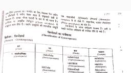
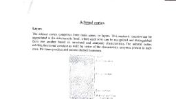
Prepared for B.Sc. Part II Botany Hons. by Prof.(Dr.) Manorma Kumari, , Lyginopteris oldhamia, Class, Order, Family, Genus, Species, , Cycadopsida, Pteridospermales, Lyginopteridaceae, Lyginopteris, L.oldhamia, , Lyginopteris is well known genus in the family Lyginopteridaceae. It was, earlier known as Calymmatotheca hoeninghausii. Williamson, Scott, Brongniart,, Binney, Potonie and Oliver and Scott described this genus in detail. It was found, in abundance in the coal ball horizon of Lancashire and Yorkshire. Various parts, of the plants were discovered and named separately. They were later found to be, different organs of the same plants Lyginopteris due to presence of similar type, of capitate gland on them (except roots). Various parts are:, Leaves, Sphenopteris hoeninghausii, Stems, Lyginopteris oldhamia, Petioles, Rachiopteris aspera, Roots, Kaloxylon hookeri, Seeds, Lagenostoma lomaxi, , Morphological Features, The stem of Lyginopteris oldhamia was long aerial and radially, symmetrical varying in diameter from 2mm to 4cm. It was frequently branched, and bore adventitious roots that were probably prop roots that supported the weak, stems. The leaves were spirally arranged and up to 0.5 metre long. The leaves, were bipinnate to tripinnate. The pinnae had pinnules or leaflets and were present, at right angles to the rachis. The rachis and petiole were studded with capitate, glands., , Lyginopteris oldhamia: A. Showing external character; B. Frond bearing pollen sac; C., Frond bearing seeds.
Page 2 :
2, , Anatomy, Stem:, The transverse sections of the stem show nearly circular outline. Outer, layer is epidermis, next to it is many layered cortex. Outer cortex contains radially, broadened fibrous strands that forms a vertical network. It provides mechanical, strength to the weak stem. The inner cortex consists of parenchymatous cells. The, pericycle present just inner to the cortex, consist of short cells and a number of, thick walled or sclerotic cells with dark contents. Five mesarch primary vascular, bundles separated by parenchymatous areas are present. The phloem is towards, the outer side in each vascular bundle and consists of irregularly arranged small, cells. Next to it, is the cambium that is well preserved in some specimens. The, xylem consists of tracheids. The protoxylem tracheids have spiral thickenings., Centripetal tracheids of the metaxylem have multiseriate bordered pits while the, centrifugal tracheids are scalariform. The pith is large and parenchymatous. Some, thick-walled cells, called sclerotic nests are scattered throughout the pith. Several, distinctly mesarch leaf traces are quite prominent. One leaf trace traverses, through the petiole and branches into two parts to supply the forking rachis., The secondary structure of the stems exhibits the presence of periderm just, inner to the two' cortical layers. The primary phloem gets crushed and inner to, this the cylinder of the secondary vascular tissue is present and it surrounds the, primary xylem. Several medullary rays are in this region. The leaf traces interrupt, the cylinder of secondary vascular tissue. Many large and small cells are, alternately present in the secondary phloem. The cambium and then secondary, xylem are present inner to the secondary phloem region. The secondary xylem, consists of large tracheids with multiseriate bordered pits arranged on the radial, walls., , (A): Lyginopteris oldhamia A.T.S., of primary stem, , (B): L.S. of Primary vascular bundle, , Leaf, The pinnules are covered with a distinct layer of upper and lower, epidermises. The upper epidermis is cutinised and the cuticle is very resistant and
Page 3 :
3, , well preserved. There are well defined palisade and spongy tissues that made up, the mesophyll tissue. The stomata are restricted to the underside., , Lyginopteris oldhamia T.S. stem showing secondary structure, , Part of T.S. Pinna of Sphenopteris hoeninghausii, , Root, The transverse section of root is roughly circular in outline. Cortex is, differentiated into outer cortex and inner cortex. Outer cortex made up of 2-3, layers of thin walled cells with scanty content. The inner cortex is 4-6 layered, whose cells were full of dense contents. These are considered to be muscilaginous, in nature. The endodermis and pericycle are clearly observed. Root possess many, primary xylem strands alternating with an equal number of phloem strands. The, xylem is exarch. The pith is scanty. The branches of root develop opposite the
Page 4 :
4, , protoxylem points. The large roots show a secondary wood. It is traversed by, large rays that always lie opposite the protoxylem groups., , T.S. Root of Lyginopteris oldhamia, , All the parts of the plant except the root are covered by numerous stalked, glandular outgrowths. These glandular outgrowths are flask-shaped and may be, up to 3mm high. They are not supplied by any vascular strand and are, therefore,, considered as mere emergences. Each outgrowth bears an apical globular head, that consists of a number of thin-walled secretory cells. It ranges in diameter from, 0.12-0.4mm. Below the secretory head is a stalk made up of several layers of, parenchyma cells. These are capitate glands., , Reproductive Structure, Lyginopteris oldhamia was heterosporous, and its ovules and seeds were, enclosed within well protective cupules. Kidston (1905) discovered the, impressions of Crossotheca in connection with the leaves of Sphenopteris, hoeninghausii and thus it was concluded that Crossotheca is the, microsporangium of Lyginopteris because the leaves of Lyginopteris were, described under the name Sphenopteris hoeninghausii. The ovules and seeds of, Lyginopteris were described under the name Lagenostoma lomaxi. Arnold (1947), stated that the microsporangiate structures of Lyginopteris belongs to Telangium., Some other workers have also shown doubts on the validity of Crossotheca being, the microsporangium of Lyginopteris.
Page 5 :
5, , Microsporangium, Crossotheca hoeninghausii has been described as the microsporangia of, Lyginopteris. Six to seven bilocular, pendant microsporangia were present on, more or less peltate fertile pinnules called microsporophylls. Each, microsporangium was about 3mm in length and its dehiscence was longitudinal., Microspores were present in the cavities of microsporangia, sometimes even in, tetrads. Each microspore had a thick rough exine and attained a diameter of 50 to, 70 micron. They have tri-radiate markings (trilete). The sporangia lack annulus, and resemble those of Cycas. There is no evidence of pollen tubes and so sperm, must have been motile., , Lyginopteris oldhamia, part of a frond bearing microsporangia, , Ovule and Seed, Olive and Scott (1903) describe Lyginopteris oldhamia seeds as, Lagenostoma lomaxii. Seed was a small, barrel-shaped structure measuring 5 to, 6mm in length and about 4mm in width. Each seed was surrounded by a cupule, present at the end of the slender rachis (Jongmans, 1930). Rachis was also, covered by capitate glands. The seeds and the cupule are known only in, petrified condition., The ovule is orthotropous with a stout integument. The integument is not, lobed and forms a single complete sheath penetrated at the apex by a narrow, micropyle. The integument is fused with the nucellus except near the micropylar, end where it is free from the nucellus and forms a canopy. This canopy part is, divided into nine loculi with a vascular bundle extending into each., The integument shows two distinct regions, the external sclerotesta which, is hard and stony and an inner fleshy endotesta. External to the integument, the, ovule is partially enclosed by a lobed cupule which is studded with numerous
 Learn better on this topic